HEAVY METAL TOLERANCE (Cr, Ag AND Hg) IN BACTERIA ISOLATED FROM SEWAGE
Agostinho A. de Lima e Silva1*, Márcia A. Ribeiro de Carvalho1, Sérgio A. L de Souza1, Patrícia M. Teixeira Dias1, Renato
G. da Silva Filho1, Carmen S. de Meirelles Saramago1, Cleonice A. de Melo Bento1, Ernesto Hofer2
1
Departamento de Microbiologia e Parasitologia, Universidade Federal do Estado do Rio de Janeiro, Rio de Janeiro, RJ, Brasil; 2
Laboratório de Zoonoses Bacterianas, Instituto Oswaldo Cruz, Rio de Janeiro, RJ, Brasil.
Submitted: September 25, 2011; Returned to authors for corrections: January 30, 2012; Approved: June 07, 2012.
ABSTRACT
Samples of sewage from a university hospital and a chemistry technical school were analysed for the
percentage of bacterial tolerance to chromium (Cr), silver (Ag) and mercury (Hg). Additionally, we
investigated the effect of these metals on pigmentation and on some enzymatic activities of the metal
tolerant strains isolated, as well as antimicrobial resistance in some metal tolerant Enterobacteriaceae
strains.Tolerance to Cr was observed mainly in Gram positive bacteria while in the case of Ag and Hg the
tolerant bacteria were predominately Gram negative. Hg was the metal for which the percentage of tolerance
was significantly higher, especially in samples from the hospital sewage (4.1%). Mercury also had the most
discernible effect on color of the colonies. Considering the effect of metals on the respiratory enzymes, one
strain of Ag-tolerant Bacillus sp. and one of Hg-tolerant P. aeruginosa were unable to produce oxidase in the presence of Ag and Hg, respectively, while the expression of gelatinase was largely inhibited in various
Gram negative strains (66% by Cr). Drug resistance in Hg-tolerant Enterobacteriaceae strains isolated from
the university hospital sewage was greater than 80%, with prevalence of multiple resistance, while the
Ag-tolerant strains from the same source showed about 34% of resistance, with the predominance of
mono-resistance. Our results showed that, despite the ability of metal tolerant strains to survive and grow in the
presence of these elements, the interactions with these metals may result in metabolic or phisiological
changes in this group of bacteria.
Key words: metal tolerance; sewage; pigment; enzymatic inhibition; antimicrobial resistance
INTRODUCTION
Heavy metals are a group of metals with density greater
than 5 g/cm3. They persist in nature and consequently tend to
accumulate in food chains. Although relatively high levels of
these elements occur in natural environment, their presence as
a contaminant in ecosystems results mainly from
anthropogenic activities (39).
Some heavy metals such as nickel, iron, copper and zinc
are essential to metabolic reactions and are required as trace
elements by the organisms. Others like mercury, silver and
cadmium have no biological role and are harmful to the
organisms, even at very low concentrations (21).
Bacteria that demonstrate the capacity of surviving in
toxic heavy metals concentrations have been isolated from
different sources (2, 6, 8, 9, 18, 26, 32).
Many bacteria have specific genetic mechanisms of
resistance to toxic metals (29, 36). In the environment metals
may select these resistant variants in a manner similar to the
selection of antibiotic resistant strains. Indeed, it is relatively
common the association of metal and antimicrobial resistance,
since both resistance genes are frequently located on the same
mobile genetic elements (12, 28). Consequently, it can be
assumed that the selective pressure exerted by heavy metals
contribute to the indirect co-selection of antibiotic resistance,
particularly in environments contaminated with the two
elements.
Concerning heavy metals, terms such as “resistance” and
“tolerance” are arbitrary and they are often used as
synonymous in literature. Gadd (15) suggested using the term
"resistance" when it is possible to characterize a specific
mechanism of bacterial detoxification for a metal. Therefore,
the term tolerance seems more appropriate to refer to the ability
of a bacterial strain to grow in the presence of high
concentrations of a metal, in all cases in which the mechanism
of this process has not been investigated.
The toxic effects of heavy metals on microorganisms are
influenced by a multitude of factors such as pH, concentration
of chelating agents, speciation, and organic matter (9, 31, 38).
The presence of those elements in the environment can result in
impacts on ecosystems, with alterations in the biomass,
diversity of microbial communities and cycling of elements
(37, 35).
Despite the large number of papers describing the action
of heavy metals on microorganisms, there are few studies on
the effects of toxic metals in the physiology of metal tolerant
bacteria, in comparison to those about their inhibitory or
deleterious effects on susceptible organisms (17, 40).
In our work we analyzed the presence of metal tolerance
(chromium, silver and mercury) in bacteria from sewage, and
some effects of heavy metals on microorganisms that are able
to survive and grow in their presence. Bacterial pigmentation
and enzymatic activities such as catalase, gelatin hydrolysis
and nitrate reduction were the characteristics chosen to study
these effects. We find it relevant to evaluate the possible
interactions between these toxic elements and tolerant bacteria
in regard to the physiological and metabolic alterations derived
from this contact, because it can be important, for example, in
situations such as in the utilization of metal tolerant
microorganisms for bioremediation of contaminated
environments. Additionally, metal tolerant Enterobacteriaceae
strains were investigated for their resistance to antimicrobial
drugs, intending to study the possible relationship between
metal tolerance and antimicrobial resistance.
This study is part of a broader investigation which aims to
obtain data about metal tolerant bacteria considering their
potential use for bioremediation, as well as about the impact
resulting from the interactions between metals and metal
tolerant bacteria. Previously we have investigated the possible
relationships between metal tolerance and the degradation of
toxic aromatic compounds (25).
MATERIALS AND METHODS
Samples
Eleven samples of sewage from a chemistry school
sewage (CSS) and nine from a university hospital sewage
(UHS) were collected, with intervals of 1 or 2 months between
each collection in the two institutions, both located in the
northern region of Rio de Janeiro city. All samples consisted of
100 mL of sewage, collected in sterile bottles and transported
in refrigeration to the laboratory.
Isolation of chromium (Cr), silver (Ag) or mercury (Hg)
tolerant bacteria
After clarifying filtration using Whatman filter paper No.
1, the sewage samples were serially diluted in
phosphate-buffered saline. For the isolation and quantification of metal
10 and 10 dilutions of each sample were inoculated on -1 -2
Nutrient Agar (Merck) containing 100 g / mL of
cycloheximide (Sigma) and one of the following metals:
K2Cr2O7 (Vetec): 60, 110, 160, 210, 260, 310, 360 g/mL;
AgNO3 (Sigma): 20, 25, 30, 35 g/mL; HgCl2 (Hoechst): 60, 80, 100, 120 g/mL. The plates were incubated at 350
C for
24h. The metal concentrations were selected according to
previous experiments (data not shown), which established the
lower concentration of each metal that determined ≥ 95%
reduction in the number of cfu/mL when compared to the total
number of cfu/mL present in the sewage samples.
This arbitrary procedure was employed to define as metal
tolerant those bacteria that were able to produce visible
colonies within 24h in the presence of the chosen metal
concentrations. In order to determine the total number of
bacteria present in each sewage sample, aliquots of 0.1 mL of
10 to 10 dilutions-2 -5 were plated on Nutrient Agar without
metal. All plates were prepared in triplicate and incubated at
35oC for 24 h. The metallic salts used were of analytical grade
and its solutions sterilized by filtration with Millipore
membranes with a 0.22 μm pore size. Some reference strains of
Gram positive and Gram negative species, with previously
determined sensitivity or tolerance to metals at the tested
concentrations, were used as controls of the metal activities:
Escherichia coli ATCC 25922, Escherichia coli K12 PCG86, Pseudomonas aeruginosa ATCC 27853, Klebsiella pneumoniae ATCC 13883, Staphylococcus aureus ATCC 29213 and Enterococcus faecalis ATCC 29212. The number of colony forming units (cfu) observed in the plates were adjusted
to cfu/mL of sewage. Cr, Ag and Hg-tolerant cfu/mL
percentages were calculated by comparison with the results
obtained in the medium without metal using the formula:
number of cfu/mL on Nutrient Agar supplemented with metal x
100 / number of cfu/mL on Nutrient Agar.
Characterization of the isolates
After counting, randomly chosen colonies from each plate
were picked and inoculated in Nutrient Broth (Merck). After
growth at 35oC for 18h the strains were streaked onto Nutrient
Agar without metal to ensure its purity and the Gram status
was determined. Then, the strains were evaluated for the ability
to grow on Nutrient Agar in the presence of the metal
concentrations detected in the primary isolation.
The Gram-negative strains were plated onto MacConkey
Agar (Merck) and inoculated in Glucose
Oxidation-Fermentation Medium. Some non-fermentative strains with
interesting characteristics, such as tolerance to high
concentrations of metals or pigment production in the presence
of metals, were identified by conventional tests and the API 20
NE identification system (bioMérieux, Marcy l’Etoile, France).
The fermentative bacteria were presumptively identified as
members of the Enterobacteriaceae family by the oxidase and
nitrate reduction tests. Conventional biochemical tests, with the
aid of BBL E/NF kit, were used to identify a set of strains
selected for antimicrobial susceptibility testing. The bacterial
isolates were maintained on Nutrient Agar slants at 4 C o for daily use, and stored in Brain Heart Infusion Broth
(BHI-Merck) with 20% glycerol at -70oC.
Determination of the effect of heavy metals on bacterial
pigmentation
After storage of samples of metal tolerant colonies for
further testing, the plates were wrapped in aluminum foil,
packed in plastic bags, and reincubated at 35 C.o After 14 days the pigmented colonies were grown in Nutrient Broth at 35 C o /24h and 10 L of cultures were placed on Nutrient Agar with
different concentrations of the respective metal. The same
media without metal was used as control.The plates were kept
under the same incubation conditions described above.Strains
that presented any degree of change on pigmentation or tone in
comparison to the controls were subjected to further
confirmatory tests, by repeating the procedure in triplicates for
each strain. A strain of P. aeruginosa was chosen for an additional test of pigment production in media containing Cr.
After growth for 5 days in minimal medium (22) containing
minimal medium without the metal the tubes were centrifuged
and the supernatants filtered with Millipore membranes with a
0.22 μm pore size. Thereafter, the filtrates were mixed with
chloroform (1:2 v/v) and the aqueous phases analyzed in
UV-visible spectrophotometer.
Effect of metals on enzymatic activity
The interference of metals on the expression of some
enzymes in metal tolerant strains was evaluated indirectly by
the catalase, oxidase, gelatin hydrolysis and nitrate reduction
tests.For each test, 30 randomily chosen strains with positive
results in the absence of the metals were analyzed. In the
oxidase tests 25 strains of Pseudomonas spp. and 5 of Bacillus spp. were analyzed while in the other tests 15 strains of
Pseudomonas spp. and 15 of Enterobacteriaceae were employed. P. aeruginosa ATCC 27853 and E. coli ATCC 25922 were used as controls in these biochemical tests. The
metal concentrations used in these tests were those that did not
show changes in the growth of the strains in comparison to the
media without metal (control). The culture media used and the
concentrations of metals were as follows: catalase and oxidase
tests - Nutrient Agar with K2Cr2O7 (60, 110, 160 g/mL);
AgNO3 (15, 20, 30 g/mL); HgCl2 (40, 60, 80 g/mL); nitrate
reduction tests - Nitrate Broth (BBL) with K2Cr2O7 (5, 10, 50 g/mL); AgNO3 (1, 5, 10 g/mL); HgCl2 (10, 20, 30 g/mL);
gelatin hydrolysis tests - Nutrient Gelatin (Merck) with
K2Cr2O7 (100, 120, 140 g/mL); AgNO3 (15, 20, 30 g/mL);
HgCl2 (20, 40, 80 g/mL). All tests were made in triplicates
and repeated at least three times. The cytocrome oxidase test
was not employed in the strains grown in the presence of
K2Cr2O7 because of the oxidative effect of this metal on the
reagent used in the test (tetramethyl-p-phenylenediamine
dihydrochloride). The conditions of inoculation, incubation and
reading of the tests were in accordance to the methods of
MacFaddin (27).
Antimicrobial susceptibility testing
Enterobacteriaceae strains that presented tolerance to
HgCl2 in concentrations ≥ 80 g/mL and to AgNO3 ≥ 30 g/mL were identified and tested for susceptibility to
antimicrobial agents by the disk diffusion method (3, 30) in
Mueller-Hinton agar (Merck). The antimicrobial agents (Cecon) used were: amikacin (30 g), chloramphenicol (30g), gentamicin (10g), sulfamethoxazole-trimethoprim (25g), tetracycline (30 g), norfloxacin (10 g), ciprofloxacin (5 g), tobramycin (10 g) and cefotaxime (30 g). Reference strains
such as E. coli ATCC 25922, S. aureus ATCC 25923 and P. aeruginosa ATCC 27853 were used as controls. The distribution of Ag and Hg-tolerant Enterobacteriaceae strains
tested for susceptibility to antimicrobial agents was as follows:
UHS - 35 Ag-tolerant strains (K. pneumoniae being the predominant specie [10 strains]) and 36 Hg-tolerant strains
(predominantly species of the genus Enterobacter [15 strains]); CSS - 32 Ag-tolerant strains (predominantly E. cloacae [21 strains]) and 33 Hg-tolerant strains (predominance of K. pneumoniae [13 strains]).
Statistical analysis
The statistical analysis of the mean values of cfu/mL of
metal tolerant bacteria from CSS and UHS was performed by a
comparison of proportions by the Student s ’ t test, with confidence levels of 5% being considered significant.
RESULTS AND DISCUSSION
Factors such as the culture media employed, growth
conditions, and incubation period, besides the various possible
forms and concentrations of metals used in the tests of
tolerance may difficult their standardization and influence the
in vitro toxicity of the metals (9, 38). Due to these facts there
are no universally accepted metal concentrations to define
bacterial tolerance or resistance.
In this context we proposed to use in this study
concentrations of each metal that determined ≥ 95% reduction
in the number of cfu/mL when compared to the total population
present in the sewage sample. Using this pattern of metal
that presented a differential behavior of metal tolerance in
comparison to the general population present in the sewage,
even though we can presume that this procedure may
underestimate the real percentage of metal tolerant bacteria.
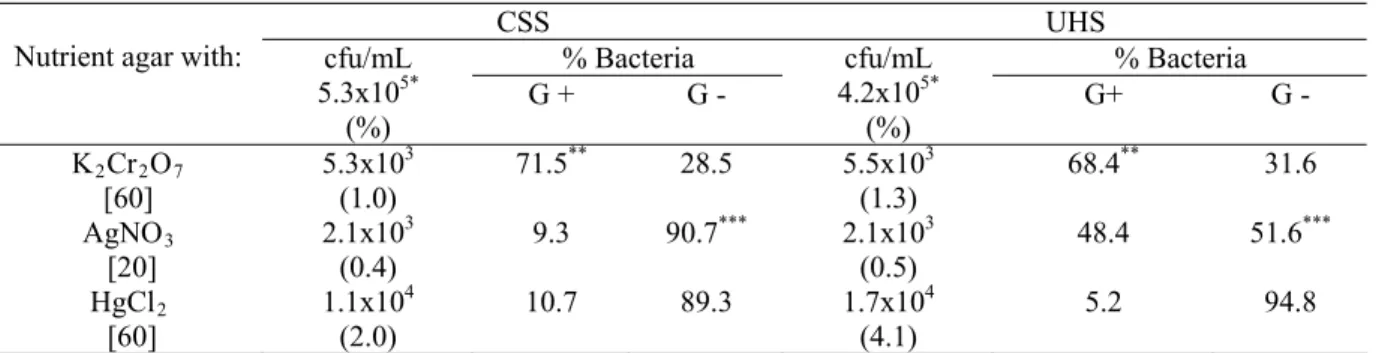
Table 1 shows the average percentage of metal tolerant
bacteria in the lowest concentrations of the metals. In the
highest concentration of each metal, colonies were still
detected, but with rates of cfu/mL lower than 0.1% (data not
shown). The average rates of cfu/mL obtained in media
containing Cr and Ag were very low in the samples from both
institutions, reflecting, perhaps, the absence of selective
pressure in these environments.
Table 1. Number of bacteria and prevalence on bacterial types obtained in the lowest concentrations of heavy metals
CSS UHS
% Bacteria % Bacteria
Nutrient agar with: cfu/mL 5.3x10⁵* (%)
G + G -
cfu/mL 4.2x10⁵* (%)
G+ G -
K2Cr2O7 [60]
5.3x10³ (1.0)
71.5** 28.5 5.5x10³
(1.3)
68.4** 31.6
AgNO3 [20]
2.1x10³ (0.4)
9.3 90.7*** 2.1x10³
(0.5)
48.4 51.6***
HgCl2 [60]
1.1x10⁴ (2.0)
10.7 89.3 1.7x10⁴
(4.1)
5.2 94.8
Abbreviations: CSS, Chemistry school; UHS, University hospital; G+, Gram positive; G-, Gram negative. [ ]: g/mL.
*mean total count of cfu/mL obtained in media without metal. **100% in the concentration of 310 g/mL of the metal. ***100% in the concentration of 35 g/mL of the metal.
The percentage of Hg-tolerant strains from both sources
were significantly higher (Student s , t test, α = 5%) when compared to the other metals tested, especially in samples from
the hospital sewage (UHS), in which the average rate of cfu/mL in the presence of 60g/mL of HgCl2 was about 4%.
This result is interesting, in view of the fact that the use of Hg
as a component of antiseptics was prohibited in Brazil since
2001 (5).
Considering that tolerance to Hg is probably due to Hg
genes
R
which are often associated with genes that confer
resistance to antimicrobial drugs (12), it is possible to admit
that the intense use of these drugs in the hospital environment
may contribute to the maintenance of genes HgR in the
bacterial population of this area.
In this study it was observed a predominance of
Gram-positive bacteria among the Cr-tolerant strains isolated from
both sewage sources in all studied concentrations of the metal (Table 1). In the concentration of 310 g/mL of the metal the
prevalence was 100% (data not shown). These results are in
accordance with those of Basu et al (2) which characterized the strains with highest tolerance to chromate as Gram-positive
bacteria.
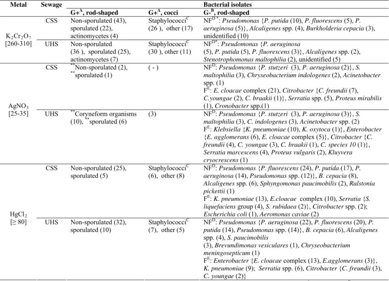
Analysing the bacterial groups isolated in the highest
concentrations of Cr, the Gram positive were predominantly
non-sporulated or sporulated rods and staphylococci, while the
Gram negative were represented exclusively by
non-fermentative microorganisms, most of them identified as
Pseudomonas spp. (Table 2).
In the case of the sewage samples collected from the
chemistry school (CSS), almost 90% of the strains that were able to grow in the presence of 20 g/mL of AgNO3 were
identified as Gram negative, in contrast to those obtained from
the hospital (UHS), in which the percentages of Gram positive
and Gram negative bacteria were similar (Table 1). Nevertheless, in the presence of 35 g/mL of this metal, 100%
of the bacteria were Gram negative in both sewage sources. It
is important to mention that in the case of Gram positive
(33).
The Gram-negative microorganisms isolated in the highest
concentrations of Ag in both sewage sources were
predominantly glucose fermentatives bacteria, which belonged
to the Enterobacteriaceae family (Table 2).
Gram negative bacteria were also predominant in the
media containing HgCl2 (Table 1). The results obtained in this
study, concerning Ag and Hg, are in accordance with other
studies that have demonstrated that Gram-negative bacteria
tend to be more tolerant to heavy metals than Gram positive(9,
10, 20). Unlike what was observed with Ag, glucose
non-fermentative Gram negative bacteria were predominant in the
highest concentrations of Hg, with a significant presence of
Pseudomonas spp. (Table 2).
The effect of heavy metals in the results of some
biochemical tests and on bacterial pigmentation was
investigated, in order to determine if the metals could induce
modifications in the physiology of tolerant bacteria, without
impairing their growth. The biochemical tests analyzed were
catalase, oxidase and nitrate reduction, which indicate
enzymatic activities involved in respiratory processes and
gelatin hydrolysis, which indicates nutritional activity.
Table 2. Distribution and identification of bacterial types in the highest concentrations of heavy metals
Abbreviations- CSS, Chemistry school; UHS, University hospital; [ ], µg/mL;( ), number of strains; ( - ), non detected; AGram positive; BGram negative; Cgrapelike clusters, catalase positive and glucose fermentative; DNF, non-fermentative; EF: fermentative; * absent at 310 µg/mL;**absent at 35 µg/mL.
Bacterial isolates Metal Sewage
G+A, rod-shaped G+A, cocci G-B, rod-shaped
CSS Non-sporulated (43), sporulated (22),
actinomycetes (4)
StaphylococciC (26 ), other (17)
NFD *: Pseudomonas {P. putida (10), P. fluorescens (5), P. aeruginosa (5)}, Alcaligenes spp. (4), Burkholderia cepacia (3), unidentified (10)
K2Cr2O7
[260-310] UHS Non-sporulated (36 ), sporulated (25), actinomycetes (7)
StaphylococciC (30 ), other (11)
NFD*: Pseudomonas {P. aeruginosa
(5), P. putida (5), P. fluorescens (3)}, Alcaligenes spp. (2),
Stenotrophomonas maltophilia (2), unidentified (5) CSS **Non-sporulated (2),
**
sporulated (1)
( - ) NFD: Pseudomonas {P. stutzeri (3), P. aeruginosa (2)}, S. maltophilia (3), Chryseobacterium indologenes (2), Acinetobacter
spp. (1)
FE: E. cloacae complex(21), Citrobacter {C. freundii (7),
C.youngae (2), C. braakii (1)}, Serratia spp. (5), Proteus mirabilis
(1), Cronobacter spp.(1) AgNO3
[25-35] UHS **Coryneform organisms (10), **sporulated (6)
(3) NFD: Pseudomonas {P. stutzeri (3), P. aeruginosa (3)}, S. maltophilia (3), C. indologenes (3), Acinetobacter spp. (2) FE: Klebsiella {K. pneumoniae (10), K. oxytoca (1)}, Enterobacter
{E. agglomerans (6), E. cloacae complex (5)}, Citrobacter {C. freundii (4), C. youngae (3), C. braakii (1), C. species 10 (1)},
Serratia marcescens (4), Proteus vulgaris (2), Kluyvera cryocrescens (1)
CSS Non-sporulated (25), sporulated (5)
StaphylococciC (6), other (8)
NFD: Pseudomonas {P. fluorescens (24), P. putida (17), P. aeruginosa (14), Pseudomonas spp.(12)}, B. cepacia (8),
Alcaligenes spp. (6), Sphyngomonas paucimobilis (2), Ralstonia pickettii (1)
FE: K. pneumoniae (13), E.cloacae complex (10), Serratia {S. liquefaciens group (4), S. rubidaea (2)}, Citrobacter spp.(2);
Escherichiacoli (1), Aeromonas caviae (2) HgCl2
[≥ 80] UHS Non-sporulated (32), sporulated (10)
StaphylococciC (7), other (5)
NFD: Pseudomonas {P. aeruginosa (22), P. fluorescens (20), P. putida (14), Pseudomonas spp.(14)}, B. cepacia (6), Alcaligenes
spp. (4), S. paucimobilis
(3), Brevundimonas vesiculares (1), Chryseobacterium meningosepticum (1)
FE: Enterobacter {E. cloacae complex (13), E.agglomerans (3)},
K. pneumoniae (9); Serratia spp. (6), Citrobacter {C. freundii (3),
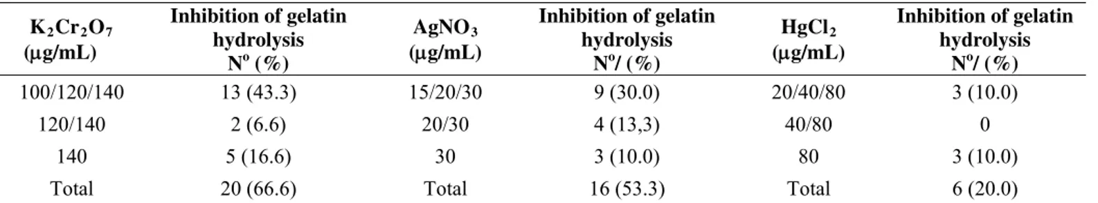
All the metals tested expressively interfered with gelatin
hydrolysis in metal tolerant strains that previously were gelatinase
positive (Table 3). Gelatin hydrolysis is a test used to determine
the ability of microorganisms to produce extracellular proteases
that degrade this substrate, a connective tissue protein that results
from the structural and chemical degradation of collagen. Some
bacteria and eucaryote organisms produce metalloproteases that
are able to degrade not only gelatin but also native collagen, and
for this reason they are called collagenases. Bacterial collagenases
may be severely inhibited by metals like Cr3+ and this
characteristic can be relevant, for example, to the stabilization of
collagen in the industrial treatment of leather (16).
Table 3. Number of Gram negative strains that presented inhibition of gelatin hydrolysis in the presence of different metal
concentrations*
K2Cr2O7 (g/mL)
Inhibition of gelatin hydrolysis
No (%)
AgNO3 (g/mL)
Inhibition of gelatin hydrolysis
No/ (%)
HgCl2 (g/mL)
Inhibition of gelatin hydrolysis
No/ (%)
100/120/140 13 (43.3) 15/20/30 9 (30.0) 20/40/80 3 (10.0)
120/140 2 (6.6) 20/30 4 (13,3) 40/80 0
140 5 (16.6) 30 3 (10.0) 80 3 (10.0)
Total 20 (66.6) Total 16 (53.3) Total 6 (20.0)
* All strains tested were positive for gelatin hydrolysis in the absence of metals
Bacterial gelatinases in the presence of metallic elements
may be affected in different manners. Pires-Bouças et al (34), studying the influence of divalent cations on the gelatinolytic
activity of Enterococcus faecalis, observed that Ca2+ and Zn2+ caused a reduction in gelatinase production, while other
common divalent cations (Fe2+ and Cu2+) inhibited it or had no
effect (Mg2+). On the other hand, considering that the authors
did not work with metal tolerant strains, it is important to
emphasize that, in the case of some of the cations tested,
gelatinase inhibition may be related to the deleterious effect of
these agents on the bacterial cell.
Kanayama and Sakai (23) observed that metals like Al, Cu
and Fe in concentrations higher than 10 mM completely
inhibited the activity of purified gelatinase, obtained from
Microbacterium liquefaciens isolated from the soil of an industrial complex of gelatin production, while Ca, Ni, Mn and
Mg slightly enhanced this enzymatic activity.
Since many gelatinases belong to the group of
metalloproteases, one possible explanation for the inhibitory
effect of heavy metals on these enzymes is that they may act by
displacing native metals from their normal binding sites (21).
The production of catalase was not affected by the
presence of heavy metals, suggesting that in tolerant bacteria
this enzyme is well protected and keeps its functional activity
in the presence of metals. This protection is important because
in the absence of this enzyme hydrogen peroxide accumulates
and may become lethal to bacteria.
The expression of oxidase was not affected by the
presence of metals in almost all tolerant strains studied, except
for one strain of Ag-tolerant Bacillus sp. that was unable to produce the enzyme in the presence of 15 and 30 g/mL of
AgNO3, and one strain of Hg-tolerant P. aeruginosa that was unable to produce the enzyme in the presence of 40 and 80 g/mL of HgCl2.
The oxidase test uses certain reagent dyes, such as
tetramethyl-p-phenylenediamine dihydrochloride, that
substitute the oxygen as artificial electron acceptors, and is
positive when bacterial cells contain cytochrome c. The negative results induced by Ag and Hg in strains of Bacillus sp. and P. aeruginosa formerly positive indicate that even though these strains are metal tolerant, the presence of these elements
may cause inactivation of some components of the respiratory
chain that determine the oxidation of the reagent that is used as
grow in the presence of the metal, these strains might have an
alternative component, additional to cytochrome c, in the final portion of the respiratory chain, which is not inactivated by the
metal and does not determine the oxidation of the reagent used
in the test.
In relation to the nitrate reduction test, no remarkable
changes were observed in the behavior of nitrate-reducing
metal tolerant bacteria, in the presence of metal. The only
exception was a strain of P. aeruginosa that in concentrations of 5 and 10 g/mL of K2Cr2O7 reduced nitrate to products
other than nitrite, as presumed by the absence of red color in
the test, even after the addition of zinc powder. But, in the presence of 50 g/mL of this metal, the production of nitrite
was detected by the production of red color, indicating that, in
this concentration, the metal impaired the reduction of nitrate
toproducts such as ammonia, molecular nitrogen, nitric oxide,
nitrous oxide and hydroxylamine.
Given the results of the biochemical tests used in this
study to evaluate the effect of metals on some enzymatic
activities of tolerant strains it is reasonable to hypothesize that
some enzymes can be affected by the presence of metals,
without any impairment in bacterial growth. So we may
suppose that the presence of metallic pollutants in the
environment, besides causing death or inhibition of susceptible
populations, can also inhibit the activity of some enzymes in
the metal tolerant populations, eliciting important changes in
the degradation of substrates and in the cycling of chemical
elements.
The presence of metals favored changes in color or hue of
some metal tolerant strains growing on solid medium. A strain
of S. aureus showed an increase in yellow pigmentation in the presence of 40 to 100 g/mL of K2Cr2 O7 , which was suppressed when the concentration increased to 160 g / mL
(Figure 1). This strain was able to grow in the presence of up to 260 g/mL of the metal.
One strain of P. aeruginosa, showing a slightly green color in media without metal, started to develop a dark green
color in the presence of 40 g / mL of K2Cr2O7 which
achieved its higher intensity in 160 g / mL, which was also
the higher concentration in which bacterial growth was
observed. Spectrophotometric tests analyzing filtrates from this
strain grown in minimal medium with and without Cr
confirmed the enhancing effect of the metal in the production
of yellow-green pigment, with maximum absorbance in the
range of 400-410 nm. Nevertheless, we could not establish a
linear relationship between the intensification of pigment
production and the different concentrations of Cr tested.
With regard to HgCl , one strain of2 Sphingomonas paucimobilis and one strain of Brevundimonas vesicularis evolved from yellow to dark brown at 60 to 100 g/mL while
an unidentified Gram-negative strain with a slight yellowish pigmentation darkened when exposed to 80 and 100 g/mL of
the same metal. Also, a Gram-positive, rod-shaped,
non-sporulated, ivory-pigmented bacterial strain developed
increasing dark color and metallic diffuse halo in the presence of 80 to 120 g/mL of HgCl2 (Figure 1).
Figure 1. Effect of heavy metals on the pigmentation of different
bacterial strains. A: S. aureus strain grown in media without metal
(control) and with K2Cr2O7. B: Gram negative rod shaped strain grown in media without metal (control) and with HgCl2. C: Gram positive rod shaped strain grown in media without metal (control)
The presence of AgNO affected only a strain of3
Chryseobacterium indologenes, which acquired a black hue when exposed to concentrations of 20 to 25 g/mL of the
metal.
Considering Ag and Hg, the concentrations of the metals
which determined the limit of bacterial growth were the same
as those in which changes in the colony colors were still noted,
suggesting that these changes may be associated to the metal
tolerance.
Bacterial pigments provide essential protection against
photooxidative damage in photosynthetic and
non-photosynthetic organisms (1). When present in low levels,
toxic metals may inhibit bacterial pigmentation (14, 19). In
some cases, the capacity to produce pigment may be directly
associated to metal tolerance. Fugimore et al (13) observed that a red pigment-deficient white mutant of Pseudomonas K-62 showed greater sensitivity to Hg2+ than the parent wild-type
reddish strain. In our study, for the strain of S. aureus that enhanced the pigment production in the presence of Cr this
association was not observed, since this strain stopped to produce pigment in the presence of 160 g / mL of this metal,
even though it was able to grow until to the concentration of 260 g / mL.
In contrast, for a strain of P. aeruginosa isolated in the present study, this association may be implied, since the degree
of pigment production showed a direct relationship with the
presence of Cr, achieving its higher intensity in the
concentration of the metal which was also the limit for the
growth of the bacteria.
With regard to Ag and Hg, the changes observed in the
colony colors are perhaps due to chemical modification of
metals when interacting with the bacteria, and not to the
induction of real pigmentation. Haefeli et al (18) observed that colonies of P. stutzeri AgR grown in medium with the metal and NaCl became dark after exposure to light, a consequence
of the formation of silver chloride, which was photo-converted
later to metallic silver.Since this study was conducted with the
plates shielded from light, it is possible that the darkening
observed in the colonies was a result of the ability of strains to
reduce Ag+ to Ago, or to precipitate the metal in its sulfide form. Similarly, the metallic brown halo observed around the
growth of the Gram positive strain when exposed to Hg may be
the result of a reaction of Hg with some substance released by
the bacteria in the environment, or bioaccumulation of the
metal, followed by its excretion into the environment in a
chemically reduced form.
In order to investigate the relationship between metal
tolerance and antimicrobial resistance we identified 67 strains
of Ag-tolerant and 69 of Hg-tolerant Enterobacteriaceae
isolated in media with high concentrations of these metals. K. pneumoniae and Enterobacter spp. were the predominant bacteria detected.
Ag-tolerant strains isolated from the UHS showed higher
overall rate of resistance to antimicrobial agents than strains
from the CSS, which was an expected result in the case of
hospital strains (Table 4). However, it is important to
emphasize that most Ag-tolerant Enterobacteriaceae strains
isolated from the hospital sewage showed susceptibility to the
drugs tested, as well as predominance of mono-resistance
profile. This result is in accordance with literature information
since the occurrence of a significant association between the
phenomenon of tolerance to Ag and antibiotic resistance has
not been reported (8,22).
It is important to consider that, differently from Hg, there
are not many reports of Ag tolerance, since this characteristic is
considered unstable and difficult to maintain and transfer (7,
33). Besides, we must consider that, in some cases, tolerance to
this metal is not a true resistance derived from genetic
expression, but a result of the production of capsular
polysaccharides, which can combine with metals and protect
the bacteria from toxicity (4).
Concerning antimicrobial resistance in Ag-tolerant strains,
the main difference between the samples obtained from the two
institutions was the multi-resistance, which was greater in
those isolated from the hospital sewage. In contrast, the
antibiotic resistance greater than 85%, with a predominance of
multi-resistance. In the Hg-tolerant strains from the CSS the
overall rate of resistance was 33.6%, a value similar to that of
Ag-tolerant strains from the hospital sewage. These results are
consistent with reports on the frequent association between
tolerance to Hg and antimicrobial resistance found in strains
isolated from different sources (22, 24, 26, 28, 41). Probably
this is due to the fact that genes that code for antibiotic
resistance and genes that code for mercury resistance are often
carried on the same plasmids or other mobile genetic elements
(41,42), favouring the occurrence of co-selective processes in
the presence of this metal or of antimicrobial drugs.
Table 4. Distribution of antimicrobial resistance in strains of metal tolerant Enterobacteriaceae
Ag-tolerant strains Source
Hg-tolerant strains Source Antimicrobial
agent UHS
[35] CSS [32] UHS [36] CSS [33] AM C TOB GEN CIP TET NFX SXT CEF 3 (8.5) 7 (20) 2 (5.7) 2 (5.7) 0 7 (20) 0 6 (17.1) 3 (8.5) 0 0 0 0 0 0 0 0 3 (9.4) 13 (36.6) 18 (50) 11 (30.5) 10 (27.7) 4 (11.1) 17 (47.2) 5 (13.8) 22 (61.1) 22 (61.1) 0 1 (3) 0 0 0 5 (15.1) 0 4 (12.1) 1 (3) AbR Total
AbR≥ 3 drugs
12 (34.2) 4 (11.4) 3 (9.4) 0 30 (83.3) 22 (61.1) 10 (33.6) 0 (0.0)
Abbreviations: AM, amikacin; C, chloramphenicol; TOB, tobramycin; GEN, gentamicin; CIP, ciprofloxacin; TET, tetracycline; NFX, norfloxacin; SXT, sulfamethoxazole-trimethoprim; CEF, cefotaxime. UHS, University Hospital Sewage; CSS, Chemistry School Sewage.
[ ], Number of strains tested ( ), Percentage of resistance AbR, Antimicrobial resistance
Analysing resistance to each antimicrobial agent in
separate, the strains of Hg-tolerant Enterobacteriaceae isolated
from the CSS were basically resistant to
trimethoprim-sulfamethoxazole and tetracycline. These results are consistent
with those obtained by Ferreira da Silva et al (11), who found a positive correlation between resistance to these drugs and Hg
tolerance in Enterobacteriaceae isolated from wastewater
treatment plant. The use of these drugs for decades probably
explains the presence of resistant strains, including those from
non-clinical origin. In the case of Ag-tolerant strains isolated
from UHS, exhibiting resistance to one or more antimicrobial
drugs, none of them was resistant to quinolones. Among the
Hg-tolerant strains from the same institution, resistance to
these drugs was present but in the lowest percentages among
all drugs tested.
In conclusion, we observed that the concentrations of Cr
employed in this study selected predominantly Gram positive
bacteria, with exclusive presence of these bacteria in the
highest concentration of the metal. In contrast, Hg and Ag
selected mainly Gram negative bacteria. These data indicate
that, depending on the pollutant metal, structural characteristics
of the cell wall may be one of the aspects determining the
survival of microorganisms in contaminated environments.
Hg tolerance was significantly higher in strains from
hospital sewage and, in contrast to Ag tolerance, this
characteristic was closely related to antimicrobial resistance
and multi-resistance in the Enterobacteriaceae tested. This
relationship suggests that the selective pressure of
antimicrobial drugs constitutes an important factor to the
persistence of Hg tolerance, considering the restriction of the
use of Hg compounds in hospitals (5).
tolerant strains with heavy metals may result in alterations on
the pigmentation, and that these alterations are not necessarily
related to the ability to survive in the presence of the metal.
Apart from this fact, we can admit that, in the environment, the
production or enhancement of pigment induced by the stress
provoked by metals on certain metal tolerant strains may
favour their survival by enhancing their resistance to
deleterious agents such as UV radiation from sun light.
Even though we did not observe inhibitory effect of
metals on catalase expression, oxidase activity was inhibited by
Hg or Ag in two metal tolerant strains, while Cr affected the
process of denitrification in one strain, inducing different final
products from nitrate degradation, depending on the
concentration of the metal. Nevertheless, in relation to
inhibition of enzymatic activity, the most intense effect
observed in our study was, doubtlessly, the loss of the capacity
to degrade gelatin in the presence of heavy metals.
Finally, considering the results obtained, we can presume
that even in the case of metal tolerant bacteria these elements
are able to determine changes in different types of metabolic
activities or physiological expressions. This fact has to be taken
into account in situations such as in the utilization of metal
tolerant microorganisms for bioremediation of contaminated
environments and in the evaluation of the impact of metals on
metal tolerant microorganisms in relation to the cycling of
chemical elements and substrate degradation.
ACKNOWLEDGEMENTS
This study was supported by FAPERJ (Fundação de
Amparo à Pesquisa do Estado do Rio de Janeiro – Support
Research Foundation of Rio de Janeiro State).
REFERENCES
1. Armstrong,G.A. (1994).Eubacteria show their true colors: genetics of carotenoid pigment biosynthesis from microbes to plants. J. Bacteriol. 176 (16), 4795-4802.
2. Basu, M.; Bhattacharya, S.; Paul, A.K. (1997). Isolation and
characterization of chromium-resistant bacteria from tannery effluents. Bull. Environ. Contam. Toxicol. 58 (4), 535-542.
3. Bauer, A.W.; Kirby, W.M.M.; Sherris, J.C.; Turck, M. (1996). Antibiotic susceptibility testing by a standardized single disc method. Amer. J. Clin. Pathol. 45, 493–496.
4. Bitton, G.; Freihofer, V. (1978). Influence of extracellular polysaccharides on the toxicity of copper and cadmium towards Klebsiella aerogenes. Microbial Ecol 4: 119-125.
5. Brasil. Resolução RE no 528, de 17 de abril de 2001. Diário Oficial [da]
República Federativa do Brasil, Brasília p.147,8 jun. 2001.
6. Castro-Silva; M. A.; Souza Lima, A. O.; Gerchenski, A. V.; Jaques, D. B.; Rodrigues, A. L.; Lima de Souza, P.; Rörig, L. R. (2003). Heavy metal resistance of microorganisms isolated from coal mining environments of Santa Catarina. Braz. J. Microbiol. (34) suppl.1. 45-47. 7. Chopra, I. (2007). The increasing use of silver-based products as
antimicrobial agents: a useful development or a cause for concern? J. Antimicrob. Chemother. 59, 587-590.
8. Choudhury, P.; Kumar, R. (1998). Multidrug- and metal-resistant strains of Klebsiella pneumoniae isolated from Penaeus monodon of the coastal waters of deltaic Sundarban. Can. J. Microbiol. 44 (2), 186-189. 9. Duxbury, T. (1986). Microbes and heavy metals: an ecological overview.
Microbiol. Sci. 3 (11), 330-333.
10. Duxbury, T.; Bicknell, B. (1983). Metal-tolerant bacterial populations from natural and metal-polluted soils. Soil. Biol. Biochem. 15 (3), 243-250.
11. Ferreira da Silva, M.; Vaz-Moreira, I.; Gonzalez-Pajuelo, M.; Nunes, O.C.; Manaia. C.M. (2007). Antimicrobial resistance patterns in Enterobacteriaceae isolated from an urban wastewater treatment plant. FEMS Microbiol. Ecol. 60 (1), 166-176.
12. Foster, T.J. (1983). Plasmid-determined resistance to antimicrobial drugs and toxic metal ions in bacteria. Microbiol. Rev. 47 (3), 361–409. 13. Fugimore, H.; Kiyono, M.; Nobuhara, K.; Pan-Hou, H. (1996). Possible
involvement of red pigments in defense against mercury in Pseudomonas K-62. FEMS Microbiol. Lett. 135 (2-3), 317-321.
14. Furman, C.R.; Owusu, V.I.; Tang, J.C. (1984). Inhibitory effect of some transition metal ions on growth and pigment formation of Serratia marcescens. Microbios 40 (159), 45-51.
15. Gadd, G.M. (1992). Metals and microorganisms: A problem of definition. FEMS Microbiol. Lett. 100 (1-3), 197-204.
16. Gayatri, R.; Rajaram, R; Ramasami, T. (2000). Inhibition of collagenase by Cr(III): its relevance to stabilization of collagen. Biochim. Biophys Acta. 1524 (2-3), 228-37.
17. Gupta, L.K.; Jindal, R.; Beri, H.K.; Chhibber, S. (1992). Virulence of silver-resistant mutant of Klebsiella pneumoniae in burn wound model. Folia Microbiol. 37 (4), 245-248.
Bacteriol. 158 (1): 389-392.
19. Hassen, A.; Saidi, N.; Cherif, M.; Boudabous, A.(1998). Effects of heavy metals on Pseudomonas aeruginosa and Bacillus thuringiensis. Bioresourse Technol. 65 (1-2), 73-82.
20. Hughes, M.N.; Poole, R.K. (1989). Metal toxicity. In: Hughes, M.N.; Poole, R.K. (eds). Metals and microorganisms. Chapman and Hall, London, p. 252-301.
21. Hughes, M.N.; Poole, R.K. (1989). The functions of metals in micro-organisms. In: Hughes, M.N.; Poole, R.K. (eds). Metals and microorganisms. Chapman and Hall, London, p. 1-38.
22. Joly, B.; Cluzel, R.; Enry, P.H.; Barjot, J. (1976). La résistance de Pseudomonas aux antibiotiques et aux métaux lourds: CMI et transferts. Ann. Microbiol. (Inst. Pasteur) 127 B, 57-68.
23. Kanayama, Y.; Sakai, Y. (2005). Purification and properties of a new type of protease produced by Microbacterium liquefaciens. Biosci. Biotechnol. Biochem. 69 (5); 916-921.
24. Lima e Silva, A.A.; Hofer, E. (1993). Resistance to antibiotics and heavy metals in Escherichia coli from marine fish. Environ. Toxicol. Water. Qual. 8 (1), 1-11.
25. Lima e Silva, A.A.; Pereira, M.P.; Silva Filho, R.G.; Hofer, E. (2007). Utilization of phenol in the presence of heavy metals by metal-tolerant nonfermentative Gram-negative bacteria isolated from wastewater. Rev. Latinoam. Microbiol. 49 (3-4), 68- 73.
26. Lima-Bittencourt, C.I.; Cursino, L.; Gonçalves-Dornelas, H.; Pontes, D.S.; Nardi, R.M.D.; Callisto, M.; Chartone-Souza, E.; Nascimento, A.M.A. (2007). Multiple antimicrobial resistance in Enterobacteriaceae isolates from pristine freshwater. Genet. Mol. Res. 6 (3), 510-521. 27. MacFaddin, J.F. (2000). Biochemical Tests for Identification of Medical
Bacteria. Willians and Wilkins, Baltimore.
28. McIntosh, D., Cunningham, M., Ji, B., Fekete, F. A., Parry, E. M., Clark, S. E., Zalinger, Z. B., Gilg, I. C., Danner, G. R., Johnson, K. A., Beattie, M., Ritchie, R. (2008). Transferable, multiple antibiotic and mercury resistance in Atlantic Canadian isolates of Aeromonas salmonicida subsp. salmonicida is associated with carriage of an IncA/C plasmid similar to the Salmonella enterica plasmid pSN254. J. Antimicrob. Chemother. 61 (6): 1221-1228.
29. Mindlin, S.; Kholodii, G.; Gorlenko, Z.; Minakhina, S.; Minakhin, L.; Kalyaeva, E.; Kopteva, A.; Petrova, M.; Yurieva, O.; Nikiforov, V. (2001). Mercury resistance transposons of Gram-negative environmental
bacteria and their classification. Res. Microbiol. 152 (9), 811-822. 30. National Committe for Clinical Laboratory Standards (2002).
Performance Standards for Antimicrobial Susceptibility Testing. 12th
Informational Supplement. NCCLS document M100. Waine, PA, USA. 31. Nwuche, C.O.; Ugoji, E.O. (2008). Effects of heavy metal pollution on
the soil microbial activity. Int. J. Environ. Sci. Tech. 5(3), 409-414. 32. Otth, L.; Solís, Gabriela; Wilson, M.; Fernández, H. (2005).
Susceptibility of Arcobacter butzleri to heavy metals. Braz. J. Microbiol. 36 (3):286-288.
33. Percival, S.L.; Bowler, P.G.; Russel, D. (2005). Bacterial resistance to silver in wound care. J. Hosp. Infect. 60, 1-7.
34. Pires-Bouças, P. D; Izumi, E.; Furlaneto-Maia, L.; Sturion, L.; Suzart, S. (2010). Effects of environmental and nutritional factors on gelatinolytic activity by Enterococcus faecalis strains isolated from clinical sources. Afr. J. Microbiol. Res. 4 (10), 969-976.
35. Roane, T.M.; Pepper, I.L. (1999). Microbial responses to environmentally toxic cadmium. Microb. Ecol. 38 (4), 358-364.
36. Silver, S.; Misra, T.K. (1988). Plasmid-mediated heavy metal resistances. Annu. Rev. Microbiol. 42, 717-743.
37. Sobolev, D.; Begonia, M.F. (2008). Effects of heavy metal contamination upon soil microbes: lead-induced changes in general and denitrifying microbial communities as evidenced by molecular markers. Int. J. Environ. Res. Public Health. 5 (5), 450-456.
38. Sterrit, R.M.; Lester, J.N. (1980). Interactions of heavy metals with bacteria. Sci. Total Environ. 14, 5-17.
39. Trevors, J.T; Oddie, K.M.; Belliveau, B.H. (1985). Metal resistance in bacteria. FEMS Microbiol. Rev.32 (1): 39-54.
40. Vaituzis, Z.; Nelson, J.D.; Wan, L.W.; Colwell, R.R. (1975). Effects of mercuric chloride on growth and morphology of selected strains of mercury-resistant bacteria. Appl. Microbiol. 29 (2), 275-286.
41. Wireman, J.; Liebert, C.A.; Smith, T.; Summers, A.O. (1997). Association of mercury resistance with antibiotic resistance in the Gram-negative fecal bacteria of primates. Appl. Environ. Microbiol. 63 (11), 4494-4503.
42. Yurieva, O.; Kholodii, G.; Minakhin, L.; Gorlenko, Z.; Kalyaeva, E.; Mindlin, S.; Nikiforov, V. (1997). Intercontinental spread of promiscuous mercury resistance transposons in environmental bacteria. Mol. Microbiol. 24 (2), 321–329.
![Table 4. Distribution of antimicrobial resistance in strains of metal tolerant Enterobacteriaceae Ag-tolerant strains Source Hg-tolerant strains Source Antimicrobial agent UHS [35] CSS [32] UHS [36] CSS [33] AM C TOB GEN CIP TET NFX SXT](https://thumb-eu.123doks.com/thumbv2/123dok_br/15799492.648438/10.892.41.829.328.595/distribution-antimicrobial-resistance-tolerant-enterobacteriaceae-tolerant-tolerant-antimicrobial.webp)
