TECHNOLOGICAL APPLICATION OF AN EXTRACELLULAR CELL LYTIC ENZYME IN XANTHAN GUM CLARIFICATION
Suresh Shastry*; Prasad M.S.
Department of Food Microbiology, Central Food Technological Research Institute, Mysore, India
Submitted: October 05, 2003; Returned to authors for corrections: September 12, 2004; Approved: March 28, 2005
ABSTRACT
An extracellular cell lytic enzyme from Pseudomonas sp. was active on heat killed cells of Xanthomonas campestris. The lytic activity caused enzymatic digestion of X.campestris xanthan gum. Digestion was effective for highly viscous native xanthan 2.0% (w/v) and 2.5% (w/v) commercial Sigma xanthan. Scanning electron microscopy and SDS-PAGE observations confirmed the cell lytic action on X.campestris cells.
Key words: Xanthan, Xanthomonas campestris, cell lytic enzyme
INTRODUCTION
Xanthan gum secreted from Xanthomonas campestris is viscous or jelly in aqueous state. The high viscosity of xanthan broth is a major problem during removal of bacterial cell biomass from the fermented broth. Centrifugation and heat treatment are either ineffective at high viscosity or lead to degradation of xanthan during isolation of cell free gum (4,11). The enzymatic lysis of bacterial cells in microbial broth is preferred for removal of cells, as structure of the gum is preserved and effective at high viscosity. The present report describes technological application of cell lytic activity against X. campestris in xanthan gum isolation.
MATERIALS AND METHODS
Microorganisms
Microbial strains of Xanthomonas campestris and
Pseudomonas spp. were local CFTRI isolates, maintained at 30ºC for 48 h on sucrose agar medium containing (g/L): sucrose 20, Na2HPO4 3, KH2PO4 2, MgSO4.7H2O 1, tryptone 5, yeast extract 1 and 2% (w/v) agar pH 7.0.
Cell lytic enzyme production
The growth medium for Pseudomonas spp. had the following composition (g/L): sucrose 30, Na2HPO4 5, KH2PO4 3, K2SO4 1,
NaCl 1, MgSO4.7H2O 0.2, CaCl2.2H2O 0.02, FeSO4.2H2O 0.002, tryptone 11, yeast extract 9, pH 7.0. The organism was grown at pH 7.0 in 500 mL Erlenmeyer flasks containing 100 mL sterilized growth medium, inoculated with 5% (v/v) overnight culture obtained at 28±2ºC on a 250 rpm rotary shaker for 18 h. The inoculated medium was incubated at 37ºC for 24 h, on a 250 rpm rotary shaker. Cell free supernatant was obtained by centrifugation at 8000 rpm for 20 min and used as crude enzyme.
Production of Xanthan gum
Xanthomonas campestris was cultivated in 500 mL Erlenmeyer flasks containing 100 ml sterilized medium composed of (g/L): sucrose 45, peptone 4, Na2HPO4 5, KH2PO4 3, MgSO4.7H2O 1, pH 7.0, and inoculated with 5%v/v overnight culture and incubated at 28±2ºC on a 250 rpm rotary shaker for 72h. The xanthan broth was pasteurized at 70ºC for 30 min. and precipitated with 2.5% (v/v) isopropanol for xanthan isolation. The precipitate was dried in an oven at 60ºC. The precipitate was dissolved in distilled water to various concentrations, referred to as hydrated Xanthan solution (CFTRI) in experiments.
Alkaline protease assay and cell lytic activity
Caseinolytic activity was determined at pH9/45ºC in 10 mM Tris-HCl buffer (9). One unit of protease activity was defined as
the amount of enzyme that released 1 µg tyrosine per min in the assay conditions.
Cell lytic activity on X. campestris cell biomass in hydrated xanthan solution was observed by decline of absorbance at 540 nm. The proteolytic activity associated with cell lytic activity was assayed by pre-incubation of crude enzyme with 5 mM PMSF for 2 h at 27ºC. The 1% (w/v) hydrated xanthan solution diluted 1:1 (v/v) with distilled water was mixed with 10% (v/v) with/without crude enzyme pre-incubated PMSF and incubated at 50ºC/pH8.0 for 3 h. Xanthan solution incubated without the enzyme was used as control.
Lipase activity
Lipolytic activity was determined at pH 7.0/37ºC in 0.1M phosphate buffer (5). One unit of lipase activity was defined as the amount of enzyme that released µmol fatty acid per min in the assay conditions.
β β β β
β1-3 Glucanase activity
β1-3 Glucanase was determined at pH 7.0/40ºC in 0.1M citrate-phosphate buffer (2). A unit of activity was defined as the amount of enzyme that released 1 µg of glucose in 20 min in the assay conditions.
Filter paper activity
Filter paper activity was determined at pH 4.8/50ºC in 0.1M citrate phosphate buffer (6). One unit of activity was defined as the amount of enzyme that released 1 µg of glucose in 60 min.
Protein determination
Protein was assayed using coomassie blue with BSA as the standard (12).
Optimization of cell lytic activity
The different dilutions of pasteurized xanthan broth were mixed with 10% (v/v) crude enzyme and incubated at pH 6.0/ 50ºC for 6h in stationary condition, to determine the minimum dilution required in highly viscous xanthan gum for effective cell lytic activity. The decline in absorbance was recorded at 540 nm (1). Hydrated xanthan (0.25% w/v, CFTRI) 2 mL was mixed with 1 mL crude enzyme at pH 6.0 and incubated at various temperatures to optimize cell lytic activity. Lytic activity was optimized at 50ºC and carried out at final pH of the enyme-xanthan mixture i.e. pH 6.0.
Application of cell lytic activity in xanthan broth and crude hydrated xanthan solution
Xanthan broth and distilled water in 1:1 (v/v) ratio were mixed with 5% (v/v) crude enzyme at pH 6.5 and incubated on 100rpm shaker incubator for 18 h at 50ºC. Commercial xanthan (Sigma, 2% and 2.5% w/v) with biomasswas hydrated and mixed with 10% (v/v) crude enzyme at pH 5.5 ± 0.5 for incubation on
100 rpm shaker incubator for 4.5 h at 50ºC. Xanthan was diluted with distilled water to a minimum of 1:3 (v/v) before measurement of absorbance, as high viscosity of xanthan broth interfered in the filling of the cuvette. The decline in absorbance was recorded at 540 nm. These experiments were performed at the final pH of the enzyme-xanthan mixture and pH adjustment was not performed.
Xanthan hydrated solution (2% w/v, CFTRI, pH8.0) mixed with 10% (v/v) crude enzyme was incubated at 50ºC for 13.5 h at stationary condition. The viscosity of xanthan solution was recorded by Brooksfield viscometer, spindle 1 (100, 50, 20 and 10 rpm) during the process of incubation. Xanthan solution was diluted with distilled water by 1:1 (v/v) before absorbance reading, as high viscosity of xanthan solution interferred in the filling of the cuvette for absorbance reading. Cell lytic activity was measured as decline in absorbance at 540 nm, due to enzymatic cell digestion of Xanthomonas campestris present in crude xanthan hydrated solution.
Detection of cell lysis by Scanning Electron Microscopy (SEM) and SDS-PAGE
The viscous fermented xanthan broth was diluted 1:5 (v/v) with distilled water and centrifuged at 10,000 rpm to pellet X. campestris cells. Harvested cells were purified by exhaustive dialysis with de-ionized water and lyophilized for storage at 4ºC. Mixture of 2 ml (0.6 mg/mL) lyophilized cell suspension and lyophilized enzyme concentrate (190U caseinolytic) was incubated for time intervals from 30-90 min at 50ºC. Treated cells were washed with 10 mM Tris-HCl buffer, pH8.0 and fixed in 2% (v/v) glutaraldehyde. Cell suspension was washed with 10 mM Tris-HCl buffer, pH8.0 and dehydrated by an acetone series. Cells were mounted on Al planchets coated with gold-palladium in a Polaron sputter coater and observed in SEM (13).
SDS-soluble membrane proteins were extracted and visualized on SDS-PAGE (10).
Cell lysis was observed on PAGE as lysis of total SDS-soluble membrane proteins. The enzymatically treated and control cells were washed with 10 mM Tris-HCl buffer pH 8.0, lyophilized, mixed with 2% (w/v) SDS in gel loading buffer and heated at 100ºC for 5 min.
RESULTS AND DISCUSSION
Cell lytic enzyme and Xanthan gum production
Enzymatic clarification of xanthan
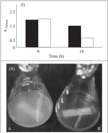
Maximum decline of absorbance of 76.7% and minimum 37.5% was observed in 1:6 and 1:1 dilutions of xanthan broth at 3h, respectively. The isolated lytic enzyme was active in fermented broth consisting of spent medium and cells (Fig. 1). Cell lytic activity in xanthan broth diluted 1:1(v/v) recorded decline of 68.3% absorbance at 18 h (Fig. 2). Clarification was effective in highly viscous 2% and 2.5% (w/v) commercial sigma xanthan as 75% and 71.5% decline in absorbance were observed respectively (Fig. 3). The present report shows that optimization of pH conditions was not a pre-requisite for enzymatic clarification of xanthan. Murofushi et al. (7) reported clarification of xanthan by an alkaline protease and lysozyme at pH 7.5-8.5, which is optimum for enzymatic action. Ernest et al. (3) reported
enzymatic clarification of xanthan by proteases from
Pellicularia sasaki or P. filamentosa in the range of pH 2-10 or 3.4-8 for optimum enzymatic action. An US Patent (8) described a method in which sodium hydroxide was added to the xanthan gum at low concentrations to adjust pH above 11 to effect clarification. These reports required pH adjustment as a pre-requisite for enzymatic clarification of xanthan, however the enzyme we isolated did not require pH optimization. Hence the scale up operation for the isolated cell lytic enzyme in xanthan clarification would be simplified.
Culture filtrate used as a source of cell lytic activity had no effect on the viscosity of xanthan broth. Xanthan solution (2% w/v, CFTRI) on clarification at the optimal condition (pH 8.0/ 50ºC) of the cell lytic enzyme recorded 64.5% decline in
Figure 2 (I).Enzymatic clarification of xanthan broth.
Control; Xanthan broth and distilled water in 1:1(v/v) ratio were mixed without crude enzyme at pH 6.5 and incubated on 100 rpm shaker incubator for 18 h at 50ºC. Test; Xanthan broth and distilled water in 1:1 (v/v) ratio was mixed with 5%v/v crude enzyme at pH 6.5 and incubated on 100 rpm shaker incubator for 18 h at 50ºC. (II) (A) Control, Xanthan broth: water (1:1v/v) composed of spent medium with cells, incubated for 18 h/50ºC. (B) Test, Xanthan broth: water (1:1v/v) incubated with crude cell lytic enzyme for 18 h/50ºC.
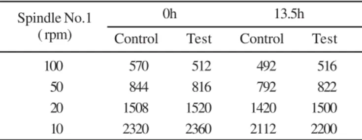
absorbance (Fig. 4). Viscosity of broth at 0h was 3040 cps and after enzymatic clarification 2960 cps, which indicated absence of xanthan gum degradation (Table 1). Xanthan (2% w/v) is gel like in appearance and its clarification at high viscosity has a potential technological application.
Role of alkaline protease in enzymatic clarification
Culture filtrate was composed of alkaline protease, lipase,
β1-3 glucanase and carboxy methyl cellulase. The PMSF inhibited 86% protease activity in the crude enzyme and its subsequent cell lytic activity recorded 18% decline in absorbance at 540 nm in hydrated xanthan solution. Lytic activity
Figure 3. (I) Enzymatic clarification of Sigma Xanthan.
Control; Commercial xanthan (Sigma, 2% and 2.5% w/v) with biomasswas hydrated and incubated at 50ºC/ pH 5.5 ± 0.5 on 100 rpm shaker incubator for 4.5 h. Test; Commercial xanthan (Sigma, 2% and 2.5% w/v) with biomasswas hydrated and incubated with 10% (v/v) crude enzyme at 50ºC/ pH 5.5 ± 0.5 on 100rpm shaker incubator for 4.5 h. (II) Sigma xanthan (2.5% v/v) clarified with crude cell lytic enzyme. (A) Control; Sigma xanthan (2.5% w/v) with biomasswas hydrated and incubated at 50ºC/ pH 5.5 ± 0.5 on 100 rpm shaker incubator for 4.5 h. (B) Test; Sigma xanthan (2.5% w/v) with biomasswas hydrated and incubated with 10% (v/v) crude enzyme at 50ºC/ pH 5.5 ± 0.5 on 100 rpm shaker incubator for 4.5 h.
Figure 4. Enzymatic clarification of the reconstituted xanthan (CFTRI) 2% w/v at optimal enzymatic conditions. Control; Xanthan hydrated solution (2% w/v,CFTRI, pH8.0) was incubated at 50ºC for 13.5 h at stationary condition. Test; Xanthan hydrated solution (2% w/v,CFTRI, pH8.0) was incubated with 10% (v/v) crude enzyme at 50ºC for 13.5 h at stationary condition.
Figure 5. Cell lytic activity with inhibited proteolytic activity of alkaline protease. Control; 1% (w/v) hydrated xanthan
solution diluted 1:1 (v/v) with distilled water was mixed incubated at 50ºC/pH8.0 for 3 h. Test; 1% (w/v) hydrated xanthan solution diluted 1:1 (v/v) with distilled water was incubated with 10% (v/v) crude enzyme at 50ºC/pH8.0 for 3 h. Proteolytic Inhibition; 1% (w/v) hydrated xanthan solution diluted 1:1 (v/v) with distilled water was incubated with 10%(v/v)crude enzyme (pre-incubated with 5mM PMSF for 2 h at 27ºC) at 50ºC/pH8.0 for 3 h.
Figure 6. Scanning electron micrograph of Xanthomonas campestris treated with crude cell lytic enzyme for 30, 60 and 90 min. a, Xanthomonas campestris cells; b1 and b2, intermediate stages of cell lysis at 30 and 60 min; c, cell debris at 90 min.
SEM and SDS-PAGE of enzymatic cell lysis
Enzymatic digestion of X. campestris cells intiated from loss of cell shape to aggregated debris as recorded in scanning electron microscopy. Enzymatic action on the cell was generalized action on the outer cell wall, as any localized action was absent. Cell debris were observed after 90 min and decline of 69% in absorbance was recorded (Fig. 6). The cell debris contributed to absorbance and as a result 100% decline in absorbance or total removal of cells during clarification was difficult to achieve.
The enzymatic action on cells was confirmed by the lysis of total SDS soluble membrane proteins. The decline in absorbance
Table 1. Viscosity measurement by Brook Field viscometer at Spindle1(100, 50, 20,10 rpm) at 50ºC, during enzymic clarification 2%(w/v) xanthan (CFTRI), pH 8.0
Spindle No.1 0h 13.5h
( rpm) Control Test Control Test
100 570 512 492 516
50 844 816 792 822
20 1508 1520 1420 1500
recorded earlier and lysis of SDS-soluble membrane proteins could be correlated as SDS-soluble membrane proteins have a role in maintenance of cell integrity (Fig. 7).
We conclude that an extracellular enzyme isolated from
Pseudomonas sp. was active on Xanthomonas campestris and could be used for X. campestris cell removal from xanthan broth in the recovery step of gum production.
RESUMO
Aplicação tecnológica de uma enzima celulolítica para clarificação de goma xantana
Uma enzima extracelular celulolítica produzida por
Pseudomonas sp. foi ativa sobre células de Xanthomonas campestris mortas pelo calor. A atividade lítica causou a digestão enzimática de goma xantana de X. campestris. A digestão foi eficiente tanto para xantana nativa altamante viscosa (2,0% w/ v) como para xantana comercial Sigma (2,5% w/v). Observações por microscopia eletrônica de varredura demonstraram a ação celulolítica sobre células de X. campestris.
Palavras-chave: xantana, Xanthomonas campestris, enzima celulolítica
REFERENCES
1. Asenjo, J.A.; Andrews, B.A.; lecorre, S. Use of a lytic enzyme system from cytophaga sp. in the lysis of Gram positive bacteria. Enzyme Microb. Tech., 7, 3-7, 1985.
2. Bull, A.T. The biochemistry of Laminarin and the nature of Laminarinase. In: Nord, F.F. (ed.): Advances in Enzymology, 28, 325-364. New York: Interscience Publishers, 1966.
3. Ernest, R.W.; Wang, J.C. Clarification of polysaccharide containing fermentation products. 1981. European Patent No. 0039962. 4. Gowthaman, M.K.; Prasad, M.S.; Karanth, N.G. Production of xanthan
gum. In: Carl A. Batt, Pradip D. Patel, (ed.). Encyclopedia of Food Microbiology, 1, 699-705. London: Academic Press 2000. 5. Kojima, Y.; Yokoe, M.; Mase, J. Purification and characterisation of
an alkaline lipase from Pseudomonas fluorescens AKL02. Biosci. Bioech. Biochem., 58(9), 1564-1568, 1994.
6. Mary Mandel, M.; Andreotti, R.; Roache, C. Measurement of saccharifying cellulose. Biotechnol. Bioeng. Symp., 6, 21-33, 1976. 7. Murofushi, K.; Hawua, T. Process for preparation of purified xanthan
gum. 1998. US Patent 5, 705, 368.
8. Patton, J.T. Modified heteropolysaccharides. 1976. US Patent No. 3.964.972.
9. Ramana Murthy, M.V.; Padmanabhan, S.; Ramakrishna, M.; Lonsane, B.K. Comparision of nine different caseinolytic assays for estimation of proteinase activity and futher improvement of the best method. Food Biotechnol., 11(1), 1-23, 1997.
10. Siekeritz and Lopez. SDS-polyacrylamide gel electrophoresis of membrane proteins: Effect of phospholipids. Anal Biochem., 53, 594-602, 1973.
11. Smith, I.H.; Garg, W.P. Recovery of microbial polysaccharides. J. Chem. Tech. Biotechnol., 32, 119-129, 1973.
12. Spector, T. Refinement of the Coomassie blue method of protein quantitation. Anal Biochem., 86, 142-146, 1978.
13. Tsujibo, H.; Miyamoto, K.; Hasehawa, T.; Yoshihiko. Purification and characterisation of two types of alkaline serine protases produced by an alkalophilic actinomycete. J. Appl. Bacteriol., 69, 520-529, 1990.
Figure 7. Enzymic action on SDS-soluble membrane proteins of Xanthomonas campestris. Lane1: Molecular mass standard corresponds to phosphorylase (97.4 KDa); Bovine serum albumin (68 KDa); Ovalbumin (43KDa); Carbonic anhydrase (29KDa); Soyabean Trypsin inhibitor (20 KDa); Lysozyme (14.3KDa); Lane 2; control; Lane 3; Test.
ACKNOWLEDGEMENTS