Clinicoradiological Session
Case 1/2010 - 15 Year-old Male Adolescent with Isolated Cleft in
Anterior Mitral Valve
Hospital Sírio Libanês, São Paulo-SP, Brazil
Edmar Atik, Patrícia O. Marques, José Lázaro Andrade
Mailing address: Edmar Atik •
InCor - Av. Dr. Enéas Carvalho de Aguiar, 44 - 05403-000 - São Paulo, SP - Brasil E-mail: conatik@incor.usp.br
Key Words
Heart defects, congenital; mitral valve / injuries; heart septal defects, atrial.
Clinical data
Patient remained asymptomatic until 5 years ago, when he presented progressively increasing fatigue on stress. Paroxysmal nocturnal dyspnea occurred twice in the last six months. Heart murmur had been heard when he was 5.
Physical examination
Eupneic, blushed and normal pulses. Weight: 53 Kg, height: 162 cm, BP: 110/80 mm Hg, Cardiac Frequency: 80 bpm. Aorta was not felt at suprasternal notch.
Chest examination showed mild impulses on the left sternal edge and ictus was palpable in the 5th intercostal space on
the midclavicular line. Heart sounds were normal and there was protomesosystolic murmur, + / + + intensity, in the mitral area, irradiating more to the left sternal edge than to the armpit.
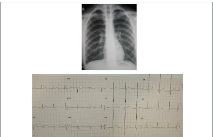
Lungs and abdomen did not present any abnormalities. Electrocardiogram revealed sinus rhythm and left ventricular hypertrophy, as well as 1st degree atrioventricular block with
PR of 0.21 mm. Ventricular repolarization was normal and there was RS morphology in V1 and R in V6. ÂQRS was at 0o,
ÂP at +30o and ÂT at +50o. No signs of left atrial overload
were found (Fig.1).
Radiographic image
Shows normal cardiac area and pulmonary vascular segment slightly increased in pulmonary hila. A slight left atrial opacity calls attention (Fig.1).
Diagnosis impression
This image is consistent with the diagnosis of heart disease that is accompanied by mitral valve injury with stenosis and/or failure, with little repercussion.
Differential diagnosis
All other acyanotic congenital heart diseases should be considered, including those with diversion of blood from left to right and the obstructive diseases, provided that these present discreet hemodynamic repercussion.
Diagnosis confirmation
Clinical elements were decisive for the diagnosis of mitral failure affecting anterior valve due to previous irradiation of the prevailing systolic murmur to the left sternal edge. The echocardiogram confirmed the existence of mitral valve defect due to full cleft on anterior valve with severe regurgitation. Increased LV with preserved function (EF-LV: 68% Ao: 27, LA: 29, LV: 58, RV: 13 mm) (Fig.2).
Management
At the operation, the full cleft on the anterior mitral valve was repaired with separate sutures and the mitral ring was plicated along the posterior commissure. As a result, mild valve regurgitation was observed in postoperative echocardiogram and decreased left ventricle (fig. 2).
Comments
The cleft on the anterior mitral valve as a congenital heart defect, even if it is isolated, is part of the context of atrioventricular septal defects. This assumption is corroborated with the presence of 1st degree atrioventricular block. This
is a very rare condition as well as the cleft on posterior mitral valve, causes of congenital mitral regurgitation. Early surgical correction by suturing the cleft edges prevents adverse developments related to left ventricular increase and dysfunction. Episodes of paroxysmal nocturnal dyspnea in this patient are related to the left atrial size close to normal size, hence its smaller complacency.
Clinico Radiological Session
Atick
Arq Bras Cardiol 2010;94 (1):138-139
Figure 1 -Chest radiography reveals normal cardiac area and increased pulmonary vascular segment on hila. Left atrium slightly opaque. Electrocardiogram reveals
PR of 0.21” with signs of left ventricular overload.
Figure 2 - Echocardiogram reveals cleft on anterior mitral valve on parasternal cross-section in A and the same defect seen on three-dimensional echocardiogram in B, both pre-operative, which is a cause of severe mitral failure on 4 camera sections in C. Proper repair of the defect is revealed in 3D, 4 days after the surgery, in D.