Mortality and Complications of Coronary Artery Bypass Grafting in
Rio de Janeiro, from 1999 to 2003
Thaís Mendonça Lips de Oliveira
1, Gláucia Maria Moraes de Oliveira
1, Carlos Henrique Klein
2, Nelson
Albuquerque de Souza e Silva
1, Paulo Henrique Godoy
1Universidade Federal do Rio de Janeiro (UFRJ)1; Fundação Oswaldo Cruz (Fiocruz)2, Rio de Janeiro, RJ - Brazil
Mailing address: Thaís Mendonça Lips de Oliveira •
Av. Ataulfo de Paiva, 944/204 - Leblon - 22440-035 - Rio de Janeiro, RJ - Brazil
E-mail: [email protected], [email protected]
Manuscript received April 29, 2009; revised manuscript received January 11, 2010; accepted February 12, 2010.
Abstract
Background: Coronary artery bypass grafting (CABG) is a consolidated procedure for the treatment of ischemic heart diseases (IHDs), which requires continuous assessment.
Objective: To assess the quality of CABG surgery by reviewing patients’ clinical characteristics, mortality rates up to one year after hospital discharge, primary causes of death and postoperative complications, at four public hospitals in Rio de Janeiro from 1999 to 2003.
Methods: CABG patient charts were randomly selected. A retrospective review was conducted to collect data on clinical characteristics, complications and deaths from patient medical charts and statements of death (SDs). Mortality rates were estimated for the hospitalization period and for up to one year after hospital discharge.
Results: The prevalence of preoperative patient characteristics were: women: 31.9%; arterial hypertension: 90.7%; dyslipidemia: 67.4%; diabetes: 37.2%; current smoking status: 22.9%; obesity: 18.3%; chronic obstructive pulmonary disease: 8.2%; prior stroke: 5.8%; extracardiac artery disease: 12.7%; elevation of creatinine levels: 4.1%; critical preoperative status: 3.7%; recent acute myocardial infarction: 23.5%; unstable angina: 40.8%; acute coronary syndrome: 50.0%; prior CABG: 2.4%; left ventricular dysfunction: 27.3%; left main coronary artery lesion: 3.9%; and associated with lesion in another system: 19.8%. In-hospital mortality rates ranged from 7.0% to 14.3%, and up to one year after hospital discharge from 8.5% to 20.2%. Ischemic heart disease (IHD) accounted for more than 80% of the deaths, and the most frequent complications after surgery were hemorrhage or post-procedural low cardiac output. Sixty percent of the patients who died had five or more complications, whereas 40% of those who survived had none.
Conclusion: Mortality and complication rates were high. Even among those patients who survived, complications were more frequent than expected. (Arq Bras Cardiol 2010; 95(3): 303-312)
Key words: Lethality; thoracic surgery; myocardial revascularization/mortality; Rio de Janeiro; Brazil.
of acute ischemic heart diseases (IHDs); 41.5% related to chronic IHDs, and 4.6% related to other diagnoses with no mention of IHD2. In-hospital CABG mortality rate was 7.8%, with a reduction from 9.2% at the beginning of the period to 5.7% at the end, and a variation of 1.9% to 11.2% among the hospitals of Rio de Janeiro state3. This long-term variation in outcomes and among hospitals indicates the need to analyze factors that might be related.
Postoperative complications can jeopardize the success of the CABG procedure. A study on complications of CABG reported that 73% of octogenarian patients, as well as 41% of the total population of the study (269 patients) experienced at least one complication4. The most frequent complications were atrial fibrillation, acute renal dysfunction, chest bleeding, need for hemotransfusion, and nosocomial infections.
We have already reported on the analysis of quality of CABG process based on data collected from patient medical charts5. The focus of this study is the quality of procedure performance, using mortality and postoperative complication rates as indicators of quality of care.
Introduction
Coronary artery bypass grafting (CABG) was introduced as a treatment for ischemic atherothrombotic coronary disease more than 40 years ago1. However, due to ongoing technological evolution of this procedure and changes introduced in the clinical treatment of the disease, a continuous assessment of the results is required by observation and critical analysis of its utilization in clinical practice
The aim of this study was to assess the quality of CABG procedures performed from 1999 to 2003 in 4 public hospitals in the city of Rio de Janeiro, by assessing baseline clinical patient characteristics, one-year mortality rates after hospital discharge, primary causes of death, and postoperative complications and the association between the latter two.
Methods
Data on CABG procedures performed from January 1999 to December 2003 were collected from patient medical charts and from authorizations for hospitalization (AIHs) at four public hospitals in the city of Rio de Janeiro. Two of these were teaching hospitals, one a federal hospital (A) and the other a state hospital (B). The other two were cardiology referral hospitals, one of them federal (C) and the other a state hospital (D).
In each one of these hospitals, CABGs were selected from the AIHs, and surgeries with valve replacement were excluded. Only the last CABG performed in each patient during the period was included; thus, each patient was selected only once5.
A decision was made to analyze randomly selected samples of charts, both of patients who had died and survivors, with a total of 150 charts per hospital. All deaths were included in the samples per hospital, except those that occurred in hospital B, in which a random selection of 75 deaths was done in order to match the 75 survivors in the sample. The selection of random samples of deaths and survivors was performed using Stata software (sample routine)6.
Data collection was performed retrospectively in 2006 by qualified researchers who where cardiologists. Groups of variables related to preoperative factors, as described in Chart 1, and postoperative complications, described in Chart 27, were selected from the data collection forms. Each variable was considered present provided it was recorded on the patient’s medical chart, with no interpretation of symptoms, signs or complementary tests by the researchers.
Information about in-hospital deaths was taken from patient medical charts and statements of death (SDs) issued by Rio de Janeiro state Health Department. The deaths that occurred after hospital discharge had already been identified by probabilistic relationship in a database of SDs and AIHs. This correlation was made in order to identify individuals who had died up to one year after post-CABG hospital discharge, using RecLink software8.
The primary cause of death was obtained by combining the information provided for the SD. According to the Training Manual for the Classification of Mortality, primary cause of death is defined as: “the disease or injury initiating the chain of events leading to the patient’s death”9. Primary causes of death have been classified in groups. The IHD group (ischemic heart diseases - I20-25) was subdivided into three subgroups: acute myocardial infarction (AMI - I21 to I23), other acute ischemic heart diseases (OutIsqAg, consisting of angina, I20, and other acute ischemic diseases, I24), and chronic ischemic heart diseases (IsqCron - I25). Other groups have also been formed consisting of the remaining causes of the circulatory system (OutApCirc - remaining part of CID-10 Chapter 1) and
Chart 1 -Deinition of preoperative factors of patients submitted to CABG at four public hospitals in the city of Rio de Janeiro, from 1999 to 2003
Preoperative factor Deinition Preoperative factor Deinition
Female Female Elevation of serum creatinine ≥ 2.3mg/dl at the preoperative stage
Age Age groups < 60 years, 60 to 64 years, 65 to 69 years, 70 to 74 years and ≥ 75 years. Preoperative critical status
Ventricular tachycardia or ibrillation detected
by electrocardiogram, or use of preoperative inotropic agents.
Diabetes
Clinical diagnosis of diabetes or ≥ 126mg/dl
fasting blood glucose at admission or use of oral hypoglycemic agents
Recent myocardial infarction Infarction < 90 days
Systemic arterial hypertension
Clinical diagnosis or systolic pressure ≥ 140
mmHg or diastolic pressure > 90 mmHg or use of antihypertensive medication
Unstable angina Recorded on the hospital admission form or as
comorbidity
Dyslipidemia levels or hypertriglyceridemia or use of medicationClinical diagnosis or elevation of Castelli I or II Acute coronary syndrome
Diagnosis of myocardial infarction up to 90 days before hospital admission or unstable angina as comorbidities recorded on the patient’s chart
Obesity Body mass index, recorded d or calculated as ≥ 30 kg/m². Prior CABG History of prior CABG
Current smoking status Smoker at admission Moderate left ventricular dysfunction 30 to 50% left ventricular ejection fraction or subjective evaluation on echocardiogram or ventriculography
Chronic obstructive
pulmonary disease Diagnosis recorded
Severe left ventricular dysfunction
30% left ventricular ejection fraction or subjective evaluation by echocardiogram or
ventriculography
Prior stroke History of stroke Left main coronary artery lesion Left main coronary artery lesion ≥ 50% detected by catheterization
Extracardiac artery disease
Diagnosis of peripheral artery disease or cerebrovascular diseases or complementary
diagnostic test
Main coronary artery lesion and other system
Left main coronary artery lesion ≥ 50% associated with lesion of vessel system ≥0%
Chart 2 -Deinition of postoperative complications in ten groups of patients undergoing CABG at four public hospitals in the city of Rio de
Janeiro, from 1999 to 2003
1. Post-procedural hemorrhage or low cardiac output:
1.1 Low cardiac output syndrome – systolic arterial pressure < 100 mmHg or need for vasoactive amines 1.2 Bleeding – bleeding requiring reoperation
1.3 Hemotransfusion – need for blood transfusion 1.4 Tamponade – diagnosis reference
1.5 Intra-aortic balloon – present if used at any moment 1.6 Circulatory assist device – present if used at any moment 2. Ischemic:
2.1 Infarction 2.2 Unstable angina 2.3 Cardiorespiratory arrest 3. Non-ischemic cardiac:
3.1 Heart failure – diagnosis of heart failure, left ventricular failure, acute pulmonary edema or pulmonary congestion
3.2 Pericarditis – diagnosis or auscultation of the pericardial knock seven days after surgery
3.3 Pulmonary embolism – diagnosis or mentioned in imaging study reports
3.4 Arrhythmia – ventricular tachycardia, ventricular ibrillation, atrial ibrillation, atrial lutter, advanced or total atrioventricular block, complete right or left bundle branch block, need for permanent or temporary cardiac pacing
4. Mechanical:
4.1 Acute mitral valve failure – clinical or echocardiographic diagnosis 4.2 Interventricular communication – clinical or echocardiographic diagnosis 4.3 Free wall rupture – clinical or echocardiographic diagnosis
5. Respiratory:
5.1 Ventilatory prosthesis for more than 24 hours 5.2 Pneumonia
5.3 Adult respiratory distress syndrome
6. Post-procedural acute renal failure - deined as the presence of serum creatinine level > 1,8mg/dl in patients whose preoperative levels were < 1,2mg/dl, or elevation > 0,5mg/dl of postoperative creatinine in those with preoperative levels of > 1,2mg/dl and < 2,0mg/dl, or elevation > 1,0mg/dl in the postoperative period of patients with preoperative serum creatinine > 2,0mg/dl, or need for postoperative dialysis7
7. Neurological:
7.1 Stroke - ischemic, hemorrhagic or embolic
7.2 Coma of any etiology 7.3 Convulsive crisis of any etiology
7.4 Psychiatric alterations - delirium, desorientarion, hallucination, psychomotor agitation in postoperative clinical course 8. Infectious:
8.1 Supericial
8.2 Soft tissues 8.3 Mediastinum 8.4 Septicemia
8.5 Septic shock
8.6 Infection in other sites 9. Peripheral vascular diseases:
9.1 Deep venous thrombosis 9.2 Amputation
9.3 Peripheral artery failure 9.4 Limb revascularization
10. Multiple organ and system failure (MOSF)
other causes of the remaining systems (Others).
Data collected were recorded in electronic files using the Epidata software.
Mortality rates were estimated for the in-hospital period up to 30, 180, and 365 days after hospital discharge, with information from AIHs and SDs combined. For death confirmation purposes, only data from patient charts or from SDs have been considered, but never from the AIH alone. In order to estimate mortality rates and other outcomes, data from each individual was weighted by the inverse of the probability of being selected for the sample. To assess the differences in distribution of variable prevalences, p values were estimated based on the effects of the sample outline of
the study. Differences associated with a p value lower than 5% were considered to be statistically significant. These analyses were performed using the Stata software6.
Results
Five hundred and forty-six hospital charts were examined, 91% of the 600 proposed for the study. Table 1 shows the prevalences of patient preoperative characteristics per hospital. Patients were predominantly over 60 years of age in all hospitals (57.8%), with inter-hospital variations, and a higher number of young patients operated at hospital C (47.1%) than at A (36.5%). Patients over 70 years of age represented 19.4% of all cases. However, no significant difference was observed in age distributions among the hospitals. Patients were predominantly men (68.1%) at all hospitals, with a 2.1-fold prevalence over females within the set of hospitals.
“Classic” cardiovascular risk factors had minor variations among hospitals. In the set of hospitals, a high prevalence of systemic arterial blood pressure (SABP) was observed (90.7%), and, in decreasing order of prevalence: dyslipidemia (67.4%), diabetes (37.2%), current smoker (22.9%), and obesity (18.3%). Acute coronary syndrome (ACS) affected half of the
patients in the set of hospitals. Among the clinical presentations of ACS, unstable angina was more frequent than AMI in three hospitals, except at hospital C, in which the prevalences of AMI and unstable angina were high and similar. It is noteworthy that hospitals C and D had the highest prevalences of recent AMI. Extracardiac artery disease was more frequent at hospitals A and C. Critical preoperative status, moderate and severe LV dysfunction, elevation of serum creatinine levels, and prior myocardial surgical revascularization were more frequent at hospital A. Left main coronary artery lesion, whether isolated or combined with injury affecting another coronary system, was more frequent at hospital D. The following preoperative factors showed statistical significance among the hospitals: arterial hypertension, extracardiac artery disease, critical preoperative status, recent AMI, unstable angina, and ACS.
Table 2 shows cumulative mortality rates per hospital up to one year after hospital discharge for patients who had undergone CABG. These rates varied among hospitals, ranging from 7.0% to 14.3% during the in-hospital period, and from
Table 1 -Estimated prevalences (%) of preoperative factors of patients undergoing CABG at four public hospitals in the city of Rio de Janeiro,
from 1999 to 2003
Preoperative factors Hospitals Total p
A B C D
Age: <60 years 36.5 44.4 47.1 38.2 42.2 0.4376
60 to 64 years 26.0 13.3 18.7 16.6 17.0 0.1781
65 to 69 years 19.2 23.0 15.0 24.8 21.4 0.3701
70 to 74 years 12.3 11.2 14.8 18.0 13.8 0.3900
>=75 years 6.1 8.1 4.3 2.4 5.6 0.1222
Female 35.2 31.8 27.2 33.7 31.9 0.2232
Diabetes 42.5 34.9 39.4 36.2 37.2 0.7004
Arterial hypertension 97.4 94.5 81.5 87.7 90.7 0.0070
Dyslipidemia 68.1 66.5 73.8 64.1 67.4 0.5817
Obesity 20.5 16.8 24.3 15.5 18.3 0.5419
Current smoking status 18.7 26.0 21.3 21.3 22.9 0.6416
Chronic obstructive pulmonary disease 5.2 5.5 8.1 13.1 8.2 0.2197
Prior stroke 7.2 5.5 9.8 3.5 5.8 0.6195
Extracardiac artery disease 24.8 6.7 24.6 10.2 12.7 0.0041
Elevation of serum creatinine levels 6.1 3.6 3.3 1.1 4.1 0.4035
Preoperative critical status 18.6 0.6 1.7 0.3 3.7 <0.0001
Recent myocardial infarction 20.8 13.3 43.1 28.3 23.5 0.0006
Unstable angina 45.9 31.6 42.3 52.0 40.8 0.0161
Acute coronary syndrome 56.4 37.4 58.2 60.2 50.0 0.0011
Prior CABG 7.2 1.7 2.1 1.3 2.4 0.1762
Ventricular dysfunction 40.5 24.8 27.8 23.8 27.3 0.1282
Moderate left ventricular dysfunction 26.7 16.6 20.8 21.3 20.0 0.5683
Severe left ventricular dysfunction 13.8 8.2 7.0 2.5 7.3 0.0522
Left main coronary artery lesion 1.5 2.6 4.0 7.0 3.9 0.0742
Table 2 -Estimated cumulative mortality rates (%) in patients undergoing CABG per period of progression and up to one year after hospital
discharge at four public hospitals in the city of Rio de Janeiro, from 1999 to 2003
Period after CABG Hospitals Total (n = 546) p
A (n = 146) B (n = 122) C (n = 144) D (n = 134)
In-hospital 14.3 13.7 7.0 7.4 10.9 <0.0001
Up to 30 days 16.0 16.5 7.0 7.4 12.3 <0.0001
Up to 180 days 19.3 19.3 9.4 8.5 14.7 <0.0001
Up to 365 days 20.2 19.3 9.4 8.5 14.9 <0.0001
8.5% to 20.2% up to one year after discharge. Significant inter-hospital differences were observed over all periods. Hospitals A and B had similar mortality rates, which was also the case of hospitals C and D. The overall in-hospital mortality rate was 10.9%, and up to one year after discharge it was 14.9%. No change in mortality was observed at hospitals C and D, from the discharge through 30 days after discharge, and from 180 days up to one year. The absolute increase in the rates recorded after discharge for up to one year were similar at hospitals A and B 5.9% and 5.6%, respectively, and lower at hospitals C and D, 2.4% and 1.1%, respectively.
Table 3 shows the distribution of relative frequencies of deaths after surgery per groups of primary causes up to one year after hospital discharge in each hospital. Groups of AMI, other acute ischemic heart diseases (OutIsqAg), and chronic ischemic diseases (IsqCron) accounted for more than 80% of the deaths in the set of hospitals. Significant differences were observed among the relative frequencies of the causes of death among the hospitals. Acute causes, AMI and other acute ischemic heart diseases (OutIsqAg) were more frequent in hospitals A and B, whereas chronic ischemic diseases (IsqCron) were more frequent in hospitals C and D, particularly in the latter. It is significant that the causes of death due to diseases affecting systems other than the circulatory system were more frequent at hospital C (15.2%).
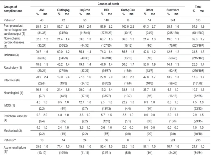
Table 4 shows the relative frequencies and the average number of postoperative complications per patient, except those who died in the operating room, per group of complications, according to primary causes of in-hospital
Tabela 3 -Relative frequencies (%) per groups of primary causes of death of patients undergoing CABG up to one year after hospital
discharge at four public hospitals in the city of Rio de Janeiro, from 1999 to 2003
Group of primary cause of death
Hospital
Total (n = 197)
A (n = 51) B (n = 65) C (n = 33) D (n = 48)
AMI 33.3 33.8 21.2 12.5 26.4
OutIsqAg 25.5 43.1 12.1 8.3 24.9
IsqCron 27.5 10.8 36.4 62.5 32.0
OutApCirc 5.9 6.2 15.2 14.6 9.6
Other 7.8 6.2 15.2 2.1 7.1
Total 100.0 100.0 100.0 100.0 100.0
p < 0.0001. AMI - acute myocardial infarction (I20-23); OutIsqAg - other acute ischemic heart diseases (I20 and I24); IsqCron - chronic ischemic heart diseases (I25); OutApCirc - other diseases of the circulatory system; Other - other causes, except ischemic heart diseases or other diseases of the circulatory system. Numbers in brackets correspond to total number of patients.
death, including those who survived after hospital discharge. All groups of postoperative complications were less frequent among the survivors than among the patients who died, except for the group of peripheral vascular diseases. The most frequent group of complications, among the patients who died and the survivors, was the hemorrhage or post-procedural low cardiac output group. It should be noted that this group includes the need for transfusions, and that among the cases of death, the average number of complications within this group was more than two complications per patient. The relative frequencies of complications in the subgroups of ischemic coronary diseases were reasonably similar, and this justified the pooling of all diseases into a single group named IHDs (ischemic heart diseases). Among these similar frequencies, exceptions were the ischemic complications, more frequent among patients who died due to a recent infarction, and the non-ischemic complications, rarer among those who died due to other acute ischemic causes. Among the patients who died in this group due to all-cause ischemic diseases (IHD), the second most frequent group of postoperative complications was that which included the diagnoses of coronary ischemia, approximately 3/4 of the cases. The IHD group of causes of death was also the only one in which mechanical complications occurred, although rare (3.6%). Non-ischemic cardiac complications were common among patients who died due to other causes of the circulatory system (66.6%) and ischemic causes (60.7%).
Table 4 -Relative frequencies (%) and average quantities (aq) of postoperative complications in patients undergoing CABG who did not die in the operating room, per groups of primary causes of in-hospital death, including the survivors, at four public hospitals in the city of Rio de Janeiro, from 1999 to 2003
Groups of complications
Causes of death
Total
% mc AMI
% mc OutIsqAg% mc % mcIsqCron % mcIHD OutApCirc% mc % mcOther Survivors% mc
Patients* 43 42 55 140 18 14 341 513
Post-procedural hemorrhage or low cardiac output (6)
88.4 2.1 85.7 2.1 89.1 2.4 87.9 2.2 100.0 2.2 64.3 2.7 38.1 1.6 54.6 1.9
(81/38) (74/36) (117/49) (272/123) (40/18) (24/9) (205/130) (541/280)
Non-ischemic cardiac diseases (4)
62.8 1.2 21.4 1.4 63.6 1.3 60.7 1.3 66.6 1.3 21.4 1.3 19.6 1.1 32.6 1.2
(33/27) (30/22) (44/35) (107/85) (16/12) (4/3) (76/67) (203/167)
Ischemic (3) 90.7 1.6 69.0 1.2 65.4 1.4 74.3 1.4 55.5 1.3 42.8 1.2 12.6 1.2 31.8 1.3
(62/39) (34/29) (49/36) (145/104) (13/10) (7/6) (50/43) (215/163)
Respiratory (3) 48.8 1.5 45.2 1.4 49.1 1.4 47.9 1.4 50.0 1.7 50.0 1.9 14.1 1.3 25.5 1.4
(29/21) (27/19) (37/27) (93/67) (15/9) (13/7) (62/48) (276/198)
Infectious (6) 20.9 2.4 19.0 2.4 27.3 1.6 22.9 2.0 33.3 2.8 42.8 1.7 13.2 1.3 17.3 1.7
(22/9) (19/8) (24/15) (65/32) (17/6) (10/6) (59/45) (151/89)
Neurological (4) 16.3 1.0 21.4 1.6 20.0 1.5 19.3 1.4 38.8 1.4 35.7 1.6 4.7 1.0 10.7 1.3
(7/7) (14/9) (17/11) (38/27) (10/7) (8/5) (16/16) (72/55)
IMOS (1) 4.6 1.0 9.5 1.0 12.7 1.0 9.3 1.0 22.2 1.0 0.3 1.0 0.3 1.0 4.5 1.0
(2/2) (4/4) (7/7) (13/13) (4/4) (1/1) (1/1) (23/23)
Peripheral vascular (4)
9.3 2.0 4.8 1.0 3.6 1.0 5.7 1.5 5.5 1.0 0.0 0.0 2.9 1.7 2.9 1.5
(8/4) (2/2) (2/2) (12/8) (1/1) (0/0) (10/6) (23/15)
Mechanical (3) 4.6 1.0 2.4 1.0 3.6 1.0 3.6 1.0 0.0 0.0 0.0 0.0 0.0 0.0 1.0 1.0
(2/2) (1/1) (2/2) (5/5) (0/0) (0/0) (0/0) (10/10)
Patients** 18 14 24 56 8 7 224 295
Acute renal failure (1)*
55.6 1.0 71.4 1.0 45.8 1.0 55.4 1.0 62.5 1.0 57.1 1.0 10.7 1.0 21.7 1.0
(10/10) (10/10) (11/11) (31/31) (5/5) (4/4) (24/24) (64/64)
AMI - acute myocardial infarction (I20-23); OutIsqAg - other acute ischemic heart diseases (I20 and I24); IsqCron - chronic ischemic heart diseases (I25); IHD - ischemic heart diseases (I20-25); OutApCirc - other diseases of the circulatory system; Other - other causes, except ischemic heart diseases or other diseases of the circulatory system. * Total number of patients who did not die in the operating room; ** Total number of patients with information about preoperative and postoperative creatinine levels. Numbers in brackets in the irst column correspond to the number of potential complications within that group of complications. Numbers in brackets in the other columns correspond to the number of complications/number of patients who experienced complications.
less frequent among survivors (14.1%). Acute renal failure was the third most common complication among patients who died due to other diseases of the circulatory system (62.5%), and the fourth most frequent complication (55.4%) in the IHD group of causes of death. Among survivors, this complication was the sixth most frequent (10.7%). The neurological complications were also present more often among patients who died due to non-ischemic causes.
Patients who died because of primary causes other than the circulatory system experienced a higher percentage of infectious complications (42.8%). Peripheral vascular complications and mechanical complications were the rarest, including in those patients who died due to IHDs.
When only those patients who had complications are taken in consideration, the average numbers of postoperative complications did not vary much among deaths and survivors. Nevertheless, the average numbers of complications related to
post-procedural hemorrhage or low cardiac output, infectious diseases and neurological diseases, were higher among those patients who died after surgery.
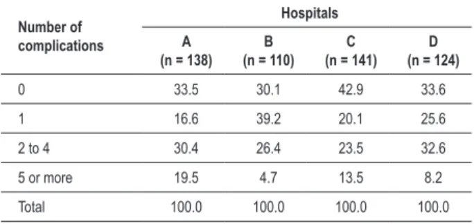
Table 5 shows the relative frequencies of the numbers of complications. There was a significant difference in the relative frequencies of the number of complications among the hospitals. The presence of two or more complications was more frequent at hospital A, in which almost half of the cases took place. The classification of hospitals in a decreasing order of two or more complications resulted in: hospital D (41.8%), C (37%), and B (31.1%). All hospitals had two or more complications in more than 30% of the cases.
Table 6 shows the significant association between the occurrence of complications and postoperative death (p
failure, critical preoperative status, prior revascularization, and lesion of the left main coronary artery or combined with other vessels. Prevalence of SABP was higher than that observed in similar studies12,13, whereas prevalences of dyslipidemia and diabetes are within those reported in literature4,12,13.
Half of the patients had a diagnosis of acute coronary syndromes (ACSs), and recent AMI was more frequent in hospitals C e D. We believe that this high prevalence of ACS reflects the recording of AMI or unstable angina in patients who already had experienced these conditions, but who were not undergoing the acute phase of the disease at admission for CABG. When the primary causes of death per hospital (Table 3) are analyzed, potential inconsistencies with the diagnoses are observed. Thus, hospitals with higher frequencies of recent AMI were those with lower frequencies of this condition as the primary cause of death. A study conducted in Rio de Janeiro public hospitals analyzed the agreement between data recorded in statements of death (SDs) and authorizations for hospitalization (AIHs), and in patient medical charts, and found that the information did not match14. A substantial disagreement was observed between the diagnoses and the IDCs recorded in patients’ charts at admission, and only 50% of the AMIs had been correctly entered with the corresponding IDC code. The in-hospital follow-up of the patient performed by different physicians in different units may result in divergent diagnoses. The rules adopted to select the primary cause of death overlook chronic diseases and those that affect older individuals, in whom death is the result of more than one condition15. It is very common that when the patient dies in the hospital, the physician who fills out and signs the statement of death is not the same professional who accompanied and/ or treated the patient. These facts entail a partial knowledge of the case, which results in an inadequate completion of the statement of death form and, consequently, incorrect selection of the primary cause of death. In our study, the causes of death classified as other diseases of the circulatory system and other causes do not include IHD as the primary cause of death. Of the 18 patients whose primary cause of death was another non-ischemic disease of the circulatory system, only 2 had IHD recorded on the statement of death form. When the primary cause was codified among the other diseases, which exclude the circulatory system, 6 statements of the 14 patients had records of IHD. We also observed that ischemic diseases were mentioned in 86% of the statements of death.
Comorbidities such as peripheral artery disease and stroke also had great variability among hospitals (from 6.7% at hospital B and 24.8% at hospital A, for peripheral atherothrombosis, and 3.5% to 9.8% in hospitals D and C, respectively, for stroke). These variances are due to different practices undertaken by the teams who ask, examine, and enter patient data into the charts. The variability in the recording of peripheral atherothrombosis and stroke could be justified by other factors of indication for surgery that could have selected patients in different stages of progression of the coronary atherothrombotic disease and, thus, with different profiles of atherothrombosis in other vascular beds.
The presence of preoperative left ventricular dysfunction was also dissimilar among the hospitals, reaching 40.5% at hospital A, nearly double the prevalence at hospital D Table 5 -Estimated relative frequencies (%) of the number of
complications in patients undergoing CABG who did not die in the operating room, at four public hospitals in the city of Rio de Janeiro, from 1999 to 2003
Number of complications
Hospitals
A
(n = 138) (n = 110)B (n = 141)C (n = 124)D
0 33.5 30.1 42.9 33.6
1 16.6 39.2 20.1 25.6
2 to 4 30.4 26.4 23.5 32.6
5 or more 19.5 4.7 13.5 8.2
Total 100.0 100.0 100.0 100.0
p = 0.0009.
Table 6 -Relative frequencies (%) of the number of complications in
patients undergoing CABG who did not die in the operating room, according to the progression to death or survival up to discharge, at four public hospitals in the city of Rio de Janeiro, from 1999 to 2003
Number of
complications Deaths (n = 158) Survivors (n = 355)
0 0.6 39.7
1 4.4 25.4
2 7.6 13.8
3 to 4 27.8 14.4
5 to 9 43.0 6.2
10 or more 16.5 0.6
Total 100.0 100.0
p =< 0.0001.
of patients who died had five or more complications, less than 7% of the survivors were affected.
Discussion
The analysis of patient medical charts provides critical data for the identification of variables that have an impact on the performance of CABG; nevertheless, its drawback lies in the quality of the records5. For the purpose of this study, we analyzed the quality of the CABG procedure as to complications and mortality up to one year after surgery at four public hospitals that performed 96% of the CABGs reimbursed by SUS in the city of Rio de Janeiro, from 1999 to 2003.
In-hospital mortality following highly complex cardiovascular procedures, including CABG, varied over time and among the institutions providing health care services3. There are accounts that in Brazil, as well as in other countries, mortality following procedures varies among hospitals and that these variances can be regional or spatial, as well as temporal10-12.
(23.8%). This fact supports the hypothesis that the patients were at different stages of atherothrombosis. Left ventricular dysfunction is associated with higher postoperative morbidity and mortality rates16-18. In a study conducted with 650 patients who had undergone CABG from July 2002 to January 2003 in the state of Rio de Janeiro, severe ventricular dysfunction was considered the second factor in order of association with mortality19. In the study on the association of preoperative factors and deaths following CABG undertaken with the same database of our study, a higher number of deaths was observed among those patients with severe left ventricular dysfunction20. Of the 23 preoperative factors analyzed, only 7 showed a significant association with the postoperative course of the patients: age, hypertension, current smoking status, dyslipidemia, stroke, ECT lesion higher than 50% combined with one or more lesions in other coronary systems, and the hospital20.
The mortality rate at the four hospitals was 10.9% during hospitalization and rose to 12.3 % up to 30 days after hospital discharge and to 14.9% within one year. University hospitals A and B had mortality rates significantly higher than reference hospitals C and D, in either one of the periods analyzed. Hospital A, the one with the highest mortality rates both in-hospital and up to one year later, had the highest prevalence of a few factors that influence surgical risk, such as LV dysfunction, presence of renal failure, diabetes mellitus, critical preoperative status, prior revascularization, and lower frequency of patients under 60 years of age. These factors must have contributed to the higher immediate postoperative mortality rate, and possibly to the worst prognosis, although other environmental factors may also have had influence. A worse clinical status may contribute to a worse diagnosis, although the aim of the surgery is to modify the course of the condition. Hospital D, which had a 7.4% mortality rate during hospitalization, similar to that of hospital C (7.0%), and the lowest increase in mortality rates within one year, had the lowest frequencies of severe preoperative prognosis characteristics (LV dysfunction, renal failure, critical status, prior revascularization). It also had the lowest preoperative rate of strokes, although it had the highest frequency of patients with left main coronary artery lesions associated with injuries in other coronary systems. These cases represented 26.4 % of the total number of operations in this hospital, against 17.1% to 19.8% at the other institutions. Thus, different criteria may have been used to select patients to undergo CABG, or there was a lack of uniformity in the information at the hospitals5. It is noteworthy that annual mortality rates over 2 to 3% in patients with stable ischemic heart disease may not represent an improvement in their prognosis, since annual mortality rates with clinical treatment range from 0.8 to 3.5%21.
In other studies conducted during the same period, in-hospital mortality rates were lower than those observed in our study. It is important, however, that mortality rates observed in random clinical trials are generally lower than those observed in clinical practice. Therefore, in a meta-analysis on CABG adverse events conducted from 1990 to 2001, mortality rates, both in-hospital and up to 30 days after surgery, were 1.7% and 2.1%, respectively22. One study conducted with 51,353 patients who had undergone CABG from 1999 and 2002
in 69 US hospitals, revealed an in-hospital mortality rate of 2.63%, representing approximately 7% of all CABG surgeries performed in that country11. The mortality rate in California, in 2005, defined as in-hospital death or up to 30 days after surgery, was 3.1%23. This can be due to differences among populations. In a meta-analysis, Nalysnyk et al22 observed a higher prevalence of men, lower rates of ventricular dysfunction, diabetes, and hypertension than in our study.22 In Iran, Karimi et al13 observed a 0.6% rate of in-hospital mortality from 2002 to 2006, among 8,890 patients submitted to CABG. This was also the case with Mack et al12, besides the lower prevalence of preoperative comorbidities, they also reported lower postoperative complication rates in all hospitals.
Clark24, from the Thoracic Surgery Society in the United States, reported on the correlation between volume and mortality of CABG: when the number of surgeries per hospital was lower than 100 procedures/year, the mortality rate was 5.0%, whereas when the number was higher, mortality was 3.2%.24 In Brazil, from 1996 to 1998, by reviewing AIHs, Noronha et al25 observed a CABG mortality rate of 7.2%. When the number of surgeries per institution was higher than 600 or lower than 151 procedures within three years, the mortality rates ranged from 5.81% to 9.00%, respectively.25
There are reports of differences in the outcomes of CABG between genders. Mortality was higher among women. Godoy et al8 reported this finding both during hospitalization and up to one year after hospital discharge in patients who had undergone surgery in the state of Rio de Janeiro. Among our patients, a subset of that group evaluated by Godoy et al8, women comprised 1/3 of the total, similar to what is reported in literature, and with no relevant differences among hospitals. Therefore, no one single factor alone can explain the differences in mortality rates.
Significant inter-hospital differences were observed regarding the primary cause of death of those patients who died up to one year after surgery. In hospitals A and B, acute ischemic causes (AMI and other acute diseases) accounted for 58.8% and 76.9% of these deaths, respectively, whereas in hospitals C and D, acute causes were just 33.3% and 20.8%, respectively, with a predominance of chronic ischemic causes (Table 3). The differences between late causes of death related to variances in surgical techniques, including type and number of bypasses used, posterior clinical treatments, and compliance with treatment, are not detected by the analysis of primary causes of death, which depends on the follow-up of patients. Besides reviewing patient clinical characteristics, mortality, and diagnoses of the primary cause of death, it is also necessary to assess postoperative complications. We observed that two or more complications affected more than 1/3 of the patients who did not die in the operating room. At hospital B, nearly 70% of the patients had at least one complication, although the frequency was lower than in those who had more complications. It is worth mentioning that this may be due to a lack of detection or poor recording, since in hospital B the quality of the information inserted in the patient charts was the worst5.
patients who died experienced five or more complications, and less than 1% had no complication. Among the survivors, 39.7% had no complication at all, and only 6.8% had five or more complications. Therefore, the record of complications is also an indicator of prognosis. A high prevalence of at least one postoperative complication (41%) was reported in another study4.
The main group of postoperative complications was the hemorrhage or low cardiac output group, which occurred in more than half of the cases, followed by the groups of non-ischemic or non-ischemic and respiratory cardiac complications, acute renal failure, infectious and neurological complications, all of them over 10%. This percentage must be underestimated, because they represent only what was recorded on the charts, but they show the need for a criterion for indicating surgery.
It could be argued that the high prevalence of hemorrhage or low cardiac output could occur due to the need for hemotransfusions. However, bleeding and surgical reexploration, associated with frequent polytransfusions, account for the increase in morbidity and mortality rates in cardiac surgery26-28. Therefore, they are important factors and must not be overlooked. Low cardiac output syndrome is not uncommon during the post-CABG period. A study evaluating 814 patients between 2002 and 2003 in São Paulo, showed that this syndrome was responsible for 54.2% of postoperative deaths29 and affected 16.1% of the patients.
A study conducted in the USA11 reported that cardiac complications, acute renal failure, and shock or hemorrhage were the most common, in 6.88%, 4.56%, and 3.41%, respectively, of all patients operated. In our study, we observed much higher relative frequencies of complications in these groups.
IDC-10, Y83 code9, which corresponds to an abnormal reaction in a patient or a late complication caused by a surgical procedure and by other surgical acts, with no mention of accidents during the intervention, should be recorded in all SDs for in-hospital deaths following CABG. However, we found it in only 57.5% of the SDs.
In a few cases of death in our study, there was a one day’s
divergence between the data recorded on the patient chart and data on the SD. To Godoy8, who did not have at hand the information from the charts, but only those abstracted from the AIHs and the SDs, these deaths apparently had occurred on the day following hospital discharge. Nevertheless, according to the information drawn from the charts, we observed that these deaths occurred during hospitalization, and were considered as in-hospital deaths.
Conclusion
This study was conducted with information taken from AIHs and SDs. Patient medical charts are expected to provide reliable information. In order to achieve this, all medical-hospital forms must be fully and correctly completed, so that actual data reflects the quality of the service provided to patients and the performance of the procedures.
Mortality and complication rates were high, and complications were frequent even among the survivors. Since mortality and complication rates were high, patients should be referred to surgery only if there are minimal levels of performance. Otherwise, the procedure would not benefit them at all. For myocardial revascularization, the in-hospital mortality or up to 30 days after the procedure in worst-prognosis cases, should be lower than 2%, better than the prognosis expected with the clinical treatment.
Potential Conflict of Interest
No potential conflict of interest relevant to this article was reported.
Sources of Funding
This study was funded by FAPERJ.
Study Association
This article is part of the thesis of master submitted by Thaís Mendonça Lips de Oliveira, from Universidade Federal do Rio de Janeiro.
References
1. Costa IA. História da cirurgia cardíaca brasileira. Rev Bras Cir Cardiovasc. 1998; 13 (1): 1-7.
2. Oliveira GMM, Klein CH, Silva NAS, Godoy PH, Fonseca TMP. Letalidade por doenças isquêmicas do coração no estado do Rio de Janeiro no período de 1999 a 2003. Arq Bras Cardiol. 2006; 86 (6): 131-7.
3. Godoy PH, Klein CH, Silva NAS, Oliveira GMM, Fonseca TMP. Letalidade na cirurgia de revascularização do miocárdio no estado do Rio de Janeiro – SIH/ SUS – no período 1999-2003. Rev SOCERJ. 2005; 18 (1): 23-9.
4. Vegni R, Almeida GF, Braga F, Freitas M, Drumond LE, Penna G, et al. Complicações após cirurgia de revascularização do miocárdio em pacientes idosos. Rev Bras Ter Intens. 2008; 20 (3): 226-34.
5. Oliveira TML, Silva NAS, Oliveira GMM, Klein CH. Qualidade de informação sobre cirurgia de revascularização do miocárdio em prontuários: o caso da abrangência - Rio de Janeiro, 1999 - 2003. Rev
SOCERJ. 2008; 21 (6): 372-81.
6. Stata Corporation; Stata – Statistics Data Analysis, version 7.0 College Station, Texas, USA, 2002.
7. Santos FO, Silveira MA, Maia RB, Monteiro MDC, Martinelli R. Insuficiência renal aguda após cirurgia de revascularização miocárdica com circulação extra-corpórea: incidência, fatores de risco e mortalidade. Arq Bras Cardiol. 2004; 83 (2): 145-9.
8. Godoy PH, Oliveira GMM, Silva NAS, Klein CH. Diferença nas taxas de letalidade e nas principais causas de óbito, entre homens e mulheres, por revascularização do miocárdio cirúrgica. Rev SOCERJ. 2008; 21 (3): 148-53.
9. Organização Mundial de Saúde. Classificação estatística internacional de doenças e problemas relacionados à saúde: Classificação Internacional de Doenças – 10a rev. São Paulo: EDUSP; 2003.
[acesso em 2008 dez 2]. Disponível em http://www.datasus.gov.br
11. Noronha SC, Martins M, Travassos C, Campos MR, Maia P, Panezzuti R. Aplicação da mortalidade hospitalar após realização de cirurgia de revascularização do miocárdio para monitoramento do cuidado hospitalar. Cad Saúde Publica. 2004; 20 (supl 2): S322-S330.
12. Mack MJ, Brown PP, Kugelmass AD, Battaglia SL, Tarkington LG, Simon AW, et al. Current status and outcomes of coronary revascularization 1999 to 2002: 148,396 surgical and percutaneous procedures. Ann Thorac Surg. 2004; 77: 761-8.
13. Karimi A, Ahmadi H, Davoovi S, Movahedi N. Factors affecting postoperative morbidity and mortality in isolated coronary artery bypass graft surgery. Surg Today. 2008; 38: 890-8.
14. Melo ECP, Travassos C, Carvalho MS. Qualidade dos dados sobre óbitos por infarto agudo do miocárdio, Rio de Janeiro. Rev Saúde Pública. 2004; 38 (3): 385-91.
15. Gaui EN, Klein CH, Oliveira GMM. Mortalidade por insuficiência cardíaca como causa básica ou contribuinte de óbito em três estados brasileiros, de 1999 a 2004. Rev SOCERJ. 2008; 21 (3): 129-37.
16. Zubiate P, Kay JH, Mendez AM. Myocardial revascularization for the patient with drastic impairment of function of the left ventricle. J Thorac Cardiovasc Surg. 1977; 73: 84-6.
17. Christakis GT, Weisel RD, Fremes SE, Ivanov J, David TE, Goldman BS, et al. Coronary artery bypass grafting in patients with poor ventricular function. Cardiovascular Surgeons of the University of Toronto. J Thorac Cardiovasc Surg. 1992; 103: 1083-91.
18. Topkara VK, Cheema FH, Kesavaramanujam S, Mercando ML, Cheema AF, Namerow PB, et al. Coronary artery bypass grafting in patients with low ejection fraction. Circulation. 2005; 112 (Suppl I): I-344–I-350.
19. Guimarães Neto JV, Romeo Filho LJM, Nunes EM. Fatores de risco para morbimortalidde hospitalar em cirurgia de revascularização do miocárdio. Rev SOCERJ. 2006; 19 (6): 487-92.
20. Carvalho MRM, Silva NAS, Oliveira GMM, Klein CH. Associação de fatores pré-operatórios e óbitos a cirurgia de revascularização do miocárdio em hospitais públicos do Rio de Janeiro: 1999-2003. Rev SOCERJ. 2008; 21 (5): 311-9.
21. Jabbour S, Young-Xu Y, Graboys TB, Blatt MC, Goldberg RJ, Bedell SE, et al. Longterm outcomes of optimized medical management of outpatients with stable coronary artery disease. Am J Cardiol. 2004; 93 (3): 294-9.
22. Nalysnyk L, Fahrbach K, Reynolds MW, Zhao SZ, Ross S. Adverse events in coronary artery bypass graft (CABG) trials: a systematic review and analysis. Heart. 2003; 89 (7): 767-72.
23. Li Z, Yeo KK, Parker JP, Mahendra G, Young JN, Amsterdam EA. Off-pump coronary artery bypass graft surgery in California, 2003 to 2005. Am Heart J. 2008; 156 (6): 1095-102.
24. Clark RE. Outcome as a function of annual coronary artery bypass graft volume. Ann Thorac Surg. 1996; 61: 21-6.
25. Noronha SC, Travassos C, Martins M, Campos MR, Maia P, Panezzuti R. Avaliação entre volume de procedimentos e a qualidade do cuidado: o caso da cirurgia coronariana no Brasil. Cad Saúde Pública. 2003; 19 (6): 1781-9.
26. Atik FA, Miana LA, Jatene FB, Auler Júnior JOC, Oliveira AS. A cirurgia de revascularização do miocárdio sem circulação extracorpórea minimiza o sangramento pós-operatório e a necessidade transfusional. Arq Bras Cardiol. 2004; 83 (4): 332-7.
27. Moulton MJ, Creswell LL, Mackey ME, Cox JL, Rosenbloom M. Reexploration for bleeding is a risk factor for adverse outcomes after cardiac operations. J Thorac Cardiovasc Surg. 1996; 111: 1037-46.
28. Unsworth-White MJ, Herriot A, Valencia O, Poloniecki J, Smith J, Murday AJ, et al. Resternotomy for bleeding after cardiac operation: a marker for increased morbidity and mortality. Ann Thorac Surg. 1995; 59 (3): 664-7.