Arq Neuropsiquiatr 2008;66(3-A):542-544
542
Clinical / Scientiic note
Primary Central nervouS SyStem lymPhoma
in an immunoComPetent hoSt
Felipe Slaviero
1, Leonardo Frighetto
2, Jairo Júnior Casali
1,
Moema Nenê Santos
3, José Ricardo Vanzin
2, Nério D. Azambuja Jr
2linFoma PrimÁrio do SiStema nervoSo Central em PaCiente imunoComPetente
Neurology and Neurosurgery Service, Hospital São Vicente de Paulo, Passo Fundo RS, Brazil: 1Medical Student, Universidade de Passo Fundo, Passo Fundo
RS, Brazil; 2Neurosurgeon, Neurology and Neurosurgery Service, Hospital São Vicente de Paulo; 3Hematologist, Hospital São Vicente de Paulo.
Received 10 March 2008, received in inal form 30 May 2008. Accepted 18 June 2008.
Dr. Felipe Slaviero – Edifício das Clínicas - Rua Teixeira Soares 885 / 10º andar - 99010-901 Passo Fundo RS - Brasil. E-mail: [email protected] Primary central nervous system lymphoma (PCNSL)
is a form of extranodal non-Hodgkin’s lymphoma, which occurs in the brain, leptomeninges, spinal cord, or eyes. PCNSL is an uncommon intracranial neoplasm, particular-ly in the immunocompetent patient population. An im-munosuppressive state is the unique established risk fac-tor for PCNSL, and the HIV-related primary lymphoma is 3600-fold more common compared with the general pop-ulation.This tumor constitutes 0.3–1.5% of all intracrani-al neoplasm in patients without AIDS1. The age-adjusted incidence in the United States has augmented 3 folds in the last three decades, passing from 0.16 per 100000 be-tween 1973–1984 to 0.48 per 100000 bebe-tween 1985–19972, although current data suggests a plateau or a slightly de-crease in the annual incidence.
The purpose of this study is to report and discuss the uncommon occurrence of PCNSL in an immunocompe-tent patient.
CaSe
The patient agreed with the publication of this study under informed consent.
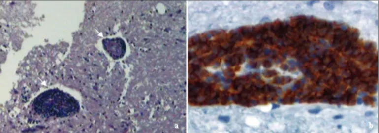
A 31-year-old caucasian woman, with no previous medical his-tory, presented with a 12 months history of personality changes, cognitive dysfunction and paroxysmal impairment of conscious-ness. Constitutional symptoms deined as “B” symptoms (loss of 10% of body weight and drenching night sweats) were present, but no fever. Neurological examination showed cognitive dysfunction, gait ataxia and absence of signs indicating intracranial hyperten-sion. There was no peripheral adenopathy or hepatosplenomegaly. Computed tomography (CT-scan) showed an expansive mass in the white matter of the left cerebral hemisphere (Fig 1A). T1-weighted magnetic resonance imaging (MRI) after gadolinium con-trast administration showed bilateral homogenous lesions (Fig 1B). HIV and hepatitis sorology were negative. Histopathological evaluation after a stereotactic biopsy of the tumor disclosed dif-fuse pleomorphic cells with large nuclei and a coarse chromatin pattern (Fig 2A). The immunohistochemical reaction was charac-teristic of diffuse large B-cell lymphoma (Fig 2B).
Further investigation with CT-scan of the chest, abdomen and pelvis, as well as bone marrow biopsy, evaluation of cerebro-spinal luid (CSF) and ophthalmologic examination were normal. An Ommaya reservoir was implanted for intraventricular chemotherapy with methotrexate (MTX) and dexamethasone.
Arq Neuropsiquiatr 2008;66(3-A)
543
Primary CNS lymphoma Slaviero et al.
Adjuvant systemic chemotherapy was performed using high-dose MTX, vincristine, procarbazine and cytarabine. The patient was submitted to fractionated whole-brain radiotherapy with 36 Gy. An eight months follow-up CT-scan showed disease control without corticosteroid therapy dependence. There was no evi-dence of neurotoxicity.
diSCuSSion
PCNSL represents 2.7% of all primary brain tumors. In 90% it is a diffuse large B-cell lymphoma. The remaining are poorly characterized low-grade lymphomas, Burkitt’s lymphoma, and T-cell lymphomas3. In immunocompetent hosts, the median age at diagnosis is 53 to 57 years, with a slight male predominance4. The classical clinical presenta-tion relates to the locapresenta-tion in the central nervous system (CNS), without “B symptoms” (weight loss, night sweats and fever). Bataille et al.5, in a study with immunocom-petent patients with PCNSL, reported that 70% had focal neurologic deicits, 43% had neuropsychiatric symptoms, 33% had increased intracranial pressure, 14% had seizures, and 4% had ocular symptoms.
Laboratory evaluation should include lactate dehy-drogenase, determination of hepatic, renal and thyroid function, HIV sorology, electrolytes analysis and reactive C protein. To determine the disease extension, a detailed ophthalmologic evaluation, CSF analysis, bone marrow biopsy, CT-scan of the chest, abdomen, and pelvis, and brain MRI are recommended. Occult extraneural disease in this screening is found in 3.9 to 12.5% of the patients6, and systemic dissemination is discovered in 10% during the course of the disease7.
If lumbar puncture can be performed safely, routine CSF analysis should be done. Pleocytosis, high protein concentrations and low glucose levels are often present, whereas the cytology typically shows clumped
pleomor-phic cells with enlarged nuclei and coarse chromatin in 26% to 31% of PCNSL patients7.
The CT-scan may reveal isodense to hyperdense im-ages. T1-weighted MRI demonstrates hypointense lesions while the T2-weighted MRI shows isointense to hyperin-tese images, secondary to the tumor’s high cell density.
Kuker et al.8, in a MRI study reported solitary lesions in two thirds of the patients. Lesions were located in the cerebral hemispheres (38%), thalamus and basal ganglia (16%), corpus callosum (14%), ventricular region (12%), and cerebellum (9%). Contrast enhancement was strong in 85% and absent in only 1% of the patients.
Stereotactic needle biopsy is the standard procedure to provide tumor specimen for histopathologic diagnosis. The role of the surgical resection alone is limited, with a median survival time of less than four months6.
The lymphomas are one of the most sensitive primary CNS tumors to a large variety of treatments. These in-clude corticosteroids, radiation therapy, chemotherapy and, combined-modality therapy.
Dexamethasone is associated with an initial response rate of 70%, usually transient. Corticosteroids should be avoided before the stereotactic procedure because it may interfere with the diagnosis.
Radiation therapy provides complete radiographic and clinical response ranging from 60% to 90% of patients treated with standard fractionation. Despite this initial response, about 90% recur within one year, with a median survival time of 10 to 18 months9.
Arq Neuropsiquiatr 2008;66(3-A)
544
Primary CNS lymphoma Slaviero et al.
response with chemotherapy alone ranges from 30% to 100% of the patients10.
Combined-modality therapy (chemotherapy plus radi-ation therapy) has achieved high response rates, improving the survival to 60 months. Because of the potential risk of severe late toxicity, which is age-related, this treatment is more indicated in young patients10. Multiagent MTX-based chemotherapy is currently the treatment of choice6. An-cillary treatment with other chemotherapeutic agents can be used, but decisions need to be made on a case-by-case basis and should be reserved to young patients without poor prognosis risk factors, as the presenting one.
Neurotoxicity manifests as ataxia, cognitive impair-ment, and incontinence. This devastating complication occurs in up to 30% of the patients and often happens in patients older than 60 years, in a mean period of 13.2 months after the treatment10.
Although currently available therapeutic regimens prolong survival, they are not curative. A complete re-sponse is deined as no brain imaging enhancing disease, corticosteroid independence, normal ophthalmologic examination, and negative CSF cytology. These purposes were successfully accomplished in this case.
Acknowledgement – We acknowledge Senair Alberto Ambros, MD for the neuroimaging descriptions and Elder Lersch, MD for the providing of histopathological images.
reFerenCeS
1. Sonstein W, Tabaddor K, Llena JF. Solitary primary CNS lymphoma: long term survival following total resection. Med Oncol 1998; 15:61-65. 2. Olson JE, Janney CA, Rao RD, et al. The continuing increase in the in-cidence of primary central nervous system non-Hodgkin lymphoma. Cancer 2002; 95:1504-1510.
3. Miller DC, Hochberg FH, Harris NI, et al. Pathology with clinical cor-relations of primary central nervous system non-Hodgkin’s lympho-ma: the Massachucetts General Hospital experience 1958-1989. Cancer 1994; 74:1383-1397.
4. Schabet M. Epidemiology of primary CNS lymphoma. J Neurooncol 1999; 43:199-201.
5. Bataille B, Delwail V, Menet E, et al. Primary intracerebral malignant lymphoma: a report of 248 cases. J Neurosurg 2000; 92:261-266.
6. Batchelor T, Loefler JS. Primary CNS lymphoma. J Clin Oncol 2006;
24:1281-1288.
7. Fitzsimmons A, Upchurch K, Batchelor TT. Clinical features and diag-nosis of primary central nervous system lymphoma. Hematol Oncol Clin N Am 2005;19:689-703.
8. Kuker W, Nagele T, Korfel A, et al. Primary central nervous system lym-phomas (PCNSL): MRI features at presentation in 100 patients. J Neu-rooncol 2005;72:169-177.
9. Nelson DF, Martz KL, Bonner H, et al. Non-Hodgkin’s lymphoma of the brain: Can high dose large volume radiation therapy improve sur-vival? Report on a prospective trial by the Radiation Therapy Oncology Group (RTOG): RTOG 8315. Int J Radiat Oncol Biol Phys 1992; 23:9-17. 10. Ferreri AJM, Abrey LE, Blay J-Y, et al. Management of primary central ner-vous system lymphoma: a summary statement from the 8th International