Brazilian Journal of Physics, vol. 35, no. 3B, September, 2005 805
Evaluation of
Escherichia Coli
Cells Damages Induced by Ultraviolet
and Proton Beam Radiation
Jaqueline Kappke1, Edilsa Rosa da Silva1, Hugo Reuters Schelin1, Sergei A. Paschuk1, Artem Pashchuk1, Analisa de Oliveira1, Nelson Carlin Filho2, Eloisa Madeira Szanto2, Jun Takahashi2, and Jairo Cavalcante de Souza2
1 Federal Center of Technological Education - CEFET/PR, Av. Sete de Setembro, 3165, CEP 80230-901, Curitiba, PR, Brazil and 2 PELLETRON Laboratory, Physics Institute of S˜ao Paulo University, Brazil
Received on 4 August, 2005
Prokaryote cells were exposed to ultra violet (UVc) radiation and to proton beams in order for the induced
effects to be studied. Morphological and physiological alterations occurred inEscherichia coli(E. coli) cells
exposed to the beams were investigated. The measurements using UVc radiation were made at the Biology Department of CEFET-PR while the measurements using proton beams were made at the Pelletron Accelerator of the Physics of the University at S˜ao Paulo. An exposition time of 3 to 15 seconds for UVc radiation and dose
ranging from 0.2 to 10.0Gyfor protons was used. A cellular survival curve versus exposition time and absorbed
dose was built for each case. After the irradiation the cells were submitted to a series of biochemical tests. It was
observed that theE.Colicells lost some basic biochemical properties when the received doses were in the range
of 0.2 to 0.7Gy. By microscopic observations it was noticed that theE.Colicells elongated after irradiation with
UVc as well as with proton beam.
I. INTRODUCTION
The prokaryotes compose an interesting group of microor-ganisms, which can be used as instruments of scientific inves-tigation. This can be explained by the fact that they possess intrinsic properties, such as reduced time of generation and relatively low cost of culture and maintenance [1], [2].
The Escherichia coli(E. coli) is a common bacterium of the intestinal tract of warm-blooded animals. It is an impor-tant biotechnological tool, which makes it possible to obtain important parameters for the metabolic and genetic characte-rization of cells of more complex organisms. For example,E. colican be used for the study of the radiation effects, which are caused in tumoral cells by therapeutical treatment [1], [2], [3].
The ionizing and not ionizing radiations can cause mutati-ons through direct or indirect action on the cellular surface. Some mutations are undesirable and even lethal, however, some can be interesting for the survival of a species [1], [2], [4].
At the present work possible transformations onE. coliare evaluated. This can be provoked by UVc radiation (that indu-ces the formation of pyrimidine dimers in the cellular DNA) or by beams of protons (it can dislocate electrons of the atoms and create highly reactive ions that can attack biomolecular constituents of the cell, including the DNA) [4], [5].
These experiments were made with UVc radiation, at the Biology Department of Federal Center of Technological Edu-cation of Parana (Curitiba, Brazil), and with beams of protons, at the Pelletron Accelerator of the Physics Department of the University of S˜ao Paulo (S˜ao Paulo, Brazil).
The occurrence of morpho-physiologic alterations in the cells ofE. coli, after-effects of irradiation and determination of the tax of cellular survival were also investigated.
II. METHODOLOGY
Cells of E. coli were cultivated in nutrient broth (pH 7.0, 36oC) for 24 hours. The cells were then centrifugated (3500r pm, 15 minutes) and resuspended in solution of NaCl 0.85%. This solution was used to make the dilutions of which 0.05mL were inoculated in Agar MacConkey, selective for gram-negative bacteria. For the irradiation with UVc radia-tion a diluradia-tion of 1 : 10000mL was used, and for the proton beam, a dilution of 1 : 1000mL was used. The Petri plates (triplicate) which contain the microorganisms in Agar Mac-Conkey were irradiated [3], [5]For ultra violet radiation, an exposition time of 3 to 15 seconds was used in a laminar flow chamber. For the radiation with proton beams the absorbed doses ranged from 0.2 to 10.0Gy.
The radiated Petri plates containing the microorganisms were cultivated during 24 hours (36oC) the number of colo-nies formed were later determined. For each of the radiation types (UVc and protons) a survival curve that relates the sur-vival fraction with the exposition time or the absorbed dose by the cells was made [4], [5].
The radiated cells were then submitted to biochemical tests and also to microscopic analysis (Gram-stain) in optical mi-croscope (magnification of 1000x). The biochemical test car-ried out was the following: Vogues-Proskauer, Indol produc-tion, descaboxilation of L-lisina, glucose fermentaproduc-tion, lac-tose fermentation, rhamnose fermentation, urea hydrolysis, H2Sproduction, citrate and ornitine [3], [5].
III. RESULTS AND DISCUSSION
ex-806 Jaqueline Kappke et al.
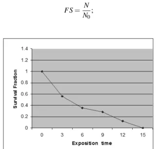
position time was calculated through equation 1 concerning survival fraction (FS), where N represents the CFU after each irradiation andN0corresponds to the CFU in plates not expo-sed to radiation [6], [7]:
FS= N N0
; (1)
FIG. 1: Survival curve of theE. colisubmitted to UVc radiation.
FIG. 2: Survival curve of theE. colisubmitted to proton radiation.
Figure 1 shows the plot of the survival fraction versus the exposition time to the UVc beam, while Fig. 2 shows the plot of the survival fraction versus the absorbed dose of the proton beams.
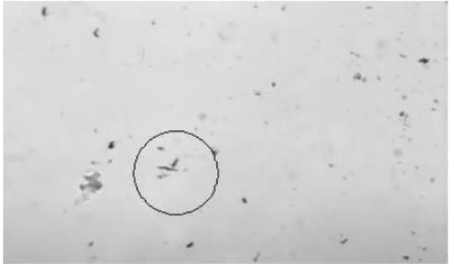
In the Gram-stain theE. coliis visible as a short and red rod [2], [3], [8] as it is shown in Fig. 3 below (E. coli not radiated). The microscopical studies showed that theE. coli cells elongated after irradiation. TheE. coli cells irradiated with UVc showed to be very elongated as can be seen in Fig. 4. As for the exposition with proton beam, it was also possible to observe that the cells were elongated as can be seen in Figs. 5 and 6.
In the literature the elongation phenomenon ofE. colicells was observed when it received a chemical treatment with cefa-lexin, cefalosphorin antibiotic of the betalactamic group. This antibiotic intervenes in the synthesis of the peptidoglycan of
FIG. 3: Gram-stain micrography of not irradiatedE. coli
(magnifica-tion of 1000x).
FIG. 4: Gram-stain micrography ofE. coliexposed to 6 seconds of
UVc radiation (magnification of 1000x).
the cell’s wall, resulting in the incapacity of the cell to divide, without provoking its death [8].
The existence of the so called hormesis phenomenon is also registered, with stimulated or benefic effect, induced by low doses of a radioactive agent. The concept of hormesis radia-tion is normally applied to radiaradia-tion doses in the range of 1 to 50Gy, which means, radiation doses that instead of provoking irreparable damage that would lead to cellular death, would provoke a radioadaptative response that would benefit the cell and its descendant [9], [10].
In the present work it was verified that theE. colicells chan-ged some characteristic biochemical properties when they re-ceived absorbed doses of 0.2, 0.4 and 0.7Gyas the loss of the capacity to descarboxilate the L-lisine aminoacid, as well as showing the cell elongated phenomenon related previously.
IV. CONCLUSIONS
With the experiments described above it was possible to conclude that:
* The 15-second exposition time with UVc radiation pro-voked a considerable reduction in theE. colipopulation;
* After the irradiation with UVc radiation, theE. colicells did not present detectable metabolic alterations through the biochemical tests used in the present work;
survi-Brazilian Journal of Physics, vol. 35, no. 3B, September, 2005 807
FIG. 5: Gram-stain micrography ofE. coliirradiated with a dose of
0.4Gywith proton beams (magnification of 1000x).
FIG. 6: Gram-stain micrography ofE. coliirradiated with a dose of
0.7Gywith proton beams (magnification of 1000x).
val fraction for the applied doses of radiation was not obser-ved. The tests should be rechecked and it would perhaps be necessary to increase the corpuscular radiation dose (proton beams) applied to theE. coli;
* The small radiation doses used (0.2, 0.4, 0.7Gy) for pro-ton beams provoked alterations in the cellular metabolism of theE. coli, detected by the loss of capacity of the bacterium in descarboxilating the l-lisine;
* TheE. coli, after exposed to a not lethal dose of radia-tion, UVc and protons, presented a change in its morphology, resulting in a well defined elongation.
Acknowledgments
The authors are very thankful to the Brazilian agencies CNPq, ANP and Fundac¸˜ao Arauc´aria for financial support.
V. REFERENCES
[1] G. J. Tortora, B. R. Funke, C. L. Case,Microbiologia. 6th ed.,
827p., Porto Alegre, Artmed, 2000.
[2] J. G. Black,Microbiologia: Fundamentos e Perspectivas. 4th
ed., 829p., Rio de Janeiro, Editora Guanabara Koogan S.A., 2002.
[3] J. Kappe, Estudo dos danos provocados emEscherichia coli
pela radiac¸˜ao eletromagn´etica. Trabalho de Diplomac¸˜ao. Curso Superior de Tecnologia em Radiologia. CEFET-PR, 2004.
[4] I. F. Heneine,Biof´ısica B´asica1st ed., p. 393, S˜ao Paulo,
Edi-tora Livraria Atheneu, 1991.
[5] A. Oliveira, J. Kappke, E. R. Silva, H. R. Schelin, M.
Soa-res, Estudo da sobrevivˆencia celular deEscherichia coliap´os a
irradiac¸˜ao ultra-violeta. Anais do I Workshop de Qu´ımica Am-biental, Curitiba, CEFET, 2003.
[6] M. F. de F. Leit˜ao, L.C.S.M. Hagler, A. N. Haglert, T.J.B.
Me-nezes, Tratado de Microbiologia, p.186, S˜ao Paulo, Editora
Menole Ltda, 1988.
[7] C. R. Silva, A. Caldeira-de-Ara´ujo, A. Amaral, M. Bernardo-Filho, Evaluation of the Cytotoxic and Mutagenic Potentiality
of Technetethium-99m inEscherichia coli, Cellular and
Mole-cular Biology TM, v. 48, 2002.
[8] M. J. Pelczar Jr, E. C. S. Chan, N. R. Krieg, Microbiologia:
Conceitos e Aplicac¸˜oes.2nd ed., S˜ao Paulo, Editora Mackron Books, 1996.
[9] S. M. J. Mortazavi, An Introduction to Radiation Hormesis. Site:
<http://www.angelfire.com/mo/radioadaptive/inthorm.html>,
access at 18/05/2004.
[10] R. Macklis, B. Bresford, Radiation Hormesis, Journal of