SUPRAPAPILLARY NEEDLE
PUNCTURE FOR COMMON
BILE DUCT ACCESS:
laboratory profi le
Everson L. A.
ARTIFON
, Paulo
SAKAI
, Guilherme Z.
CARDILLO
and Shinichi
ISHIOKA
ABSTRACT – Background - Biliary cannulation to perform endoscopic retrograde cholangiopancreatography may be diffi cult due to technical reasons and often is necessary to perform papillotomy, where complications as pancreatitis and perforation may occur. Aim - To show minimal complications by a new model of biliary access by means of the suprapapillary needle puncture and its laboratory profi le. Patients and methods - After the approval of the protocol by the Scientifi c Ethics Committee of the institution a free and informed consent was signed by all patients participating in the study. From July 2003 to August 2004, fulfi lling the inclusion and exclusion criteria, 30 patients were selected for endoscopic retrograde cholangiopancreatography, using the suprapapillary puncture technique. All patients remained hospitalized, fasting and with basal hydroelectrolytic replacement, were clinically followed up and samples for the determination of serum amylase, lipase and C-RP (C-reactive protein) were collected before and 4 h, 12 h and 24 h after the procedure and reevaluated 60 days after the procedure. Laboratory parameters were submitted to statistical study using analysis of variance for repeated measurements. Multiple comparisons were made based on Wald’s statistics. Results - The technique was successful in 93.4% (28/30) of the patients. No statistically signifi cant difference regarding to the laboratory profi le were observed. Complications related to the technique of papillary puncture occurred in 1/28 patients by not using the guide wire and in 1/28 where mild hemorrhage after dilation of the papillary fi stula occurred. Regarding complications related to therapeutic procedures, there were 2/28 retroduodenal perforations, with one (1/30) following unsuccessful puncture and another due to the passage of Dormia’s basket through the dilated fi stula path. All patients submitted to diagnostic puncture and evaluated 60 days after the procedure presented with the major duodenal papilla of normal aspect. The patients with dilation of the suprapapillary fi stula showed the fi stula continuing to drain clear bile. Conclusion - Suprapapillary puncture allows investigative and therapeutic procedures without signifi cant increases in amylase, lipase and C-RP. Patients submitted to diagnostic puncture present complete recovery of the papilla, while dilation of the fi stula maintains it pervious later on, but without complications.
HEADINGS – Cholangiopancreatography, endoscopic retrograde. Pancreatitis. Catheterization. Hemorrhage.
Work conducted at the Gastrointestinal Endoscopy Unit, “Hospital das Clínicas”, University of São Paulo, Medical School, São Paulo, SP, Brazil.
Address for correspondence: Dr. Everson L.A. Artifon – R. Castro Alves, 373 – apt.84 – Aclimação – 01532-001 – São Paulo, SP, Brazil. E-mail: eartifon@hotmail.com
ARTIGO ORIGINAL
/ ORIGINAL
ARTICLE
INTRODUCTION
Biliary cannulation by means of the transpapillary access is the initial and fundamental step to perform endoscopic retrograde cholangiopancreatography (ERCP), but on cannulation attempts of the major duodenal papilla complications such as pancreatitis, hemorrhage and perforation may occur. Acute pancreatitis after ERCP occurs in 4% to 27% of the cases and is the most frequent complication(5, 8, 11).
During attempts to cannulate the biliary duct through the papillary ostium a mechanical injury may occur leading to edema of the pancreatic duct ostium next to
the ampulla of Vater and thus its obstruction and acute pancreatitis(8). In cases of diffi cult cannulation of the biliary
duct, alternative techniques as pre-cut or fi stulotomy may be used. However, there is involvement of an electric current in these techniques which may cause thermal injury and acute pancreatitis.
In this study the authors show the laboratory profi le of a new model of biliary access(1, 2, 3) with the possibility of using
diagnostic and therapeutic procedures of minimal mechanic and thermal injury. The technique consists of the use of a catheter with a large-caliber needle in order to puncture the major duodenal papilla directed towards the common bile
PATIENTS AND METHODS
Patients
Thirty patients were selected for ERCP from July 2003 to August 2004, using the suprapapillary puncture technique, but in two patients with intradiverticular papilla the procedure could not be carried out. The protocol was authorized by the Scientifi c Ethics Committee of the institution and a free and informed consent was signed by all patients participating in the study. All procedures were performed by an experienced endoscopist. In order to perform ERCP with a minimally invasive technique patients at high risk for pancreatitis (previous history of acute biliary pancreatitis, young and female patients) and who presented a distal choledochus of at least 8 mm diameter on computerized abdominal tomography (CT-scan) were included. Patients with severe coagulopathy and/or those who refused to participate in the study, as well as those with a previous history of gastric surgery such as total gastrectomy or Billroth II gastrectomy were excluded.
All patients remained hospitalized, fasting and with basal hydroelectrolytic replacement, were clinically followed up by the staff and fellows of the Department of Gastroenterology; samples for the determination of serum amylase, lipase and C-RP (C-reactive protein) were collected before and 4 h, 12 h and 24 h after the procedure.
Complications
Pancreatitis after ERCP was characterized by a three-fold increase of the maximum value of the limit of normality, clinical fi ndings of abdominal pain, continuous nausea/vomiting up to 6 hours after the procedure and complemented by helical CT-scan with double contrast in order to evaluate the presence of pancreatic edema.
Retroperitoneal perforation was considered when there was an image of leakage of contrast medium in the retroperitoneum after injection through a false route obtained on biliary cannulation attempts.
Exteriorized blood due to hematemesis or enterorrhagia with serum hemoglobin (Hb) levels less than 8 mg/dL and needing blood transfusion characterized a hemorrhagic complication.
Accessories
A therapeutic videoduodenoscope Olympus® TJF-140 model
and papillary puncture needle catheter model Artifon® catheter
(SCITECH®,Goiânia, Goiás, Brazil) were used. This catheter
is made of polyethylene, with an 18-gauge needle covered by a fl exible metallic sheath at the distal end. Usual accessories of biliary-pancreatic manipulation including guide thread with a hydrophilic 0.025/0.0018-inch extremity, catheter, biliary balloon dilator, plastic and expandable metal stents were used.
Technique
With the duodenoscope positioned and rectifi ed at the second duodenal portion, the papillary puncture catheter is exteriorized to the duodenal lumen and in a position that allows craniolateral direction corresponding to the normal position of the biliary axis. Puncture is performed at a point
corresponding to the proximal third of the line between the transversal fold and the papillary ostium. Biliary aspiration is then performed followed by passage of the 0.025/0.0018-inch guide wire. At this moment an easy ascension of the used guide wire parallel to the spine is important. In the case this does not occur, gentle lateralization movements should be carried out with simultaneous attempts to pass the guide wire. Diagnostic procedures were considered when
the puncture occurred without suprapapillary dilation and
if its occurred with or without endobiliary procedures we considered a therapeutic procedure. In this study the number of fi ve unsuccessful biliary access attempts was considered as failure of the method. In this case classical fi stulotomy was indicated.
Injection of a medium contrast allows to obtain a cholangiogram. Therapeutic procedures included placement of plastic and metal biliary prostheses, removal of gallstone, dilation of the suprapapillary fi stula.
Late follow-up
All patients were evaluated 60 days after the procedure. Videoduodenoscopy was performed and persistence of the suprapapillary fi stula pathway was verifi ed.
During the late follow-up, duodenoscopy was complemented using cholangiopancreatography in the presence of pain with alterations in canalicular enzymes.
Statistics
The study of amylase, lipase and C-RP along the evaluations (before and 4, 12 and 24 hours after the procedures) was performed using analysis of variance for repeated measurements. Multiple comparisons were made based on Wald’s statistics. Assumption of normality of the data was analyzed with the Shapiro-Wilk test and the probability normal graph. Because of lack of normal distribution of the variables, the data were transformed using logarithmic function.
RESULTS
The results of this study show the laboratory and technical profi le of the suprapapillary puncture with special interest of the authors to characterize the microinvasive aspect of the method through the evidence of responses of pancreatic enzymes and serum markers of infl ammation. The success of the technique occurred in 93.4% (28/30) of the patients. The mean age was 47.23 years and the female/male ratio was 18/10.
Technical data
Regarding number of punctures, stratifi ed from 1 to 3 and 4 or 5, correlating the complication rate, no statistical difference
was observed (P = 0.445). Diameter of the choledochus
(8 mm–12 mm; mean 8.91 mm ± 0.72) did not present signifi cant
correlation with the number of punctures (P = 0.5635).
Presence of choledocolithiasis did not signifi cantly interfere in amylase determination and C-RP but serum lipase determination performed 24 h after the procedures presented a signifi cant
Laboratory data
Amylase - Serum determinations, ranging from 21 to 383 U/L (mean: 91.37 ± 87.18), remained within normality standards at times 4, 12 and 24 h after the procedure. But there occurred signifi cant proportional increases at 4 and 12 h (Pbefore – 4 h)/P4 h–12 - = 0,0297) (Table 1).
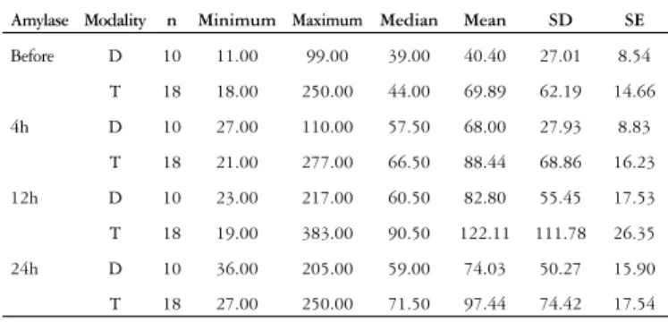
Regarding the diagnostic (D) or therapeutic (T) procedures, a statistical difference was observed only in the 12-h sample (P4-12 h = 0.04801) (Table 2).
Amylase n Minimum Maximum Median Mean SD SE IC95% Mean
Before 28 11.00 250.00 42.00 59.36 53.72 10.15 38.53 80.19
4h 28 21.00 277.00 60.50 81.14 57.84 10.93 58.72 103.57
12h 28 19.00 383.00 78.50 108.07 96.23 18.19 70.76 145.38
24h 28 27.00 250.00 61.50 89.08 66.78 12.62 63.19 114.98
TABLE 1 – Serum amylase determinations at times 4, 12 and 24 h after
the procedure
Amylase Modality n Minimum Maximum Median Mean SD SE
Before D 10 11.00 99.00 39.00 40.40 27.01 8.54
T 18 18.00 250.00 44.00 69.89 62.19 14.66
4h D 10 27.00 110.00 57.50 68.00 27.93 8.83
T 18 21.00 277.00 66.50 88.44 68.86 16.23
12h D 10 23.00 217.00 60.50 82.80 55.45 17.53
T 18 19.00 383.00 90.50 122.11 111.78 26.35
24h D 10 36.00 205.00 59.00 74.03 50.27 15.90
T 18 27.00 250.00 71.50 97.44 74.42 17.54
TABLE 2 – Serum amylase determinations at times 4, 12 and 24 h after
the procedure, diagnostic (D) or therapeutic (T)
GRAPHIC 1 – Serum determinations in amylase levels at times 4, 12 and
24 h after the procedure. As concerns, the frequency of punctures stratifi ed in 1-3 and 3 or 4, no statistical difference was observed for all times after the procedures
0 20 40 60 80 100 120 140 160 180
before 4h 12h 24h
Time
M
ean
+
ep
am
ila
se
not yes
Lipase n Minimum Maximum Median Mean SD SE IC95% Mean
Before 28 25.00 718.00 53.50 84.21 126.84 23.97 35.03 133.40
4h 28 34.00 750.00 68.50 111.79 139.75 26.41 57.60 165.98
12h 28 30.00 823.00 77.00 129.57 163.48 30.89 66.18 192.96
24h 28 27.00 628.00 73.50 107.46 117.55 22.21 61.88 153.05
TABLE 3 – Serum lipase determinations at times 4, 12 and 24 h after
the procedure
As concerns the frequency of punctures stratifi ed in 1-3 and 3 or 4, no statistical difference was observed for all times after the procedures (Graph 1).
Lipase - After the procedure, values ranged from 27 to 823 U/L (mean: 116.27 ± 121.39); proportional increase in mean lipase occurred only at 4 h (Pbefore - 4 h = 0.0004) (Table 3).
Regarding the diagnostic (D) or therapeutic (T) procedures, there was no statistical difference between the groups (Table 4). The same occurred as regards puncture frequency (Graph 2).
Lipase Modality n Minimum Maximum Median Mean SD SE
Before D 10 25.00 117.00 47.50 58.30 31.39 9.93
T 18 30.00 718.00 71.00 98.61 156.26 36.83
4h D 10 44.00 117.00 59.50 69.82 25.71 8.13
T 18 34.00 750.00 80.50 135.11 170.46 40.18
12h D 10 35.00 319.00 69.00 89.40 82.27 26.02
T 18 30.00 823.00 92.50 151.89 193.35 45.57
24h D 10 29.00 101.00 55.00 60.40 24.33 7.69
T 18 27.00 628.00 81.00 133.61 140.02 33.00
TABLE 4 – Serum determinations in lipase levels at times 4, 12 and 24 h
after the procedure, diagnostic (D) or therapeutic (T)
GRAPHIC 2 – Serum determinations in lipase levels at times 4, 12 and
24 h after the procedure. As concerns, the frequency of punctures stratifi ed in 1-3 and 3 or 4, no statistical difference was observed for all times after the procedures
0 20 40 60 80 100 120 140 160 180 200
antes 4h 12h 24h
Time
M
ean
±
ep
li
p
ase
1, 2 ou 3 4 ou 5
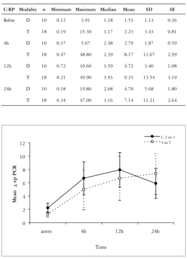
C-reactive protein - The mean of serum C-RP ranged from 0.18 to 49.99 µg/mL (mean: 6.71 ± 5.18); a signifi cant increase was observed regarding the determinations at 4 h
(P before - 4 h < 0.001) and 12 h after the procedure
(P 4-12 h = 0.0173), with a signifi cant decrease in the levels
GRAPHIC 3 – Serum determinations in C-reactive protein (C-RP) levels at times 4, 12 and 24 h after the procedure. As concerns, the frequency of punctures stratifi ed in 1-3 and 3 or 4, no statistical difference was observed for all times after the procedures
0 2 4 6 8 10 12
antes 4h 12h 24h
Time
Mean
+
ep
PCR
1, 2 ou 3 4 ou 5
C-RP n Minimum Maximum Median Mean SD SE IC95% Mean
Before 28 0.12 15.30 1.17 1.98 2.82 0.53 0.88 3.07
4h 28 0.37 48.80 2.39 6.25 10.45 1.97 2.20 10.30
12h 28 0.21 49.90 3.93 7.62 11.14 2.11 3.30 11.94
24h 28 0.18 47.00 3.16 6.26 9.56 1.81 2.56 9.97
TABLE 5 – Serum C-reactive protein (C-RP) determinations at times 4,
12 and 24 h after the procedure
C-RP Modality n Minimum Maximum Median Mean SD SE
Before D 10 0.12 3.91 1.18 1.53 1.13 0.36
T 18 0.19 15.30 1.17 2.23 3.43 0.81
4h D 10 0.37 5.67 2.48 2.79 1.87 0.59
T 18 0.47 48.80 2.39 8.17 12.67 2.99
12h D 10 0.72 10.60 3.59 4.72 3.40 1.08
T 18 0.21 49.90 3.93 9.23 13.54 3.19
24h D 10 0.18 19.80 2.68 4.70 5.68 1.80
T 18 0.34 47.00 3.16 7.14 11.21 2.64
TABLE 6 – Serum C-reactive protein (C-RP) determinations at times
4, 12 and 24 h after the procedure, diagnostic (D) or therapeutic (T)
Complications
Regarding complications related to the technique of papillary puncture, they occurred in 1 of 28 patients with injection in papillary submucosa due to technical negligence by not using the guide wire and in this patient conventional ERCP was repeated after 7 days. In 1/28 mild hemorrhage after dilation of the papillary fi stula occurred. Hemostasis was obtained with millesimal adrenaline solution.
Concerning complications related to therapeutic procedures, there were 2/28 retroduodenal perforations, with one (1/30) following unsuccessful puncture and another due to the passage of Dormia’s basket through the dilated fi stula path. These patients were treated conservatively and discharged after 5 and 7 days, respectively, without late complications.
Late follow-up
All patients submitted to diagnostic puncture and evaluated 60 days after the procedure presented with the major duodenal papilla of normal aspect.
The patients with dilation of the suprapapillary fi stula showed the fi stula continuing to drain clear bile.
DISCUSSION
The procedure which demands manipulation of the ostium of the major duodenal papilla is a determinant of complications, among which pancreatitis predominates.
The occurrence of pancreatitis after ERCP is directly correlated with the mechanical and thermal injuries during
cannulation and papillary section attempts, respectively(6).
Post-ERCP pancreatitis occurs on average in 7% of the cases and includes several predictors which may be superposed on thermal and mechanical traumas increasing the incidence up to 27%(5, 6, 8).
On application of the procedure to the fi rst cases, papillary submucosal injection occurred but with extremely careful passage of the guide wire after puncture, this complication did not occur in the subsequent cases in our initial experience. In fact, a very important point is that to perform this new procedure achieving success in the common bile duct access, experience and appropriate knowledge about pancreatic biliary endoscopy are required.
With the exception of very rare cases of biliary-pancreatic anatomic alterations, directed puncture towards the biliary axis of usual placement allows safe biliary access without complications as shown in this study where a successful procedure occurred in 28/30 patients. So that, the safe and effi cient possibility of accessing the distal choledochus without requiring EUS and without thermal trauma rendering the procedure feasible for diagnostic and therapeutic purposes. In the future a thorough profi le of the cost involved in the different alternative methods of biliary access should be established, including access through papillary puncture.
The number of punctures required to obtain the biliary access, the diameter of the choledochus and presence of choledocholithiasis did not signifi cantly interfere in the complication rates. The diagnostic or therapeutic procedures presented proportional
Artifon ELA, Sakai P, Cardillo GZ, Ishioka S. Punção suprapapilar por agulha para acesso ao ducto biliar comum: perfi l laboratorial. Arq Gastroenterol. 2006;43(4):299-304.
RESUMO – Racional - A cateterização para acesso às vias biliares na colangiopancreatografi a retrógrada pode apresentar difi culdades técnicas, sendo necessário freqüentemente efetuar-se papilotomia, procedimento não isento de complicações como perfuração e pancreatite.
Objetivos - Demonstrar menor incidência de complicações a partir do perfi l laboratorial, através de nova técnica desenvolvida, a punção suprapapilar. Material e métodos - Após aprovação pelo Comitê de Ética em Pesquisa da instituição, 30 pacientes foram selecionados no período de julho de 2003 a agosto de 2004. Preenchidos os critérios de inclusão e exclusão, os pacientes, após explicação do protocolo e a assinatura do consentimento livre e esclarecido, foram submetidos a colangiopancreatografi a retrógrada pela técnica de punção suprapapilar. Após o procedimento, foi feito seguimento com o paciente internado para avaliar possíveis complicações, bem como determinação dos níveis séricos da amilase, lipase e proteína C reativa nas 4 h, 12 h e 24 h subseqüentes e reavaliados 60 dias após. O estudo estatístico foi feito por análise de variância para medidas múltiplas e comparações múltiplas foram feitas por meio do teste de Wald. Resultados
- O sucesso da técnica ocorreu em 93,4% (28/30) dos pacientes. Não foram observadas alterações estatisticamente signifi cantes no perfi l laboratorial. Complicações relacionadas à técnica de punção ocorreram em dois pacientes: um pelo não uso do fi o guia e em outro por hemorragia, após dilatação da papila. Relacionadas ao procedimento, ocorreram duas perfurações retroduodenais: uma decorrente de punção e outra após passagem do cesto de Dormia pela fístula dilatada. Após seguimento de 60 dias, nenhuma complicação foi observada.
Conclusão - Punção suprapapilar permite procedimentos investigativos e terapêuticos sem aumento signifi cativo da amilase, lipase e proteína C reativa. Na punção diagnóstica ocorre reepitelização completa da papila, enquanto na dilatação da fístula mantém-se a perviedade, porém sem complicações.
DESCRITORES – Pancreatocolangiografi a retrógrada endoscópica. Pancreatite. Cateterismo. Hemorragia.
The investigative and therapeutic procedures present similar profi les regarding mean of amylase, lipase and C-RP normality. This justifi es the conclusion that thermal trauma is one of the most important factors causing post-ERCP pancreatitis.
The number of punctures did not signifi cantly increase amylase, lipase and C-RP values. Thus the exact number of punctures which would limit the method is not yet well-established and requires further controlled studies. In most patients (75%, 21/28), the bile duct access occurred with 1 to 3 puncture attempts.
In the present study no previous conventional cannulation was attempted and thus normal mean serum amylase, lipase and C-RP levels were obtained. Therefore we believe that the papilla manipulated through unsuccessful biliary cannulation attempts may overestimate those cases submitted to later fi stulotomy. With the technical and laboratory profi le obtained by this study, the safe and rational application of papillary puncture to patients at risk for post-ERCP pancreatitis becomes clear.
Historically, transpapillary biliary access occurs through the ostium, but the anatomic detail of the hepatopancreatic junction at the level of the major duodenal papilla is an important factor
in determining obstruction of the pancreatic duct followed by
mechanical trauma due to biliary access attempts(4, 9, 11). Thus,
papillary puncture is presented as an alternative procedure for biliary access without manipulation of the ampulla of Vater and consequently leading to less occurrence of post-ERCP pancreatitis. In this study we emphasize the lack of occurrence of pancreatitis and evidence that the mean of serum amylase, lipase and C-RP levels remained normal in the serially collected samples after the procedure(2, 7, 10).
On the other hand, it would be adequate to perform a randomized and comparative study between classic endoscopic fi stulotomy and suprapapillary puncture regarding technical success, complications, laboratory profi le and late follow-up.
CONCLUSION
REFERENCES
1. Artifon ELA, Furuya CK, Sakai P, Hondo FY, Ishioka S. Fistulopapilotomia por punção suprapapilar – um novo método de acesso biliar microinvasivo. Gastren. 2004;16(3):223.
2. Artifon ELA, Hondo FY, Sakai P, Ishioka S. A new approach of bile duct through papillary roof needle puncture. Endoscopy. 2005;37:1138.
3. Demling L, Koch H, Classen M. [Endoscopic papillotomy and removal of gallstones: animal experiments and fi rst clinical results]. Dtsch Med Wochenschr. 1974;99:2255-7.
4. Freeman ML. Adverse outcomes of endoscopic retrograde cholangiopancreatography: avoidance and management. Gastrointest Endosc Clin N Am. 2003;13:775-98. 5. Freeman ML, Guda NM. Prevention of post-ERCP pancreatitis: a comprehensive
review. Gastrointest Endosc. 2004;59:845-64.
6. Kawai K, Akasaba Y, Murakami K. Endoscopic sphincterotomy of the ampulla of Vater. Gastrointest Endosc. 1974;20:148-51.
7. Masci E, Mariani A, Cuioni S, Testoni A. Risk factors for pancreatitis following endoscopic retrograde cholangiopancreatography: a meta-analysis. Endoscopy. 2003;35:830-4.
8. Mavrogiannis C, Liatsos C, Romanos A. Needle-knife fi stulotomy versus needle-knife precut papillotomy for the treatment of common bile duct Stones. Gastrointest Endosc. 1999;50:334-9.
9. McCune WS, Shorb PE, Moscovitz H. Endoscopic cannulation of the ampulla of Vater: a preliminary report. Ann Surg. 1968;167:752-6.
10. Schapira L, Khawaja FL. Endoscopic fi stulo-sphincterotomy: an alternative method of sphincterotomy using a new sphincterotome. Endoscopy. 1982;14:58-60. 11. Vandervoort J, Soetikno RM, Tham TC. Risk factors for complications after performance
of ERCP. Gastrointest Endosc. 2002;56:652-6.