UNIVERSIDADE FEDERAL DO CEARÁ
FACULDADE DE FARMÁCIA, ODONTOLOGIA E ENFERMAGEM
PROGRAMA DE PÓS-GRADUAÇÃO EM ODONTOLOGIA
NARA LHAYS TEIXEIRA NUNES
EFEITOS DA ADMINISTRAÇÃO LOCAL DO ÁCIDO TILUDRÔNICO NA
PERIODONTITE EXPERIMENTAL EM RATOS DIABÉTICOS
FORTALEZA
NARA LHAYS TEIXEIRA NUNES
EFEITOS DA ADMINISTRAÇÃO LOCAL DO ÁCIDO TILUDRÔNICO NA
PERIODONTITE EXPERIMENTAL EM RATOS DIABÉTICOS
Dissertação de Mestrado apresentada à coordenação do Programa de Pós-Graduação em Odontologia da Faculdade de Farmácia, Odontologia e Enfermagem da Universidade Federal do Ceará, como requisito parcial para obtenção do título de Mestre em Odontologia
Área de concentração: Clínica Odontológica
Orientadora: Prof.ª Dr.ª Flávia Aparecida Chaves Furlaneto
FORTALEZA
AGRADECIMENTOS
Meu especial agradecimento à minha orientadora, professora Flávia Furlaneto, pelos incentivos e dedicação ao nosso projeto, bem como pela excelência de suas orientações científicas, sem as quais a realização deste trabalho não seria possível. Momentos adversos e de correria aconteceram, mas os resultados foram gratificantes. Obrigada por me proporcionar essa experiência ímpar, que muito contribuiu para o meu crescimento profissional e pessoal nesses últimos dois anos.
Ao professor Michel Messora pela inestimável colaboração científica neste trabalho, bem como pela calorosa recepção na Faculdade de Odontologia de Ribeirão Preto-FORP, quando da realização das fases experimentais da pesquisa.
Ao professor Rodrigo Rêgo por sua valiosa orientação nas disciplinas de clínica. Agradeço-lhe a sua significativa contribuição.
Às professoras Mônica Studart e Paula Goes pelas valorosas sugestões tão úteis a este trabalho.
À doutoranda Nicolly Frota pelo auxílio na parte experimental, pela amizade, pela carinhosa acolhida na sua residência em Ribeirão Preto e por dividir comigo grandes aflições e emoções, agora recompensadas!
Ao doutorando Mário Lisboa pelas contribuições imensuráveis para este trabalho, bem como por sua disponibilidade e paciência no ofício de ensinar. Obrigada por seu apoio, amizade e motivação em todos os momentos em que precisei de sua ajuda. A ele a minha especial gratidão.
Ao doutorando Gustavo Vieira e o mestrando Luiz Fernando Ferreira pelo auxílio na parte experimental, especialmente ao último por sua disponibilidade e paciência em repassar seus conhecimentos.
Aos meus colegas de Mestrado pela amizade ao longo desse curso. Em especial ao mestrando Haniery Alves por dividir comigo horas de estudo, caronas e por sua amizade verdadeira.
Às alunas de Iniciação Científica da Faculdade de Odontologia de Ribeirão Preto-FORP, Giselle Silva e Marília Vischi, que muito contribuíram para a realização da parte experimental deste trabalho.
Às técnicas de laboratório, Adriana Almeida (Lab 3D Bio – FORP/USP) e Milla Ricoldi (Laboratório de Biologia Molecular – FORP/USP). À especialista de Laboratório, Fabíola Singaretti (Laboratório de Biologia Molecular – FORP/USP), pelo tempo cedido e pela valorosa ajuda.
À Universidade Federal do Ceará, na pessoa do seu Magnífico Reitor Jesualdo Pereira Farias,
por me possibilitar desenvolver e concluir com êxito o Curso de Mestrado, fornecendo-me todo o suporte acadêmico necessário.
Ao Programa de Pós-Graduação em Odontologia da Universidade Federal do Ceará, na pessoa da Coordenadora Professora Lidiany Karla Azevedo Rodrigues.
Aos professores do Programa de Pós-Graduação em Odontologia – UFC pela exímia dedicação ao ministrar as disciplinas.
Às funcionárias da secretaria do PPGO, Lúcia e Janaíne; e da FORP, Dulce, Tatiana e Daniela pelo prestigioso suporte prestado.
Aos funcionários da Pró-Reitoria de Pesquisa e Pós-Graduação da UFC, que me receberam pronta e cordialmente e me forneceram muito mais do que suporte técnico.
À parceria do Conselho de Aperfeiçoamento de Pessoal de Nível Superior (CAPES) com a Fundação Cearense de Apoio ao Desenvolvimento Científico e Tecnológico (Funcap) por possibilitar a obtenção de financiamento pelo Programa Áreas Estratégicas.
AGRADECIMENTOS ESPECIAIS
A Deus e nossa Senhora por terem me iluminado e concedido serenidade e força para concluir com êxito este curso de Mestrado.
Aos meus amados pais, exemplos de fé, amor, união e força, por me orientarem nas minhas decisões e por me encorajarem para enfrentar os percalços do caminho. Não mediram esforços e me apoiaram integralmente durante toda a minha vida, possibilitando minha formação pessoal e profissional. A eles a minha mais profunda gratidão.
À minha irmã, Marília, por toda força e amor, por sempre me encorajar, principalmente com seus belos textos. Ao meu irmão, Elias Petruço e minha cunhada Rafaela, por terem me acolhido com tanto amor em muitos momentos deste Mestrado. À minha prima, Isadora, por sempre me incentivar em todos os aspectos.
Ao meu noivo, Alexandre Parente, por todo amor, companheirismo, motivação e incentivo nesses dois anos de Mestrado. Obrigada por ter me compreendido e permanecido ao meu lado em todos os momentos.
A todas as minhas amigas, em especial Sarah Félix, que entenderam minha ausência nos momentos especiais e ainda me apoiaram.
RESUMO
O bisfosfonato ácido tiludrônico (TIL) apresenta propriedades antirreabsortivas e anti-inflamatórias e ainda não foi estudado na associação periodontite-diabetes mellitus (DM). O objetivo deste estudo foi avaliar os efeitos da administração local do TIL na periodontite experimental (PE) em ratos com DM induzido por streptozotocina (STZ). No 1º dia, trinta e dois ratos receberam injeção de STZ. Os animais foram divididos nos grupos (n = 8): DM/C (Controle), DM/PE, DM/PE/TIL1 e DM/PE/TIL3. Nos grupos PE, uma ligadura foi colocada na área cervical dos primeiros molares inferiores no 8º dia. Nos grupos DM/PE/TIL1 e DM/PE/TIL3, soluções de TIL (1 e 3 mg/kg de peso corporal, respectivamente) foram injetadas na margem gengival vestibular dos primeiros molares inferiores em dias alternados. Os animais foram submetidos à eutanásia no 18° dia. Análises histomorfométricas foram realizadas. Os dados foram estatisticamente analisados (p<0,05). O grupo DM/PE/TIL3 apresentou perda óssea alveolar e perda de inserção reduzidas quando comparado com o grupo DM/PE (p<0,05). Dentro dos limites deste estudo, pode-se concluir que i) a administração local de soluções de TIL apresentou um efeito protetor na destruição tecidual
na PE em ratos diabéticos e ii) a dosagem de TIL pode influenciar seus efeitos.
ABSTRACT
The bisphosphonate tiludronic acid (TIL) presents anti-resorptive and anti-inflammatory properties and it has not been evaluated in the association periodontitis-diabetes mellitus
(DM) to date. The purpose of this study was to evaluate the effects of local administration of TIL on experimental periodontitis (EP) in rats with streptozotocin (STZ)-induced DM. On day 1, thirty two rats received STZ injection. The animals were divided into groups (n=8): DM/C (Control), DM/EP, DM/EP/TIL1 and DM/EP/TIL3. In groups EP, a ligature was placed around the cervical area of mandibular first molars at day 8. In groups DM/EP/TIL1 and DM/EP/TIL3, TIL solutions of 1 and 3 mg/kg body weight, respectively, were injected into the buccal gingival margin of mandibular first molars every other day. Animals were euthanized at day 18. Histomorphometric analyses were performed. Data were statistically analyzed (p<0.05). Group DM/EP/TIL3 presented reduced alveolar bone loss and attachment loss when compared with group DM/EP (p<0.05). Within the limits of this study, it can be concluded that i) the local administration of TIL solutions presented a protective effect on tissue destruction in EP in diabetic rats and ii) the dosage of TIL may influence its effects.
SUMÁRIO
RESUMO……… 09
ABSTRACT……… 10
1. INTRODUÇÃO GERAL……….. 12
2. PROPOSIÇÃO……….. 17
3. DESENVOLVIMENTO……… 18
4. CONCLUSÕES GERAIS……….. 40
1. INTRODUÇÃO GERAL
A periodontite é uma doença multifatorial que envolve biofilmes bacterianos e a geração de respostas inflamatórias.1,2 Ela é caracterizada principalmente pela reabsorção óssea alveolar, perda de inserção e formação de bolsas periodontais.3 Em um recente levantamento epidemiológico realizado nos Estados Unidos, foi demonstrado que um em cada dois americanos com 30 anos de idade ou mais possui periodontite.4 Neste estudo, 47% da amostra examinada, representando 64,7 milhões de adultos, apresentavam periodontite nas formas leve (8,7%), moderada (30%) e severa (8,5%). Para adultos com 65 anos de idade ou mais, o percentual de ocorrência de periodontite moderada ou severa foi de 64%.4 Embora o biofilme dentário periodontopatogênico seja o fator etiológico primário da doença periodontal, existem evidências de que a resposta do hospedeiro e outras condições, incluindo o fumo e o diabetes mellitus (DM), estão associadas com a progressão e severidade da periodontite.5-9
O DM é um grupo de desordens metabólicas caracterizado pela hiperglicemia resultante de defeitos na secreção e/ou na ação da insulina.10 A hiperglicemia crônica do DM
está associada a disfunções em diferentes órgãos a longo prazo, especialmente olhos, rins, nervos, coração e vasos sanguíneos.10 Segundo a Associação Americana de Diabetes,10 o DM pode ser classificado em quatro tipos clínicos: (1) diabetes tipo 1, o qual resulta da destruição das células β do pâncreas, levando à deficiência absoluta de insulina; (2) diabetes tipo 2, que é o resultado de um defeito progressivo na secreção da insulina; (3) diabetes associado a defeitos genéticos na função das células β ou na ação da insulina, à insuficiência pancreática exócrina (fibrose cística) ou induzido por drogas ou outros agentes químicos e (4) diabetes gestacional, desenvolvido durante a gravidez.
O DM é considerado um fator de risco para a periodontite, sendo esta considerada a sexta complicação mais comum do DM.14-16 A presença de DM está intimamente associada ao desenvolvimento, progressão e severidade da doença periodontal17-19 e vários mecanismos tem sido estudados para compreender melhor essa associação.20,21 A hiperglicemia pode acelerar a destruição periodontal, inibindo a função de leucócitos polimorfonucleares, alterando o metabolismo do colágeno e a permeabilidade vascular, reduzindo a viabilidade e a diferenciação celular nos tecidos periodontais e modificando a composição da microbiota bucal.22-24 Como consequência do estado hiperglicêmico, a glicação não enzimática e a oxidação de lipídeos induzem à formação e ao acúmulo de produtos finais da glicação avançada (advanced glycation end products – AGEs) no plasma e tecidos.25 AGEs podem modificar a ligação cruzada de moléculas da matriz, prejudicar a eficiência de fatores de crescimento, elevar o estresse oxidativo tecidual e intensificar a inflamação por meio de interação com receptores celulares para AGEs.25,26 O DM pode também afetar a microarquitetura do osso.27 A condição resultante, conhecida como osteopatia diabética, é caracterizada pelo desequilíbrio entre a síntese de matriz e formação de cristais de hidroxiapatita devido à diminuição no número e atividade de osteoblastos.27,28 Na presença de
DM, os tecidos periodontais são caracterizados pela microangiopatia, hiperplasia epitelial e acentuada inflamação.27,29-31 Progressiva e prolongada inflamação gengival, com uma desorganização mais evidente da matriz colágena, podem ser observadas em animais com DM na presença de fatores retentivos de placa.32
Como indivíduos com DM e pobre controle metabólico são mais susceptíveis à periodontite, apresentando cicatrização tecidual e resposta imunoinflamatória comprometidas, o uso de terapias adjuvantes (por exemplo, antimicrobianos e moduladores da resposta do hospedeiro) pode atender às necessidades terapêuticas específicas desse grupo de pacientes.7 Três categorias principais de modulação da resposta do hospedeiro vêm sendo estudadas na terapia periodontal: antiproteinases (representadas pelas tetraciclinas), fármacos anti-inflamatórios não esteroidais (AINEs) e fármacos que inibem a reabsorção óssea, representados por agentes antirreabsortivos, como os bisfosfonatos (BFs).17,33
(HbA1c) após 3 meses, o que não ocorreu com a administração da dose antimicrobiana de doxiciclina associada à RAR.36 Além disso, Ozdemir et al.17 observaram que a administração local de DSD, associada ou não ao bisfosfonato (BF) clodronato, promoveu a redução da expressão de metaloproteinase de matriz (MMP)-9 e interleucina (IL)-1β em ratos com DM e periodontite experimental. Outro agente modulador em potencial para o tratamento da periodontite em pacientes diabéticos é a vitamina D3, um hormônio esteroide modulador da resposta imunológica e inflamatória.37,38 A vitamina D3 pode influenciar o desenvolvimento de doenças inflamatórias crônicas, inibindo a via fator de transcrição nuclear kappa B39 (NF-kB) e é capaz de inibir a expressão de fator de necrose tumoral (TNF)-α de monócitos em pacientes diabéticos.40 Demonstrou-se também que, quando a principal forma circulante da vitamina D3 no organismo, a 25-hidroxi-vitamina D3 (25(OH)D3), foi administrada em camundongos diabéticos com periodontite experimental, houve redução nos níveis de TNF-α e diminuição da perda óssea alveolar.41
Os BFs são fármacos sintéticos químicos muito eficazes no tratamento de algumas patologias ósseas, como osteoporose, doença de Paget, mieloma múltiplo, hipercalcemia de malignidade e metástases ósseas, diminuindo o risco de fraturas.42,43 Existem três gerações de
BFs conhecidas, sendo que a potência dos fármacos aumenta da primeira àterceira geração.33 A primeira geração possui cadeias laterais alquila (por exemplo, o ácido tiludrônico), a segunda geração inclui os aminobisfosfonatos com uma cadeia lateral amino-terminal (como exemplo, o alendronato) e a terceira geração possui uma cadeia lateral cíclica (por exemplo, o zoledronato).44
Especificamente em relação aos estudos clínicos em humanos, benefícios adicionais foram demonstrados quando os BFs foram associados ao debridamento mecânico, em comparação com o debridamento mecânico isoladamente.46,54,56,64,66-68 Esses benefícios caracterizam-se principalmente pela redução da perda óssea alveolar, aumento da densidade mineral óssea e redução de profundidades de sondagem.46,47,54,57,64,66,68
O ácido tiludrônico (4-clorofenil tiometileno-1,1-bisfosfonato), um BF não-nitrogenado de 1ª geração, foi caracterizado por exercer atividade inibitória dose-dependente na reabsorção óssea em diversos estudos pré-clínicos in vivo, incluindo modelos de ratos tiroparatireoidectomizados,69 neurectomizados70 e ratas ovariectomizadas.69 Estudos in vitro
demonstraram que esse BF também possui ação anti-inflamatória, podendo inibir a liberação de IL-6 por osteoblastos71 e a secreção de IL-1ß, IL-6, óxido nítrico (NO) e TNF-α por macrófagos ativados, de maneira dose-dependente.72 Também foi demonstrada a ação inibitória do ácido tiludrônico (TIL) sobre enzimas importantes no processo de degradação de componentes da matriz extracelular na periodontite, a MMP-1 e a MMP-3, em cultura de células de ligamento periodontal humano.73 Além disso, foi sugerido que o TIL suprime a síntese de fator de crescimento endotelial vascular (VEGF), fator que possui um papel
importante na mediação da vasculopatia diabética e nos níveis de glicose,74,75 a partir de osteoblastos.76 Por não conter nitrogênio em sua formulação, o TIL não apresenta os efeitos adversos comumente associados ao uso de BFs nitrogenados, como lesões oculares,77 irritação gastrointestinal, desenvolvimento da resposta de fase aguda78 e osteonecrose dos maxilares. 79-83
O TIL é um composto seguro, com margens terapêuticas apreciáveis.84 Recentemente, constatamos que a aplicação local de TIL no tecido gengival levou àdiminuição da expressão de osteoclastos e da perda óssea alveolar na periodontite experimental em ratos normosistêmicos.85 Observamos também que a terapia levou à diminuição da expressão gênica de alguns mediadores inflamatórios, como TNF-α, IL-1β, MMP-8 e ciclooxigenase (COX)-2 (dados não publicados).
2.PROPOSIÇÃO
3.DESENVOLVIMENTO
Esta dissertação de Mestrado baseia-se no Artigo 46º do Regimento Interno do Programa de Pós-Graduação em Odontologia da Universidade Federal do Ceará, que regulamenta o formato alternativo para dissertações de Mestrado e teses de Doutorado. Este capítulo consta de uma cópia do artigo científico de autoria da candidata, redigido de acordo com as normas da revista científica escolhida para publicação (“Journal of Periodontology”).
Artigo Científico:
"Effects of Local Administration of Tiludronic Acid on Experimental Periodontitis in Diabetic Rats."
Nara L. T. Nunes, DDS*
Flávia A. C. Furlaneto, DDS, PhD†
Corresponding author:
Flávia Aparecida Chaves Furlaneto
E-mail address: flafurlaneto@hotmail.com (e-mail can be published)
Department of Oral & Maxillofacial Surgery and Periodontology, School of Dentistry of Ribeirao Preto, University of Sao Paulo –USP
Av. do Cafe, s/n 14040-904 Ribeirao Preto, SP, Brazil
Fax: +55 16 3602-4788 (fax number can be published)
Source of support: Foundation for Support in Scientific and Technological Development of Ceara (FUNCAP, Fortaleza, CE, Brazil) and Coordination for the Improvement of Higher Education Personnel (CAPES, Brasilia, DF, Brazil) - Process 23038.009502/2013.
There is no relationship between any author and commercial firms that may pose a conflict of interest.
Word count: 2940 Number of figures: 2 Number of tables: 1
Running title: Effects of Tiludronic Acid on Periodontitis in Diabetic Rats.
ABSTRACT
Background and Purpose: The bisphosphonate tiludronic acid (TIL) presents anti-resorptive and anti-inflammatory properties and it has not been evaluated in the association periodontitis-diabetes mellitus (DM) to date. The purpose of this study was to evaluate the effects of local administration of TIL on experimental periodontitis (EP) in rats with streptozotocin (STZ)-induced DM. Methods: On day 1, thirty two rats received STZ injection. The animals were divided into groups (n=8): DM/C (Control), DM/EP, DM/EP/TIL1 and DM/EP/TIL3. In groups EP, a ligature was placed around the cervical area of mandibular first molars at day 8. In groups DM/EP/TIL1 and DM/EP/TIL3, TIL solutions of 1 and 3 mg/kg body weight, respectively, were injected into the buccal gingival margin of mandibular first molars every other day. Animals were euthanized at day 18. Histomorphometric analyses were performed. Data were statistically analyzed (p<0.05).
Results: Group DM/EP/TIL3 presented reduced alveolar bone loss and attachment loss when compared with group DM/EP (p<0.05). Conclusions: Within the limits of this study, it can be concluded that i) the local administration of TIL solutions presented a protective effect on tissue destruction in EP in diabetic rats and ii) the dosage of TIL may influence its effects.
INTRODUCTION
Although the periodontopathogenic biofilm is considered the primary etiologic factor of periodontal diseases, there are evidences that the host response and other conditions, including smoking and diabetes mellitus (DM), are associated with the progression and severity of periodontitis.1-5
Diabetes is a group of metabolic diseases characterized by hyperglycemia resulting from defects in insulin secretion and/or action.6 The chronic hyperglycemia of DM is associated with long-term dysfunction of different organs, especially the eyes, kidneys, nerves, heart and blood vessels.6 DM is a risk factor for periodontitis, which is considered the sixth most common complication of DM.7-9 Several mechanisms have been reported to explain the greater incidence and severity of periodontal disease in patients with DM.10,11 Diabetic periodontium is characterized by microangiopathy, increasing inflammation as well
as alterations of collagen and bone metabolisms.12-15 As a result, uncontrolled diabetes in patients with periodontitis leads to more severe bone resorption, attachment loss and impaired bone formation.10,16-18 It is also important to emphasize that periodontitis adversely affects glycemic control in patients with DM and increases the risk of development of diabetic complications.19
Since poorly controlled diabetic patients are more susceptible to periodontitis, with impaired tissue healing and immuno-inflammatory response, the use of adjuvant therapies, such as antimicrobials and host response modulators, may attend to specific therapeutic needs of this group of patients.3 Bisphosphonates (BPs) are synthetic chemical drugs very efficient in the treatment of some bone diseases.20,21 The proven efficacy of BPs to inhibit osteoclastic bone resorption22 has led to their use in the management of periodontitis.23,24 Tiludronic acid (TIL; chloro-4-phenyl-thiomethylene-1,1-bisphosphonate), a non–nitrogen-containing bisphosphonate (BP) from the first generation, was characterized by dose-dependently inhibiting bone resorption in several in vivo preclinical studies.25,26 In vitro studies demonstrated that this BP also presents anti-inflammatory actions, as it can dose-dependently inhibit interleukin (IL)-6 synthesis by osteoblasts27 and the secretion of IL-1ß, IL-6, nitric oxide (NO) and tumor necrosis factor (TNF)-α by activated macrophages.28 Furthermore, it was suggested that TIL suppresses the synthesis of vascular endothelial growth factor
tissues decreased osteoclasts expression and alveolar bone loss in experimental periodontitis (EP) in non-diabetic rats.32 We also observed that this therapy reduced the genic expression of some pro-inflammatory mediators, such as TNF-α, IL-1β, matrix metalloproteinases (MMP)-8 and cyclooxygenase (COX)-2 (data not published).
Considering the antiresorptive and anti-inflammatory properties of TIL, it may be a promising therapeutic strategy for periodontitis treatment in diabetic patients. To the best of the authors’ knowledge, there are no studies evaluating the effects of the BP TIL in the association periodontitis-DM. The purpose of this study was to analyze the effects of local administration of TIL on EP in rats with streptozotocin (STZ)-induced DM.
METHODS
Sample
This study was conducted in compliance with the ethical principles of animal experimentation, as well as standards for the didactic-scientific practice of vivisection and the Universal Declaration of Animal Rights by the United Nations Educational, Scientific and
Cultural Organization. The present study was conducted after review and approval by the Ethics Committee on Animal Experimentation at School of Dentistry of Ribeirao Preto, University of Sao Paulo –FORP/USP (protocol 2014.1.442.58.0).
A power calculation was performed to determine the sample size. The animal was considered the study unit. The sample size was determined to provide 80% power to recognize a significant difference of 20% among groups and the standard deviation of 15% with a 95% confidence interval (α= 0.05), considering the change in the alveolar bone in the furcation area (ANBL-area of no bone or periodontal ligament) as the primary outcome variable. Therefore, a sample size of eight animals per group was required.
Experimental model
Induction of DM and evaluation of fasting plasma glucose (FPG)
On day 1, DM was induced by intraperitoneal injection of STZ‡ (60 mg/kg body weight) dissolved in 0.2 mL citrate-buffered solution (0.01 M, pH 4.5) after 16 hours fasting.33 Blood samples were drawn from the retro-orbital venous plexus in anesthetized animals at the day of periodontitis induction (day 8) and euthanasia (day 18) to determine FPG levels. Rats with FPG levels higher than 250 mg/dL34 on day 8 were considered diabetic and included in the study.
Induction of periodontitis
On day 8, all animals were anesthetized by an intramuscular injection of xylazine§ (6 mg/kg body weight) and ketamine|| (70 mg/kg body weight). A cotton ligature was placed around their right mandibular first molars,35 except for the rats of group DM/C. In DM/EP/TIL1 and DM/EP/TIL3 groups, 40-µl TIL¶ solutions (1 and 3 mg/kg body weight, respectively) were injected into the buccal gingival margin adjacent to right mandibular first molars on days 8, 10, 12, 14 and 16. Throughout the experimental period, the animals were
weighed every other day, and the doses of TIL were adapted accordingly.
On day 18, the animals were anesthetized as already described and euthanized by carbon dioxide inhalation.36 The right mandibles were excised, fixed in 4% paraformaldehyde for 24 hours and rinsed with water.
Histopathological and histometric analyses
The specimens were decalcified in 10% EDTA solution. After complete decalcification, they were processed and embedded in paraffin. Serial sections, 5 µm thick, were obtained in a mesio-distal direction. The sections were stained with hematoxylin and eosin. Sections representing the most central buccal-lingual portion in the furcation area of right mandibular first molars were selected for histopathological and histometric analyses. The histopathological analysis was performed by a certified histologist using a light microscope.# The parameters analyzed and the scores were based on the study by Lisboa et al.35 (Table 1).
the first molar. In order to assess the attachment loss (AL), the linear distance between the CEJ and the epithelial attachment was measured on the distal root of the mandibular first molar. Histometric analysis was performed by one blinded and calibrated examiner (N.L.T.N.).
Examiner calibration
To estimate the intra-examiner error, the same sample was measured again one week after the first measurement. Examiner calibrations were assessed by Intraclass Correlation Coefficient (ICC >0.8).
Statistical analysis
Data were grouped and presented as means and standard deviations. Normality and homoscedasticity of the data were verified. The significance of differences among groups were assessed by analysis of variance (ANOVA) followed by post-hoc Tukey test. Paired t-tests were used for intra-group comparisons. The significance level was set at 5% in all t-tests.
RESULTS
All animals tolerated the experimental procedures well. No significant differences in relation to body weight were observed among groups (ANOVA, Tukey; p>0.05; Fig. 1A). When intra-groups comparisons regarding body weight were performed, a significant difference between the beginning and the end of the experiment was observed in all groups (paired t-test; p<0.05). All animals presented FPG levels higher than 250 mg/dL throughout the study. No significant differences regarding FPG levels were observed in intra or inter-group comparisons (ANOVA, Tukey; paired t-test; p>0.05; Fig. 1B).
Histopathological analysis
distributed in the alveolar bone. The little medullary spaces harbored a loose and vascularized connective tissue and/or bone marrow.
The destruction of all periodontal tissues prevailed in group DM/EP. The connective tissue presented a severe inflammatory infiltrate extending to the boundaries of the bone tissue. A great number of neutrophils and macrophages and a few lymphocytes and fibroblasts were sustained by a poorly organized extracellular matrix. The cementum presented many areas of active resorption. The alveolar bone presented thin trabeculae and a very irregular contour due to the presence of many resorption lacunae with a great number of active osteoclasts. In most of the specimens, the inflammatory infiltrate reached bone medullary spaces. Some specimens presented necrotic bone spicules surrounded by inflammatory cells, specially in the furcation region.
Animals of group DM/EP/TIL1 presented less intense inflammatory response and better periodontal repair when compared with the ones of group DM/EP. The inflammatory process was restricted to the connective tissue and did not extend to the bone tissue. The cementum presented areas with resorption in some specimens. The bone tissue of the interradicular septum presented thin bone trabeculae and irregularities on its surface. Besides,
trabeculae were coated with a great number of active osteoclasts.
In group DM/EP/TIL3, although some inflammation was present in the connective tissues, they were clearly restructured. These tissues presented a moderate amount of fibroblasts and collagen fibers. Cementum resorption level was very discreet. The bone tissue presented trabeculae of moderate thickness and few active osteoclasts. The contour of the alveolar bone was more regular than that present in the animals of group DM/EP/TIL1.
Histometric analysis
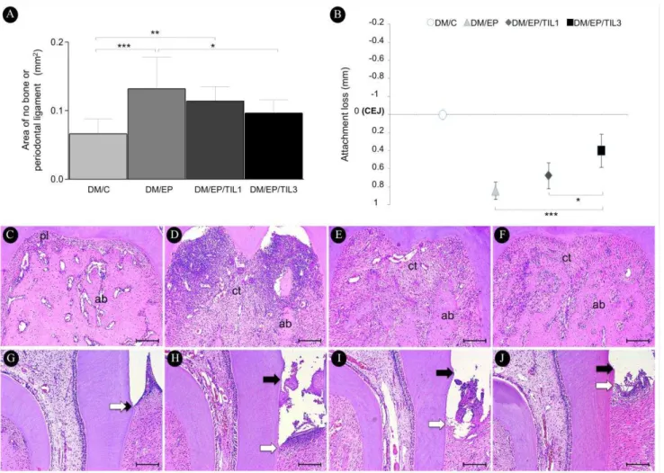
Group DM/EP/TIL3 presented lower ANBL than group DM/EP (p<0.05; Figs. 2A, 2C-F). There were no differences in ANBL when groups DM/C and DM/EP/TIL3 were compared (p>0.05). Group DM/EP/TIL3 presented reduced AL when compared with groups DM/EP (p<0.001) and DM/EP/TIL1 (p<0.05; Figs. 2B, 2G-J).
DISCUSSION
The diabetogenic agent used, streptozotocin, causes selective destruction of pancreatic beta cells37 and, thereby, decreases insulin secretion.38,39 In fact, the general metabolism of STZ-induced diabetic rats is similar to that in human DM.40 In this study, the FPG levels remained elevated throughout the experimental period and there was a significant reduction in body weight.
Since we have previously investigated the effects of TIL in normoglycemic rats,32 that was not the purpose of the present study. Besides, it is well established that DM increases the severity of periodontitis in the experimental model used.14,41 Diabetic rats with EP present abscess formations14,42 and augmented alveolar bone loss,14,39,41-45 higher degree of inflammation14,36,39,41,42,46 and worsen organization of the gingival connective tissue14,36 when compared with normoglycemic rats with periodontitis. Furthermore, the level of pro-inflammatory mediators is increased41,45,46 and the reparative capacity of periodontal tissues is reduced in the diabetic animals.33
With the purpose of evaluating the effects of TIL on periodontal bone loss, the alveolar bone level was measured in diabetic animals’ first molars. The animals treated with TIL 3mg/kg presented less alveolar bone resorption than the animals not treated (group
parameters and alveolar bone resorption than the placebo therapy. It has been also verified that the local treatment of periodontal intrabony defects with alendronate resulted in significant clinical attachment gain, reduction of probing depths and improved bone fill compared to the defects treated with placebo.55
An interesting finding of this study is that, when the parameters of area of no bone or periodontal ligament and attachment loss were analyzed, group DM/EP/TIL3 presented results statistically different from group DM/EP while group DM/EP/TIL1 did not show significant difference when compared with the animals not treated. Furthermore, group DM/EP/TIL3 was the only one that did not present significant difference in relation to group DM/C in histometric analysis in the furcation area. These data may suggest that the dosages of TIL influenced its properties, indicating a dose-dependent effect. In fact, some studies demonstrated that the effects of BPs on decreasing cellular infiltration, number of osteoclasts and alveolar bone loss in periodontal tissues are dose-dependent.56,57 The dose-dependent effect of TIL was observed in a previous study of our group, when it was locally administered in non-diabetic rats.32 In this preceding study, dosages of 0.1, 0.3 and 1 mg/kg body weight were evaluated and the last was the only one to provide a significant decrease in alveolar bone
loss. In the present study, since the dosage of 3 mg/kg presented a trend towards better results than the dosage of 1 mg/kg in some parameters, it is possible that, depending on the effects on the periodontium to be considered, greater dosages are required in diabetic than in non-diabetic animals. It might occur due to the more pronounced periodontal inflammatory process usually observed in diabetic when compared with normoglycemic rats.14,36,39,41,42,46
It is essential that the effects of TIL in EP in diabetic rats be studied in cellular and molecular levels, in order to evaluate the inflammatory mediators that might be influenced by the drug and its pathways of action. The findings of the present study need to be confirmed with more advanced experimental models in the phylogenetic scale and in clinical trials, including type-2 diabetic patients. It is also mandatory to analyze if the local administration of TIL could provide additional benefits to scaling and root planing, which is the conventional periodontal therapy currently. More studies are required also to generate dose-response curves and evaluate different therapeutic regimens.
CONCLUSIONS
Within the limits of this study, it can be concluded that i) the local administration of TIL solutions presented a protective effect on tissue destruction in EP in diabetic rats and ii) the dosage of TIL may influence its effects.
FOOTNOTES
*
Graduate Program in Dentistry, Federal University of Ceara, Fortaleza, CE, Brazil.
†Department of Oral & Maxillofacial Surgery and Periodontology, School of Dentistry of
Ribeirao Preto, University of Sao Paulo –USP, Ribeirao Preto, SP, Brazil.
‡Streptozotocin, Amresco®
Life Science Research, Solon, OH, USA.
§
Rompum®, Bayer Saude Animal, Sao Paulo, SP, Brazil.
||
Dopalen®, Agribands, Paulinia, SP, Brazil.
¶
Tiludronic acid, Tildren®, Ceva Saude Animal Ltda, Paulinia, SP, Brazil. #
Axiovision 4.8.2, Carl Zeiss MicroImaging GmbH, Jena, Germany. **
DC300F, Leica Microsystems, Wetzlar, Germany.
††
DMLB, Leica Microsystems, Wetzlar, Germany.
‡‡ImageJ®, National Institutes of Health, Washington, DC, USA.
ACKNOWLEDGEMENTS
Improvement of Higher Education Personnel (CAPES, Brasilia, DF, Brazil) - Process 23038.009502/2013. All authors declare that there is no conflict of interest.
REFERENCES
1. Bissada NF, Manouchehr-Pour M, Haddow M, et al. Neutrophil functional activity in juvenile and adult onset diabetic patients with mild and severe periodontitis. J
Periodontal Res 1982;17:500-502.
2. Tervonen T, Knuuttila M. Relation of diabetes control to periodontal pocketing and alveolar bone level. Oral Surg Oral Med Oral Pathol 1986;61:346-349.
3. Grossi SG, Skrepcinski FB, DeCaro T, et al. Response to periodontal therapy in diabetics and smokers. J P eriodontol 1996;67(10):1094-1102.
4. Kornman KS, Crane A, Wang HY, et al. The interleukin- 1 genotype as a severity factor in adult periodontal disease. J Clin Periodontol 1997;24:72-77.
5. Kornman KS. Mapping the pathogenesis of periodontitis: a new look. J P eriodontol
2008;79(8):1560-8.
6. American Diabetes Association. Diagnosis and classification of diabetes mellitus.
Diabetes Care 2010;33:S62-S69.
7. Löe H. Periodontal disease. The sixth complication of diabetes mellitus. Diabetes Care
1993;16(1):329-34.
8. Mealey B; American Academy of Periodontology. Diabetes and periodontal diseases. Committee on Research, Science and Therapy. J P eriodontol 2000;71:664-678.
9. Mealey BL, Oates TW; American Academy of Periodontology. Diabetes mellitus and periodontal diseases. J P eriodontol 2006;77:1289-1303.
10. Taylor GW, Burt BA, Becker MP, et al. Non-insulin dependent diabetes mellitus and alveolar bone loss progression over 2 years. J Periodontol 1998;69:76-83.
11. Tsai C, Hayes C, Taylor GW. Glycemic control of type 2 diabetes and severe periodontal disease in the US adult population. Community Dent Oral Epidemiol 2002;30:182-192. 12. Retzepi M, Donos N. The effect of diabetes mellitus on osseous healing. Clin Oral
Implants Res 2010;21(7):673-681.
13. Chang PC, Chung MC, Wang YP, et al. Patterns of diabetic periodontal wound repair: A study utilizing micro-computed tomography and immunohistochemistry. J Periodontol
14. Chang PC, Chien LY, Yeo JF, et al. Progression of periodontal destruction and the roles of advanced glycation end products in experimental diabetes. J Periodontol
2013;84(3):379-88.
15. Tesseromatis C, Kotsiou A, Parara H, et al. Morphological changes of gingiva in streptozotocin diabetic rats. Int J Dent 2009;2009:725628.
16. Emrich LJ, Shlossman M, Genco RJ. Periodontal disease in non-insulin-dependent
diabetes mellitus. J Periodontol 1991;62:123-131.
17. Sandberg GE, Sundberg HE, Fjellstrom CA, et al. Type 2 diabetes and oral health: A comparison between diabetic and non-diabetic subjects. Diabetes Res Clin Pract
2000;50:27-34.
18. Campus G, Salem A, Uzzau S, et al. Diabetes and periodontal disease: a case-control study. J P eriodontol 2005;76:418-425.
19. Lalla E, Papapanou PN. Diabetes mellitus and periodontitis: a tale of two common interrelated diseases. Nat Rev Endocrinol 2011;7:738-748.
20. Kornman KS. Host modulation as a therapeutic strategy in the treatment of periodontal disease. Clin Infect Dis 1999;28:520-526.
21. Reddy MS, Geurs NC, Gunsolley JC. Periodontal host modulation with antiproteinase, anti-inflammatry, and bone-sparing agents. A systematic review. Ann Periodontol
2003;8(1):12-37.
22. Wang J, Stern PH. Dose-dependent differential effects of risedronate on gene expression in osteoblasts. Biochem Pharmacol 2011;81(8):1036-42.
23. Lane N, Armitage GC, Loomer P, et al. Bisphosphonate therapy improves the outcome of conventional periodontal treatment: results of a 12-month, randomized, placebo controlled study. J Periodontol 2005;76(7):1113-22.
24. Sharma A, Pradeep AR. Clinical efficacy of 1% alendronate gel as a local drug delivery system in the treatment of chronic periodontitis: a randomized, controlled clinical trial. J
Periodontol 2012;83(1):11-8.
25. Ammann P, Rizzoli R, Caverzasio J, et al. Effects of the bisphosphonate tiludronate on bone resorption, calcium balance, and bone mineral density. J Bone Miner Res
1993;8(12):1491-8.
26. Murakami H, Nakamura T, Tsurukami H, et al. Effects of tiludronate on bone mass, structure, and turnover at the epiphyseal, primary, and secondary spongiosa in the proximal tibia of growing rats after sciatic neurectomy. J Bone Miner Res
27. Tokuda H, Kozawa O, Harada A, et al. Tiludronate inhibits interleukin-6 synthesis in osteoblasts: inhibition of phospholipase D activation in MC3T3-E1 cells. J Cell Biochem
1998;69(3):252-9.
28. Mönkkönen J, Similä J, Rogers MJ. Effects of tiludronate and ibandronate on the secretion of proinflammatory cytokines and nitric oxide from macrophages in vitro. Life Sci 1998;62(8):PL95-102.
29. Duh E, Aiello LP. Vascular endothelial growth factor and diabetes: the agonist versus antagonist paradox. Diabetes 1999;48(10):1899-906.
30. Aiello LP, Wong JS. Role of vascular endothelial growth factor in diabetic vascular complications. Kidney Int Suppl 2000;77:S113-9.
31. Yoshida M, Tokuda H, Ishisaki A, et al. Tiludronate inhibits prostaglandin F2α-induced vascular endothelial growth factor synthesis in osteoblasts. Mol Cell Endocrinol
2005;236:59–66.
32. Furlaneto FA, Nunes NL, Oliveira Filho IL, et al. Effects of locally-administered tiludronic acid on experimental periodontitis in rats. J Periodontol 2014;85(9):1291-301. 33. de Oliveira Diniz CK, Corrêa MG, Casati MZ, et al. Diabetes mellitus may increase bone
loss after occlusal trauma and experimental periodontitis. J Periodontol
2012;83(10):1297-303.
34. Samarghandian S, Borji A, Delkhosh MB, et al. Safranal treatment improves hyperglycemia, hyperlipidemia and oxidative stress in streptozotocin-induced diabetic rats. J Pharm Pharm Sci 2013;16(2):352-362.
35. Lisboa MR, Gondim DV, Ervolino E, et al. Effects of electroacupuncture on experimental periodontitis in rats [published online ahead of print March 05, 2015]. J Periodontol; doi:10.1902/jop.2015.140630.
36. Silva JA, Lorencini M, Reis JR, et al. The influence of type I diabetes mellitus in periodontal disease induced changes of the gingival epithelium and connective tissue.
Tissue Cell 2008;40(4):283-292.
37. Akbarzadeh A, Norouzian D, Mehrabi MR, et al. Induction of diabetes by streptozotocin in rats. Indian J Clin Bioch 2007;22:60-64.
38. Lalla E, Lamster IB, Feit M, et al. A murine model of accelerated periodontal disease in diabetes. J P eriodontal Res 1998;33(7):387-99.
39. Kim JH, Lee DE, Choi SH, et al. Diabetic characteristics and alveolar bone loss in streptozotocin- and streptozotocin-nicotinamide-treated rats with periodontitis. J
40. Shirakata Y, Eliezer M, Nemcovsky CE, et al. Periodontal healing after application of enamel matrix derivative in surgical supra/infrabony periodontal defects in rats with streptozotocin-induced diabetes. J P eriodontal Res 2014;49(1):93-101.
41. Nishikawa T, Naruse K, Kobayashi Y, et al. Involvement of nitrosative stress in experimental periodontitis in diabetic rats. J Clin Periodontol 2012;39(4):342-9.
42. Chang PC, Chien LY, Chong LY, et al. Glycated matrix up-regulates inflammatory signaling similarly to P orphyromonas gingivalis lipopolysaccharide. J P eriodontal Res
2013;48(2):184-93.
43. Doxey DL, Cutler CW, Iacopino AM. Diabetes prevents periodontitis-induced increases in gingival platelet derived growth factor-B and interleukin 1-beta in a rat model. J
Periodontol 1998 Feb;69(2):113-9.
44. Toker H, Ozdemir H, B ı H, et al. N-acetylcysteine decreases alveolar bone loss on experimental periodontitis in streptozotocin-induced diabetic rats. J P eriodontal Res
2012;47(6):793-9.
45. Jiang ZL, Cui YQ, Gao R, et al. Study of TNF-α IL-1β and LPS levels in the gingival crevicular fluid of a rat model of diabetes mellitus and periodontitis. Dis Markers
2013;34(5):295-304.
46. Silva JA, Ferrucci DL, Peroni LA, et al. Sequential IL-23 and IL-17 and increased MMP8 and MMP14 expression characterize the progression of an experimental model of periodontal disease in type 1 diabetes. J Cell Physiol 2012;227(6):2441-50.
47. Blakytny R, Spraul M, Jude EB. Review: The diabetic bone: a cellular and molecular perspective. Int J Low Extrem Wounds 2011;10(1):16-32.
48. Bain S, Ramamurthy NS, Impeduglia T, et al. Tetracycline prevents cancellous bone loss and maintains near-normal rates of bone formation in streptozotocin diabetic rats. Bone
1997;21(2):147-153.
49. Davi H, Tronquet C, Caix J, et al. Disposition of tiludronate (Skelid) in animals. Xenobiotica 1999;29(10):1017-31.
50. Roodman GD. Interleukin-6: an osteotropic factor? J Bone Miner Res 1992;7(5):475-8. 51. Ishimi Y, Miyaura C, Jin CH, et al. IL-6 is produced by osteoblasts and induces bone
resorption. J Immunol 1990;15;145(10):3297-303.
53. Ozdemir SP, K ş B, THYPERL, et al. Effects of low-dose doxycycline and bisphosphonate clodronate on alveolar bone loss and gingival levels of matrix metalloproteinase-9 and interleukin-1beta in rats with diabetes: a histomorphometric and immunohistochemical study. J P eriodontol 2012;83(9):1172-82.
54. Rocha M, Nava LE, Vázquez de la Torre C, et al. Clinical and radiological improvement of periodontal disease in patients with type 2 diabetes mellitus treated with alendronate: a randomized, placebo-controlled trial. J Periodontol 2001;72(2):204-9.
55. Pradeep AR, Sharma A, Rao NS, et al. Local drug delivery of alendronate gel for the treatment of patients with chronic periodontitis with diabetes mellitus: a double-masked controlled clinical trial. J Periodontol 2012;83:1322-1328.
56. Brunsvold M.A, Chaves ES, Kornman KS, et al. Effects of a bisphosphonate on experimental periodontitis in monkeys. J P eriodontol 1992;63(10):825-830.
57. Alencar VB,lBezerra MM,eLima V, et al. Disodium chlodronate prevents bone resorption in experimental periodontitis in rats. J Periodontol 2002 Mar;73(3):251-6.
58. Mitsuta T, Horiuchi H, Shinoda H. Effects of topical administration of clodronate on alveolar bone resorption in rats with experimental periodontitis. J Periodontol
2002;73(5):479-486.
59. Reddy GT, Kumar TM, Veena. Formulation and evaluation of alendronate sodium gel for the treatment of bone resorptive lesions in Periodontitis. Drug Deliv 2005;12(4):217-22. 60. Goya JA, Paez HA, Mandalunis PM. Effect of topical administration of monosodium
olpadronate on experimental periodontitis in rats. J Periodontol 2006;77(1):1-6.
61. Adami S, Zamberlan N. Adverse effects of bisphosphonates. A comparative review. Drug Saf 1996;14(3):158-70.
62. Diel IJ, Fogelman I, Al-Nawas B, et al. Pathophysiology, risk factors and management of bisphosphonate-associated osteonecrosis of the jaw: Is there a diverse relationship of amino- and non-aminobisphosphonates? Crit Rev Oncol Hematol 2007;64(3):198-207 63. Wang HL, Weber D, McCauley LK, et al. Effect of long-term oral bisphosphonates on
implant wound healing: literature review and a case report. J Periodontol 2007;78(3):584-94.
65. Reid IR. Osteonecrosis of the jaw: who gets it, and why? Bone 2009;44(1):4-10.
66. Janovska Z. Bisphosphonate-related osteonecrosis of the jaws. A severe side effect of bisphosphonate therapy. Acta Medica (Hradec Kralove) 2012; 55(3):111-5.
67. Vasconcelos AC, de Azambuja Berti-Couto S, Figueiredo MA, et al. Laboratory methods and biomarkers in the evaluation of bisphosphonate effects on body tissues: a literature review. J Oral Pathol Med 2013;42(8):577-86.
68. Bonjour JP, Ammann P, Barbier A, et al. Tiludronate: bone pharmacology and safety.
FIGURE LEGENDS
Figure 1. Means and standard deviations of body weight (g) (A) and fasting plasma glucose (mg/dL) (B) in groups DM/C, DM/EP, DM/EP/TIL1 and DM/EP/TIL3; *p= 0.0098; **p< 0.0001; ***p= 0.0018.
Figure 2. Histomorphometric analysis of periodontal tissues. Means and standard deviations of area of no bone or periodontal ligament (mm2) (A; furcation area) and attachment loss (mm) (B; interproximal area) of the specimens, with comparisons among groups. Photomicrographs of periodontal tissues in the furcation (C - F) and interproximal (G - J) areas of mandibular first molars: group DM/C (C and G); group DM/EP (D and H); group DM/EP/TIL1 (E and I); group DM/EP/TIL3 (F and J).
Abbreviations and symbols: ab= alveolar bone; CEJ= cementoenamel junction; ct= connective tissue; pl= periodontal ligament; black arrows= cementoenamel junction; white
TABLE
Table 1. Histopathological analysis of the furcation and interproximal areas of mandibular first molars: percentage of animals per score evaluated.
HISTOPATHOLOGICAL ANALYSIS
PARAMETERS AND RESPECTIVE SCORES EXPERIMENTAL GROUPS
DM/C n=8 DM/EP n=8 DM/EP/TI L1 n=8 DM/EP/T L3 n=8
% animals/
Score
% animals/
score
% animals/
score
%
animals /score
Intensity of local inflammatory infiltrate
(0)Absence of inflammation 100 0 0 0
(1)Small amount of inflammatory cells 0 0 50 100
(2)Moderate amount of inflammatory cells 0 0 50 0
(3)Large amount of inflammatory cells 0 100 0 0
Extension of inflammatory infiltrate
(0)Absence of inflammation 100 0 0 0
(1)Extending to part of the connective tissue of the furcation/interproximal areas
0 0 50 100
(2)Extending to the whole connective tissue of the furcation/interproximal areas
0 0 50 0
(3)Extending to the whole connective tissue and to the bone tissue of the furcation/interproximal areas
0 100 0 0
External root resorption (cementum e dentin)
(0)Absent 100 0 0 0
(1)Only inactive resorption areas 0 0 50 50
(2)Few active resorption areas 0 0 50 50
Alveolar bone resorption
(0)Within normality patterns 100 0 0 75
(1)Small amount of resorption areas 0 0 100 25
(2)Moderate amount of resorption areas 0 0 0 0
(3)Large amount of resorption areas 0 100 0 0
Connective tissue pattern
(0)Moderate amount of fibroblasts and large amount of collagen fibers (dense connective tissue)
100 0 0 0
(1)Moderate amount of both fibroblasts and collagen fibers
0 0 50 100
(2)Small amount of both fibroblasts and collagen fibers; presence of interstitial edema
0 37.5 50 0
(3)Severe tissue destruction with interstitial edema and necrotic areas
0 62.5 0 0
Alveolar bone pattern
(0)Bone trabeculae with regular contour coated with active osteoblasts, including areas of new bone formation
100 0 0 0
(1)Bone trabeculae with irregular contour coated with active osteoblasts and osteoclasts
0 0 50 100
(2)Bone trabeculae with irregular contour coated with active osteoclasts
0 62.5 50 0
(3)Areas of necrotic bone and bone trabeculae with irregular contour coated with active osteoclasts
FIGURES
Figure 1. Means and standard deviations of body weight (g) (A) and fasting plasma glucose (mg/dL) (B) in groups DM/C, DM/EP, DM/EP/TIL1 and DM/EP/TIL3; *p= 0.0098; **p< 0.0001; ***p=
Figure 2. Histomorphometric analysis of periodontal tissues. Means and standard deviations of area of no bone or periodontal ligament (mm2) (A; furcation area) and attachment loss (mm) (B; interproximal area) of the specimens, with comparisons among groups. Photomicrographs of periodontal tissues in the furcation (C - F) and interproximal (G - J) areas of mandibular first molars: group DM/C (C and G); group DM/EP (D and H); group DM/EP/TIL1 (E and I); group DM/EP/TIL3 (F and J).
4.CONCLUSÕES GERAIS
REFERÊNCIAS
1. Löe H, Theilade E, Jensen SB, et al. Experimental gingivitis in man. J P eriodontol
1965;36:177-187.
2. Lindhe J, Nyman S. The effect of plaque control and surgical pocket elimination on the establishment and maintenance of periodontal health. A longitudinal study of periodontal therapy in cases of advanced disease. J Clin. Periodontol 1975;2(2):67-79.
3. Socransky SS, Haffajee AD. The bacterial etiology of destructive periodontal disease: current concepts. J Periodontol 1992;63(4 Suppl):322-331.
4. Eke PI, Dye BA, Wei L, et al. Prevalence of periodontitis in adults in the United States: 2009 and 2010. J Dent Res 2012;91:914-20.
5. Bissada NF, Manouchehr-Pour M, Haddow M, et al. Neutrophil functional activity in juvenile and adult onset diabetic patients with mild and severe periodontitis. J
Periodontal Res 1982;17:500-502.
6. Tervonen T, Knuuttila M. Relation of diabetes control to periodontal pocketing and alveolar bone level. Oral Surg Oral Med Oral Pathol 1986;61:346-349.
7. Grossi SG, Skrepcinski FB, DeCaro T, et al. Response to periodontal therapy in diabetics and smokers. J P eriodontol 1996;67(10 Suppl):1094-1102.
8. Kornman KS, Crane A, Wang HY, et al. The interleukin- 1 genotype as a severity factor in adult periodontal disease. J Clin Periodontol 1997;24:72-77.
9. Kornman KS. Mapping the pathogenesis of periodontitis: a new look. J P eriodontol
2008;79(8 Suppl):1560-8.
10. American Diabetes Association. Diagnosis and classification of diabetes mellitus.
Diabetes Care 2010;33:S62-S69.
11. Shaw JE, Sicree RA, Zimmet PZ. Global estimates of the prevalence of diabetes for 2010 and 2030. Diabetes Res Clin Pract 2010;87(1):4-14.
12. Danaei G, Finucane MM, Lu Y, et al. National, regional, and global trends in fasting plasma glucose and diabetes prevalence since 1980: systematic analysis of health examination surveys and epidemiological studies with 370 country-years and 2.7 million participants. Lancet 2011;378:31–40.
14. Löe H. Periodontal disease. The sixth complication of diabetes mellitus. Diabetes Care
1993;16(1):329-34.
15. Mealey B; American Academy of Periodontology. Diabetes and periodontal diseases. Committee on Research, Science and Therapy. J P eriodontol 2000a;71:664-678.
16. Mealey BL, Oates TW; American Academy of Periodontology. Diabetes mellitus and periodontal diseases. J P eriodontol 2006;77:1289-1303.
17. Ozdemir SP, K ş B, Tüter G, et al. Effects of low-dose doxycycline and bisphosphonate clodronate on alveolar bone loss and gingival levels of matrix metalloproteinase-9 and interleukin-1beta in rats with diabetes: a histomorphometric and immunohistochemical study. J P eriodontol 2012;83(9):1172-82.
18. Thorstensson H, Hugoson A. Periodontal disease experience in adult long-duration insulin-dependent diabetics. J Clin Periodontol 1993;20(5):352-8.
19. Grossi SG. Treatment of periodontal disease and control of diabetes: an assessment of the evidence and need for future research. Ann Periodontol 2001;6:138-145.
20. Taylor GW, Burt BA, Becker MP, et al. Non-insulin dependent diabetes mellitus and alveolar bone loss progression over 2 years. J Periodontol 1998;69:76-83.
21. Tsai C, Hayes C, Taylor GW. Glycemic control of type 2 diabetes and severe periodontal disease in the US adult population. Community Dent. Oral Epidemiol 2002;30:182-192. 22. Mealey BL. Diabetes and periodontal disease: two sides of a coin. Compend Contin Educ
Dent 2000b;21(11):943-946, 948, 950, passim; quiz 956.
23. Cramer C, Freisinger E, Jones RK, et al. Persistent high glucose concentrations alter the regenerative potential of mesenchymal stem cells. Stem Cells Dev Dec 2010;19(12):1875-1884.
24. Desta T, Li J, Chino T, et al. Altered fibroblast proliferation and apoptosis in diabetic gingival wounds. J Dent Res 2010;89(6):609-614.
25. Lalla E, Lamster IB, Stern DM, et al. Receptor for advanced glycation end products, inflammation, and accelerated periodontal disease in diabetes: mechanisms and insights into therapeutic modalities. Ann Periodontol 2001;6(1):113-118.
26. Hudson BI, Bucciarelli LG, Wendt T, et al. Blockade of receptor for advanced glycation endproducts: a new target for therapeutic intervention in diabetic complications and inflammatory disorders. Arch Biochem Biophys 2003;419(1):80-88.
27. Retzepi M, Donos N. The effect of diabetes mellitus on osseous healing. Clin Oral
28. Blakytny R, Spraul M, Jude EB. Review: The diabetic bone: a cellular and molecular perspective. Int J Low Extrem Wounds 2011;10(1):16-32.
29. Chang PC, Chung MC, Wang YP, et al. Patterns of diabetic periodontal wound repair: A study utilizing micro-computed tomography and immunohistochemistry. J Periodontol
2012;83(5):644-52.
30. Chang PC, Chien LY, Yeo JF, et al. Progression of periodontal destruction and the roles of advanced glycation end products in experimental diabetes. J Periodontol
2013;84(3):379-88.
31. Tesseromatis C, Kotsiou A, Parara H, et al. Morphological changes of gingiva in streptozotocin diabetic rats. Int J Dent 2009;2009:725628.
32. Silva JA, Lorencini M, Reis JR, et al. The influence of type I diabetes mellitus in periodontal disease induced changes of the gingival epithelium and connective tissue.
Tissue Cell 2008;40(4):283-292.
33. Kirkwood KL, Cirelli JA, Rogers JE, et al. Novel host response therapeutic approaches to treat periodontal diseases. Periodontol 2000 2007;43:294-315.
34. Deo V, Gupta S, Bhongade ML, et al. Evaluation of subantimicrobial dose doxycycline as
an adjunct to scaling and root planing in chronic periodontitis patients with diabetes: a randomized, placebo-controlled clinical trial. J Contemp Dent Pract 2010;11(3):9-16. 35. Gilowski L, Kondzielnik P, Wiench R, et al. Efficacy of short-term adjunctive
subantimicrobial dose doxycycline in diabetic patients – randomized study. Oral Diseases
2012;18:763–770.
36. Engebretson SP, Hey-Hadavi J. Sub-antimicrobial doxycycline for periodontitis reduces hemoglobin A1c in subjects with type 2 diabetes: a pilot study. P harmacol Res
2011;64:624–629.
37. Maruotti N, Cantatore F. Vitamin D and the immune system. J Rheumatol 2010;37:491– 5.
38. Alshouibi EM, Kaye EK, Cabral HJ, et al. Vitamin D and periodontal health in older men.
J Dent Res 2013; 92(8):689-693.
39. D’Ambrosio D, Cippitelli M, Cocciolo M, et al. Inhibition of IL-12 production by 1,25-dihydroxyvitamin D3. Involvement of NF-kappaB downregulation in transcriptional repression of the p40 gene. J Clin Invest 1998;101:252–62.
40. Giulietti A, van Etten E, Overbergh L, et al. Monocytes from type 2 diabetic patients have a pro-inflammatory profile. 1,25-Dihydroxyvitamin D(3) works as anti-inflammatory.
41. Li H, Xie H, Fu M, Li W, et al. 25-hydroxyvitamin D3 ameliorates periodontitis by modulating the expression of inflammation-associated factors in diabetic mice. Steroids
2013;78:115–120.
42. Kornman KS. Host modulation as a therapeutic strategy in the treatment of periodontal disease. Clin Infect Dis 1999;28:520-526.
43. Reddy MS, Geurs NC, Gunsolley JC. Periodontal host modulation with antiproteinase, anti-inflammatry, and bone-sparing agents. A systematic review. Ann Periodontol
2003;8(1):12-37.
44. Badran Z, Kraehenmann MA, Guicheux J, et al. Bisphosphonates in periodontal treatment: a review. Oral Health Prev Dent 2009;7:30-12.
45. Wang J, Stern PH. Dose-dependent differential effects of risedronate on gene expression in osteoblasts. Biochem Pharmacol 2011;81(8):1036-42.
46. Lane N, Armitage GC, Loomer P, et al. Bisphosphonate therapy improves the outcome of conventional periodontal treatment: results of a 12-month, randomized, placebo controlled study. J Periodontol 2005;76(7):1113-22.
47. Sharma A, Pradeep AR. Clinical efficacy of 1% alendronate gel as a local drug delivery
system in the treatment of chronic periodontitis: a randomized, controlled clinical trial. J Periodontol 2012a;83(1):11-8.
48. Menezes AM, Rocha FA, Chaves HV, et al. Effect of sodium alendronate on alveolar bone resorption in experimental periodontitis in rats. J Periodontol 2005;76(11):1901-9. 49. Zhao L, Marquis A, La VD, et al. Effects of biphenyl sulfonylamino methyl
bisphosphonic acids on Porphyromonas gingivalis and cytokine secretion by oral epithelial cells. Med Chem 2013;9(6):855-60.
50. O'uchi N, Nishikawa H, Yoshino T, et al. Inhibitory effects of YM175, a bisphosphonate, on the progression of experimental periodontitis in beagle dogs. J P eriodontal Res
1998;33(4):196-204.
51. Alencar VB, Bezerra MM, Lima V, et al. Disodium chlodronate prevents bone resorption in experimental periodontitis in rats. J Periodontol 2002;73(3):251-6.
52. Buduneli E, Vardar S, Buduneli N, et al. Effects of combined systemic administration of low dose doxycycline and alendronate on endotoxin-induced periodontitis in rats. J
Periodontol 2004;75(11):1516-23.
53. Mitsuta T, Horiuchi H, Shinoda H. Effects of topical administration of clodronate on alveolar bone resorption in rats with experimental periodontitis. J Periodontol
54. Reddy GT, Kumar TM, Veena. Formulation and evaluation of alendronate sodium gel for the treatment of bone resorptive lesions in Periodontitis. Drug Deliv 2005;12(4):217-22. 55. Goya JA, Paez HA, Mandalunis PM, et al. Effect of topical administration of
monosodium olpadronate on experimental periodontitis in rats. J Periodontol
2006;77(1):1-6.
56. Veena HR, Prasad D. Evaluation of an aminobisphosphonate (alendronate) in the management of periodontal osseous defects. J Indian Soc Periodontol 2010;14(1):40-5. 57. Sharma A, Pradeep AR. Clinical efficacy of 1% alendronate gel in adjunct to
mechanotherapy in the treatment of aggressive periodontitis: a randomized controlled clinical trial. J Periodontol 2012b;83(1):19-26.
58. Duarte PM, de Assis DR, Casati MZ, et al. Alendronate may protect against increased periodontitis-related bone loss in estrogen-deficient rats. J Periodontol 2004;75(9):1196-202.
59. Buduneli E, Buduneli N, Vardar-Sengül S, et al. Systemic low-dose doxycycline and alendronate administration and serum interleukin-1beta, osteocalcin, and C-reactive protein levels in rats. J P eriodontol 2005;76(11):1927-33.
60. Buduneli E, Vardar-Sengül S, Buduneli N, et al. Matrix metalloproteinases, tissue inhibitor of matrix metalloproteinase-1, and laminin-5 gamma2 chain immunolocalization in gingival tissue of endotoxin-induced periodontitis in rats: effects of low-dose doxycycline and alendronate. J Periodontol 2007;78(1):127-34.
61. Cetinkaya BO, Keles GC, Ayas B, et al. Effects of risedronate on alveolar bone loss and angiogenesis: a stereologic study in rats. J Periodontol 2008;79:1950-1961.
62. Rogers MJ, Gordon S, Benford HL, et al. Cellular and molecular mechanisms of action of bisphosphonates. Cancer 2000;88:2961-2978.
63. Shibutani T, Inuduka A, Horiki I, et al. Bisphosphonate inhibits alveolar bone resorption in experimentally-induced peri-implantitis in dogs. Clin Oral Implants Res
2001;12(2):109-14.
64. Rocha M, Nava LE, Vázquez de la Torre C, et al. Clinical and radiological improvement of periodontal disease in patients with type 2 diabetes mellitus treated with alendronate: a randomized, placebo-controlled trial. J Periodontol 2001;72(2):204-9.
65. Tani-Ishii N, Minamida G, Saitoh D, et al. Inhibitory effects of incadronate on the progression of rat experimental periodontitis by Porphyromonas gingivalis infection. J
66. Rocha ML, Malacara JM, Sánchez-Marin FJ, et al. Effect of alendronate on periodontal disease in postmenopausal women: a randomized placebo-controlled trial. J Periodontol
2004;75(12):1579-85.
67. Takaishi Y, Miki T, Nishizawa Y, et al. Clinical effect of etidronate on alveolar pyorrhoea associated with chronic marginal periodontitis: report of four cases. J Int Med Res 2001;29(4):355-65.
68. El-Shinnawi UM, El-Tantawy SI. The effect of alendronate sodium on alveolar bone loss in periodontitis (clinical trial). J Int Acad Periodontol 2003;5(1):5-10.
69. Ammann P, Rizzoli R, Caverzasio J, et al. Effects of the bisphosphonate tiludronate on bone resorption, calcium balance, and bone mineral density. J Bone Miner Res
1993;8(12):1491-8.
70. Murakami H, Nakamura T, Tsurukami H, et al. Effects of tiludronate on bone mass, structure, and turnover at the epiphyseal, primary, and secondary spongiosa in the proximal tibia of growing rats after sciatic neurectomy. J Bone Miner Res
1994;9(9):1355-64.
71. Tokuda H, Kozawa O, Harada A, et al. Tiludronate inhibits interleukin-6 synthesis in
osteoblasts: inhibition of phospholipase D activation in MC3T3-E1 cells. J Cell Biochem
1998;69(3):252-9.
72. Mönkkönen J, Similä J, Rogers MJ. Effects of tiludronate and ibandronate on the secretion of proinflammatory cytokines and nitric oxide from macrophages in vitro. Life Sci 1998;62(8)PL95-102.
73. Nakaya H, Osawa G, Iwasaki N, et al. Effects of bisphosphonate on matrix metalloproteinase enzymes in human periodontal ligament cells. J Periodontol
2000;71(7):1158-66.
74. Duh E, Aiello LP. Vascular endothelial growth factor and diabetes: the agonist versus antagonist paradox. Diabetes 1999;48:1899–906.
75. Aiello LP, Wong JS. Role of vascular endothelial growth factor in diabetic vascular complications. Kidney Int Suppl 2000;77:S113–9.
76. Yoshida M, Tokuda H, Ishisaki A, et al. Tiludronate inhibits prostaglandin F2α-induced vascular endothelial growth factor synthesis in osteoblasts. Mol Cell Endocrinol 2005; 236: 59–66.
77. Adami S, Zamberlan N. Adverse effects of bisphosphonates. A comparative review. Drug
78. Diel IJ, Fogelman I, Al-Nawas B, et al. Pathophysiology, risk factors and management of bisphosphonate-associated osteonecrosis of the jaw: Is there a diverse relationship of amino- and non-aminobisphosphonates? Crit Rev Oncol Hematol 2007;64(3):198-207. 79. Wang HL, Weber D, McCauley LK, et al. Effect of long-term oral bisphosphonates on
implant wound healing: literature review and a case report. J Periodontol 2007;78(3):584-94.
80. Oizumi T, Yamaguchi K, Funayama H, et al. Necrotic actions of nitrogen-containing bisphosphonates and their inhibition by clodronate, a non-nitrogen-containing bisphosphonate in mice: potential for utilization of clodronate as a combination drug with a nitrogen-containing bisphosphonate. Basic Clin Pharmacol Toxicol 2009;104(5):384-92.
81. Reid IR. Osteonecrosis of the jaw: who gets it, and why? Bone 2009;44(1):4-10.
82. Janovska Z. Bisphosphonate-related osteonecrosis of the jaws. A severe side effect of bisphosphonate therapy. Acta Medica (Hradec Kralove) 2012; 55(3):111-5.
83. Vasconcelos AC, de Azambuja Berti-Couto S, Figueiredo MA, et al. Laboratory methods and biomarkers in the evaluation of bisphosphonate effects on body tissues: a literature
review. J Oral Pathol Med 2013;42(8):577-86.
84. Bonjour JP, Ammann P, Barbier A, et al. Tiludronate: bone pharmacology and safety.
Bone 1995;17(5 Suppl);473S-477S.
85. Furlaneto FA, Nunes NL, Oliveira Filho IL, et al. Effects of locally-administered tiludronic acid on experimental periodontitis in rats. J Periodontol 2014;85(9):1291-1301. 86. Pradeep AR, Sharma A, Rao NS, et al. Local drug delivery of alendronate gel for the treatment of patients with chronic periodontitis with diabetes mellitus: a double-masked controlled clinical trial. J Periodontol 2012;83:1322-1328.
87. Tonetti MS, Chapple IL, Working Group 3 of Seventh European Workshop on Periodontology. Biological approaches to the development of novel periodontal therapies-consensus of the Seventh European Workshop on Periodontology. J Clin Periodontol