Targeting Atp6v1c1 Prevents Inflammation
and Bone Erosion Caused by Periodontitis
and Reveals Its Critical Function in
Osteoimmunology
Sheng Li1,2☯, Liang Hao1☯, Lin Wang2, Yun Lu1, Qian Li3, Zheng Zhu1,2, Jian-Zhong Shao3, Wei Chen1*
1Department of Pathology, University of Alabama at Birmingham, Birmingham, AL, 35294, United States of America,2College of Stomatology, Nanjing Medical University, Nanjing, 210029, People’s Republic of China,3Life Science College, Zhejiang University, 388 Yuhang Road, Hangzhou, 310058, People's Republic of China
☯These authors contributed equally to this work.
Abstract
Periodontal disease (Periodontitis) is a serious disease that affects a majority of adult Amer-icans and is associated with other systemic diseases, including diabetes, rheumatoid arthri-tis, and other inflammatory diseases. While great efforts have been devoted toward
understanding the pathogenesis of periodontitis, there remains a pressing need for devel-oping potent therapeutic strategies for targeting this pervasive and destructive disease. In this study, we utilized novel adeno-associated virus (AAV)-mediated Atp6v1c1 knockdown gene therapy to treat bone erosion and inflammatory caused by periodontitis in mouse model. Atp6v1c1 is a subunit of the V-ATPase complex and regulator of the assembly of the V0 and V1 domains of the V-ATPase complex. We demonstrated previously that Atp6v1c1 has an essential function in osteoclast mediated bone resorption. We hypothesized that Atp6v1c1 may be an ideal target to prevent the bone erosion and inflammation caused by periodontitis. To test the hypothesis, we employed AAV RNAi knockdown of Atp6v1c1 gene expression to prevent bone erosion and gingival inflammation simultaneously. We found that lesion-specific injection of AAV-shRNA-Atp6v1c1 into the periodontal disease lesions protected against bone erosion (>85%) and gingival inflammation caused byP.gingivalis W50infection. AAV-mediated Atp6v1c1 knockdown dramatically reduced osteoclast num-bers and inhibited the infiltration of dendritic cells and macrophages in the bacteria-induced inflammatory lesions in periodontitis. Silencing of Atp6v1c1 expression also prevented the expressions of osteoclast-related genes and pro-inflammatory cytokine genes. Our data suggests that AAV-shRNA-Atp6v1c1 treatment can significantly attenuate the bone erosion and inflammation caused by periodontitis, indicating the dual function of AAV-shRNA-Atp6v1c1 as an inhibitor of bone erosion mediated by osteoclasts, and as an inhibitor of inflammation through down-regulation of pro-inflammatory cytokine expression. This study demonstrated that Atp6v1c1 RNAi knockdown gene therapy mediated by AAV-shRNA-a11111
OPEN ACCESS
Citation:Li S, Hao L, Wang L, Lu Y, Li Q, Zhu Z, et al. (2015) Targeting Atp6v1c1 Prevents Inflammation and Bone Erosion Caused by Periodontitis and Reveals Its Critical Function in Osteoimmunology. PLoS ONE 10(8): e0134903. doi:10.1371/journal. pone.0134903
Editor:Michael Glogauer, University of Toronto, CANADA
Received:January 6, 2015
Accepted:July 16, 2015
Published:August 14, 2015
Copyright:© 2015 Li et al. This is an open access article distributed under the terms of theCreative Commons Attribution License, which permits unrestricted use, distribution, and reproduction in any medium, provided the original author and source are credited.
Data Availability Statement:All relevant data are within the paper and its Supporting Information files.
Funding:This study was supported by NIH grant R01DE023813 (YPL), and the UAB Department of Pathology Start-Up funding (WC). The funders had no role in study design, data collection and analysis, decision to publish, or preparation of the manuscript.
Atp6v1c1 is a promising novel therapeutic approach for the treatment of bone erosion and inflammatory related diseases, such as periodontitis and rheumatoid arthritis.
Introduction
The ultimate outcome of periodontitis is alveolar bone and tooth loss, which results from the interaction between oral biofilm microorganisms and the host immune response in the peri-odontitis lesion area. Different cell types have been demonstrated to participate in this inflam-matory progress, such as T lymphocytes, macrophages, and dendritic cells [1,2]. Besides oral disease, periodontal diseases are associated with many systemic diseases such as rheumatoid arthritis, diabetes, infective endocarditis, and scorbutus (scurvy) [3,4]. This is due to the fact that immune cells in the lesion areas activate nuclear factor kappa-B ligand (RANKL), lead to osteoclast activation [5], and result in bone and tooth loss in a state of inflammation [6]. Previ-ous studies revealed that osteoclasts induced in this inflammatory response are the leading cause of tooth and alveolar bone loss [7]. During the process of inflammation, a multi-unit vac-uolar-type H+-ATPase (V-ATPase) complex decreases the pH at the bone surface. This extra-cellular acidification was induced by inflammatory cytokines, followed by the activated osteoclasts’resorption of bone around the root or teeth. The decrease of pH is important for bone erosion related to osteoclasts [8,9].
TheATP6V1C1gene encodes V-type proton ATPase subunit C1 [10,11]. This gene is responsible for encoding the enzyme vacuolar ATP (V-ATP enzyme) and acidifying compo-nents within the multi-subunit enzyme-mediated eukaryotic cellular compartments. V-ATPase-dependent acidification is an important step for intracellular processes, including zymogen activation, receptor-mediated endocytosis, and synaptic vesicle proton gradient gen-eration [12,13]. Our previous research determined that Atp6v1c1 is mainly expressed in oste-oclasts, whereas subunits Atp6v1c2a (C2a) and Atp6v1c2b (C2b) are not [9]. C1 expression is highly induced by RANKL during the process of osteoclast differentiation. C1 interacts with Atp6v0a3 (a3), and is mainly localized on the ruffled border of activated osteoclasts [9]. A pre-vious study showed that in addition to being an essential component of V-ATPases, Atp6v1c1 may regulate filament actin arrangement in breast cancer cells [14]. Silencing of Atp6v1c1 prevents breast cancer growth and bone metastasis [15], indicating the potential multiple functions of Atp6v1c1 in normal cell functions and diseases. Since cancer growth and metas-tasis are related to immune response, we hypothesize that inhibition of Atp6v1c1 may also prevent the immune response and the following bone erosion. As a subunit of Atp6i that is expressed in both osteoclasts and immune cells such as macrophages and dendritic cells [9,
16], Atp6v1c1 should have an osteoimmune function during the development of periodontitis.
Materials and Methods
For complete Materials and Methods, please seeS1 File.
Animals
Adult (8 wks) female wild-type (WT) BALB/cJ mice (from Jackson Laboratory) were used for current study [9,23,24]. All of the mice were divided into 3 groups: (1) Normal group (mice without bacteria) (n = 7); (2) Bacteria infection treated with AAV-shRNA-Atp6v1c1 (AAV-sh-Atp6v1c1) (n = 7); (3) Bacteria infection treated with AAV-sh-luc-YFP (n = 7). The experi-ments were performed three times on three different occasions, generating a total sample num-ber 21 for each group. All mice were maintained under a 12-hour light–dark cycle with ad libitum access to regular food and water at the University of Alabama at Birmingham (UAB) Animal Facility. The Protocol used in our current experiment was approved by the Institu-tional Animal Care and Use Committee of UAB (Animal Protocol Number 131209236).
Infection with
Porphyromonas gingivalis
strains
Porphyromonas gingivalisW50 (ATCC: 53978) were seeded and cultured on sheep’s blood agar plates supplemented with hemin and vitamin K (BAPHK) for 3 days in an oxygen-free environ-ment. Next a single clone was harvested and transferred to Trypticase Soy Broth supplemented with hemin and vitamin K. Bacteria were harvested on the 4thday and centrifuged for 30min and then resuspended. The bacteria concentrations of each species were determined via optical density readings at 600nm (One OD unit equals 6.67108bacteria). The bacteria density of each species was adjusted to 1010cells/ml in PBS containing 2% carboxymethylcellulose (CMC: Sigma-Aldrich). The procedure of periodontal infection was conducted following the previously described protocol with some modifications [25]. In brief, all animals received antibiotic treat-ment for 3 days to reduce the original oral confounders, followed by 3 days without antibiotic, prior to oral inoculation with 0.2mlP.gingivalisin CMC in 20μl with a dental micro-brush
once per day for 4 consecutive days. To verify bacterial effectiveness, the sample from the mice oral cavity was taken on day 14 after infection. The sterile cotton swab was used to collect the bacteria plaque and samples were incubated anaerobically to identifyP.gingivalis[25].
AAV-shRNA-Atp6v1c1 transduction of
P
.
gingivalis
W50 infected mice
In the present study, we injected AAV-sh-Atp6v1c1 in specific sites as described previously [19]. On the 4thday after initial infection, the mice were anesthetized via peritoneal injection with 62.5mg/kg ketamine and 12.5mg/kg xylazine. Next, AAV-sh-Atp6v1c1 was injected approximately 0.3–0.5mm above the gingival margin of the maxillary molars on the right and left palatal aspects with 3ul containing 2 x10⁹packaged genomic particles in either AAV-sh-Atp6v1c1 or AAV-sh-luc-YFP viral vector using 5-μl Hamilton syringe attached to a
microin-fusion pump (World Precision Instruments, Sarasota, FL). The injection was repeated every day for 7 consecutive days. In the negative control group (normal), mice were maintained with-out any treatment.
Sample harvest and preparation
8 weeks after initial infection, all animals were sacrificed by CO2inhalation and the maxillae
were fixed in 4% paraformaldehyde and according to standard protocol. Samples for paraffin sections were fixed in 4% formaldehyde for 24 hours and washed with PBS, decalcified in 10% EDTA in 0.1M TRIS solution (pH = 7.0) for 10 days. After washing with 1XPBS three times, samples were embedded into paraffin after series dehydration. Another three samples from the right side in every independent experiment were obtained for Real-Time quantitative PCR (qRT-PCR) analyses and Enzyme-linked immunosorbent assays (ELISAs). Gingival tissues and/ or alveolar bone were isolated under a stereo microscope. Gingival tissues and alveolar bone from three samples were pooled for qRT-PCR, and gingival tissues from another three samples were pooled for ELISAs for cytokines. All of these experiments were repeated three times.
Bone resorption measurements
The imaging protocol was described previously [27,28]. The picture of molar teeth, roots and maxillary bone were captured by using Stereomicroscope. The bone loss area was calculated by using image software (Adobe, San Jose, CA, USA). In this procedure, the bone resorption area began at the cemento-enamel junction, including the areas of the root, to the alveolar ridge, which was measured using ImageJ (Bethesda, USA). The results were expressed in mm2.
Histological and immunohistochemical analysis
Hematoxylin & eosin (H&E) staining and calculation were done as described previously [29,
30]. Anti-ATP6v1c1 (H-300) (Santa Cruz, CA), rat monoclonal anti-F4/80 (eBioscience, San Diego, CA) and rabbit anti-mouse CD11c (Santa Cruz, Dallas, Texas, USA) were used for Immunohistochemical analysis (IHC) separately. The IHC analyses in the present study were repeated three times separately.
Real-time quantitative PCR (qRT-PCR)
The tissues in the lesion area were isolated and stored at -80°C for determination of the effect of Atp6v1c1 on the levels of nucleic acid in inflammatory periodontal tissues as described pre-viously [27,28,31,32]. qRT-PCR was carried out using TaqMan probes (Applied Biosystems, Life Technologies, Grand Island, NY, USA) as listed inS1 Table.
Enzyme-linked immunosorbent assay (ELISA)
ELISA was applied to determine the effect of Atp6v1c1 on the levels of protein in lesion areas as previously described [28,32,33]. The following ELISA kits have been used: IL-1α (Biole-gend, San Diego, CA, USA), IL-6 (Biole(Biole-gend, San Diego, CA, USA), and IL-17A (ebioscience, San Diego, CA). The results were expressed as pg cytokine/mg tissue.
Statistical analysis
All of the experimental data were presented as mean ± SD. For the parametric data, the two-tailed Student's t-test and one-way ANOVA test were used for analysis. For non-parametric data, Mann-Whitney U test was used for analysis (Pvalues<0.05 orUvalues>1.96).
Results
The expression of Atp6v1c1 was knocked down efficiently by
AAV-shRNA Atp6v1c1 as well as bone erosion related to osteoclasts in vitro
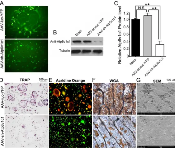
YFP in target cells to determine whether AAV-sh-luc-YFP and AAV-sh-Atp6v1c1 can trans-duce target cells successfully. Osteoclasts were intrans-duced by using M-CSF and RANKL in mouse bone marrow (MBM), which was isolated from WT mice with BALB/cJ background, which were then transduced by AAV-sh-Atp6v1c1 or AAV-sh-luc-YFP. Fluorescence of cultured cells showed that transduction of pre-osteoclasts and osteoclasts with AAV-sh-Atp6v1c1 or AAV-sh-luc-YFP was successful (Fig 1A). Western blot analysis was then applied to confirm transduction, and we found that the expression of Atp6v1c1 in osteoclasts treated with AAV-sh-Atp6v1c1 decreased almost 70% compared to osteoclasts treated with AAV-sh-luc-YFP (Fig 1B and 1C). The results indicated that the application of AAV-sh-Atp6v1c1 can efficiently reduce Atp6v1c1 protein expressionin vitro. Therefore the function of Atp6v1c1 is indispens-able for extracellular acidification mediated by osteoclast in bone resorption. We then per-formed tartrate-resistant acid phosphatase (TRAP) staining, which showed that TRAP-positive osteoclasts decreased significantly in the AAV-sh-Atp6v1c1 group (Fig 1D). The acridine orange staining also showed that the osteoclasts treated with AAV-sh-Atp6v1c1 had weak extracellular acidification (Fig 1E). We accordingly anticipated that knockdown of Atp6v1c1 would inhibit bone resorption as well. Compared to the control group, AAV-mediated
Fig 1. The expression of Atpv1c1 was efficiently knocked down by AAV-sh-Atp6v1c1.(A-G) M-CSF/RANKL was applied for 3 days to allow
differentiation of osteoclasts, which were then transduced with AAV-sh-Atp6v1c1, AAV-sh-luc-YFP, or untreated (mock). (A) Transduction of osteoclasts was successful indicated by fluorescence. (B, C) Atpv1c1 protein level by Western blot showed that AAV-sh-Atp6v1c1 treated osteoclasts have reduced Atp6v1c1 expression significantly (P<0.01). (D) TRAP staining revealed that the number of osteoclasts was decreased in the AAV-sh-Atp6v1c1 treatment group. (E) AAV-sh-luc-YFP (control) or AAV-sh-Atp6v1c1 transduced osteoclasts were stained with acridine orange to show extracellular acidification. (F) Wheat germ agglutinin (WGA) was applied for visualizing resorption lacunae and (G) scanning electron microscopy (SEM) showed that osteoclast-mediated bone resorption was inhibited by ATP6v1c1 knockdown.**:P<0.01, N.S: No Significance.
knockdown of Atp6v1c1 completely inhibited bone resorption (Fig 1F and 1G). These data clearly showed that AAV-sh-Atp6v1c1 can inhibit acidification and bone resorption mediated by osteoclastsin vitro.
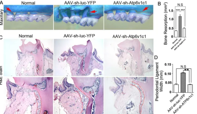
Inhibition of Atp6v1c1 prevents bone erosion in periodontitis lesion areas
caused by
P
.
gingivalis
W
50
It has been indicated that thePorphyromonas gingivalis W50(P.gingivalis W50) strains can cause severe bone loss in experimental animal models of periodontal disease [27]. We first veri-fied bone resorption induced by this bacteria strain. The results showed that significant bone loss can be caused byP.gingivalis W50compared to a normal group (P<0.05) (Fig 2A and 2B).
There was also a significant difference in bone resorption between bacteria infected mice treated with AAV-sh-Atp6v1c1 and with AAV-sh-luc-YFP (Fig 2B). Alternatively, the differ-ence in bone resorption between the normal group and the AAV-sh-Atp6v1c1 treated group was not significant (Fig 2B). The AAV-sh-luc-YFP group infected by bacteria had much more bone loss when compared to the group treated with AAV-sh-Atp6v1c1 (p<0.0001) and the
normal group (p<0.0001) (Fig 2B). These results indicate thatP.gingivalisW50-stimulated
bone erosion can be inhibited by local application of AAV-sh-Atp6v1c1 in the experimental periodontal disease mouse model. The periodontal ligament width between the group with AAV-sh-Atp6v1c1 treatment, normal group and the AAV-sh-luc-YFP group was then exam-ined by H&E sections. Results showed that the periodontal ligament width is significantly higher in the AAV-sh-luc-YFP treated group compared to the normal and AAV-sh-Atp6v1c1
Fig 2. Bone resorption in experimental periodontitis was reduced by Atp6v1c1 knockdown.(A) In the AAV-sh-luc-YFP group with infection, marked bone resorption can be seen and all teeth roots are exposed near the tip of the root in the maxillary (red dotted area). (B) There is almost twice the amount of bone resorption in the AAV-sh-luc-YFP group when compared to the AAV-sh-Atp6v1c1 group. (C) The red dot area shows a widened periodontal ligament area in the AAV-sh-luc-YFP treated disease group. (D) The periodontal ligament width is almost doubled in the AAV-sh-luc-YFP treatment group compared to the AAV-sh-Atp6v1c1 treatment group, indicating that alveolar bone loss was prevented by AAV-sh-Atp6v1c1.**:P<0.01,***:P<0.001, N.S: No Significance.
treated group (Fig 2C and 2D), indicating that AAV-sh-Atp6v1c1 prevents the periodontal lig-ament from damage and reduces alveolar bone resorption.
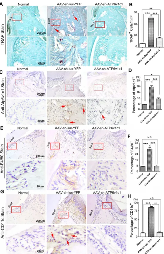
AAV-mediated Atp6v1c1 knockdown decreased TRAP positive
osteoclasts as well as immune cells in the periodontitis lesion area
To further explore the possible effect of AAV-sh-Atp6v1c1 on activation of osteoclasts in the lesion area, TRAP staining was applied to histological sections to detect osteoclasts (Fig 3A and 3B). The TRAP positive osteoclasts in the lesion area (periodontal ligament area) were reduced in the AAV-sh-Atp6v1c1 treated group significantly (Fig 3A and 3B) when compared to the group with AAV-sh-luc-YFP treatment (Fig 3B). We then confirmed the expression of Atp6v1c1 in osteoclastsin vivo, which is also a confirmation of whether AAV-sh-Atp6v1c1 successfully transduced in the local lesion area. Tooth root sections from bacteria infected mice treated with AAV-sh-Atp6v1c1 or AAV-sh-luc-YFP were applied to IHC using anti-Atp6v1c1. It was found that AAV-sh-Atp6v1c1 reduced expression of Atp6v1c1in vivonotably (Fig 3C and 3D). Further IHC staining of F4/80 and CD11c in different groups showed that AAV-sh-Atp6v1c1 decreased the number of immune cells such as macrophages and dendritic cells (Fig 3E–3H).
The expression of osteoclast marker genes and cytokines were
prevented by AAV-sh-Atp6v1c1 in the periodontitis lesion area
To confirm ourin vivohistological results, the effect of AAV-sh-Atp6v1c1 on inflammation, ELISA and qRT-PCR was applied to quantify inflammation-related cytokine expression in the periodontitis lesion tissues (Fig 4). Compared to the normal group without infection, we found that some important osteoclast differentiation-related genes [i.e.RANKL,OPGand Interleu-kin-6 (IL-6)] and specific osteoclast functional gene [i.e. Cathepsin K (Ctsk)] were increased in theP.gingivalisW50 infection group treated with AAV-sh-luc-YFP (Fig 4A). Most impor-tantly, AAV-sh-Atp6v1c1 treatment reduced the expression ofRANKL,IL-6 andCtskin peri-odontal tissues from theP.gingivalisW50 group and restored the expression level in the normal group (Fig 4A). Furthermore, the expression of proinflammatory cytokines such as TNFα, IL-6 and IL-1α, which is critical for osteoclast differentiation, were also decreased in the AAV-sh-Atp6v1c1 treated group (Fig 4B). All of these results indicated that Atp6v1c1 knock-down reduces the expression level of genes related to osteoclast and inflammation, and reduces bone erosion and inflammation in periodontitis lesion areas.
Discussion
Periodontitis is an infectious disease with a bacterial complex pathogen, which has been con-sidered a major cause of periodontitis. In periodontal diseases, the immune response of the host plays an important role [25,34], by inducing gingival soft tissue damage and osteoclast-mediated bone erosion [35]. In our current study, we found that knockdown of the targeted subunit (Atp6v1c1) of Atp6i by an AAV vector can inhibit inflammation and reduce periodon-tal disease progression significantly. Our lab also discovered other important genes that are found important in immune response recent years [36–38].
AAV-sh-Atp6v1c1 is a promising therapeutic agent for periodontitis by
inhibiting both osteoclasts and immune cells
in the V1 domain of V-ATPase, and is intimately involved in the reversible dissociation of the V1 and V0 domains, and is considered to be directly responsible for regulating the dissociative mechanism of V-ATPase [39,40]. AAV-sh-Atp6v1c1 inhibited bone destructionin vitro
through inhibition of acidification mediated by osteoclasts and also reduced pro-inflammatory cytokine expression. Additionally, it reduced the number of osteoclasts and protected mice from bacteria induced bone loss in periodontitis lesion area. These results strongly indicated that application of AAV-sh-Atp6v1c1 gene therapy locally can inhibit alveolar bone loss and tissue damage in periodontitis lesion areas, and may be a promising new strategy for treating periodontal diseases.
Application of AAV-sh-Atp6v1c1 locally for periodontal disease is
effective
Current therapies for periodontal diseases are focused mainly on anti-microbial treatments, which generally have limited application. Gene therapy, on the other hand has great possibili-ties to achieve an outcome with a sustained therapeutic gene product [35,41]. In order to achieve a practical therapeutic method that is relatively safe and non-invasive, we should address which genes are best to be studied and which vectors can be employed safely [35]. Our group previously characterized Atp6i [42] and found that it plays important functions in extra-cellular acidification, which is necessary for immune response and bone resorption. Atp6v1c1 is specific to the proton pump in osteoclasts [8,9], and the deletion of Atp6v1c1 can result in loss of osteoclast function. It has been shown that AAV2 is relatively safe and effective in differ-ent disease models. Another study reported that a relatively long period of sustained expression can be achieved by injection of AAV2 intra-articularly [43]. Osteoprotegerin (OPG) gene transfer can attenuate the osteoclast activity and number in an osteolysis animal model [44]. AAV-IL-4 can also reduce the collagen-induced arthritis (CIA) and inflammatory responses [20]. Sara et al. demonstrated the potential forin vivoAAV2-mediated immune gene therapy and provided data on the inter-relationship between tumor vasculature and immune cell recruitment [45,46]. In our current study, we applied injections of AAV-sh-Atp6v1c1 to treat periodontitis-mediated bone resorption and inflammation. The efficiency of Atp6v1c1 inhibi-tion may reflect the important funcinhibi-tion of the molecules in the development of periodontitis, in osteoclastic resorption and inflammatory processes that result in tissue damages, as well as in the formation of osteoclasts. Delivery of gene therapy locally is much more efficient and more effective than systemic administration since the synthesis of therapeutic gene products was restricted to the lesion specific area, which can minimize the side effects and the costs of treatment [35]. In our current study, the results showed that AAV-sh-Atp6v1c1 not only pre-vents inflammation and bone erosion in periodontitis, but it may also be useful for other inflammatory diseases.
Our qRT-PCR and ELISA results also demonstrated that inflammation in the sh-Atp6v1c1 treatment group decreased significantly. The inflammation reduction in the AAV-of AAV-sh-Atp6v1c1 knockdown in periodontal tissues (Red arrows). (D) Statistical analysis indicates that in the AAV-sh-Atp6v1c1 treatment group, the number of Atp6v1c1 positive cells was reduced significantly. (E) Immunohistochemical staining of F4/80 was applied to the different groups in the periodontitis lesion area. (F) Statistical analysis indicates that the number of F4/80 positive cells was reduced significantly in the AAV-sh-Atp6v1c1 treatment group. (G) Immunohistochemical staining of CD11c was applied to the different groups in the periodontitis lesion area (Red arrows). (H) Statistical analysis indicates that the number of CD11c positive cells was reduced significantly in the AAV-sh-Atp6v1c1 treatment group.*:P<0.05,**:P<0.01,***:
P<0.001, N.S: No Significance.
Fig 4. The expression of osteoclast marker genes and cytokines in the periodontal tissues were reduced by AAV-sh-Atp6v1c1.(A) osteoclast-specific genes (i.e.Ctsk), osteoclast differentiation genes (i.e.
RANKLandOPG), and cytokines (i.e.IL-6,IL-17A and TNFα) in different groups. Expression levels were normalized to the housekeeping gene hypoxanthine-guanine phosphoribosyl transferase (Hprt). (B) 6, IL-1αand TNFαlevels in the periodontitis lesion area were detected by ELISA.*:P<0.05,**:P<0.01,***: P<0.001, N.S:P>0.05.
sh-Atp6v1c1 treatment group may be due to the down regulation ofIL-6(a pro-inflammatory cytokine associated with periodontitis). It is reported that IL-6, which is expressed by osteo-blasts, promotes inflammation as well as bone resorption [47]. According to the results in the present study, AAV-sh-Atp6v1c1 not only reduced the expression of Atp6v1c1, but also reduced inflammatory cytokine expression. After extraction of protein from tissue samples from different groups, the ELISA assays were performed and the results have been analyzed to find more mechanistic information. The expression of T-cell mediated cytokine IL-6 as well as TNF-αand IL-1αwas decreased by application of AAV-sh-Atp6v1c1. In all of the cytokines analyzed by ELISA in our present study, IL-1αis a close relative with bone resorption. Our results indicate that in theP.gingivalis-infected group treated with AAV-sh-Atp6v1c1, the expression level of IL-1αdecreased significantly. Because it has been reported that IL-1α knockdown may inhibit the circuits between inflammatory processes and bone resorption, AAV-sh-Atp6v1c1 treatment can interfere with this positive feedback also and reverse bone resorption in the progression of periodontitis.
Our present findings possess a critical insight into Atp6v1c1 osteoimmune function between bone resorption and inflammatory host responses mediated by bacterial pathogens in periodontitis. These observations highlight a critical pathogenic role for Atp6v1c1 in these set-tings and identify Atp6v1c1 as a logical target for the suppression of periodontal bone destruc-tion and inflammadestruc-tion.
Supporting Information
S1 File. Supplemental Materials and Methods.
(DOCX)
S1 Table. qRT-PCR probe Numbers.
(DOCX)
Acknowledgments
We thank Dr. Yi-Ping Li for his extensive suggestions, discussion, and support for the research of the project, and for his review of our manuscript. We also thank Drs. Sonoko Ogawa and Sergei Musatov for their kind support for providing AAV-H1 vectors, AAV-H1-shRNA-luc-YFP, and important suggestions. We thank Ms. Christie Paulson and Ms. Suzy Newton for their assistance with the manuscript. We also thank the Small Animal Phenotyping Core, Neu-roscience Image Core and NeuNeu-roscience Molecular Detection Core Laboratory (P30
NS0474666), Histomorphometry and Molecular Analyses Core, and the Center for Metabolic Bone Disease (P30 AR046031). This study was supported by NIH grants R01DE023813 (Y.P.L.), and the UAB Department of Pathology Start-Up funding (Wei Chen). The authors declare no potential conflicts of interest with respect to the authorship and/or publication of this article.
Author Contributions
Conceived and designed the experiments: WC. Performed the experiments: SL LH YL QL ZZ WC. Analyzed the data: SL LH LW YL QL ZZ JZS WC. Contributed reagents/materials/analy-sis tools: SL LH YL WC. Wrote the paper: WC LH SL.
References
http://www.nature.com/srep/2014/140604/srep05171/abs/srep05171.html#supplementary-information.
2. Fischer CL, Walters KS, Drake DR, Dawson DV, Blanchette DR, Brogden KA, et al. Oral mucosal lipids are antibacterial against Porphyromonas gingivalis, induce ultrastructural damage, and alter bacterial lipid and protein compositions. In J Oral Sci. 2013; 5(3):130–40. doi:10.1038/ijos.2013.28
3. Seymour GJ, Ford PJ, Cullinan MP, Leishman S, Yamazaki K. Relationship between periodontal infec-tions and systemic disease. Clinical microbiology and infection: the official publication of the European Society of Clinical Microbiology and Infectious Diseases. 2007; 13 Suppl 4:3–10. Epub 2007/11/06. doi:10.1111/j.1469-0691.2007.01798.xPMID:17716290.
4. Kim J, Amar S. Periodontal disease and systemic conditions: a bidirectional relationship. Odontology. 2006; 94(1):10–21. doi:10.1007/s10266-006-0060-6PMID:16998613
5. Chen W, Zhu G, Hao L, Wu M, Ci H, Li YP. C/EBPalpha regulates osteoclast lineage commitment. Proc Natl Acad Sci U S A. 2013; 110(18):7294–9. Epub 2013/04/13. doi:10.1073/pnas.1211383110PMID: 23580622; PubMed Central PMCID: PMCPmc3645589.
6. Kawai T, Matsuyama T, Hosokawa Y, Makihira S, Seki M, Karimbux NY, et al. B and T lymphocytes are the primary sources of RANKL in the bone resorptive lesion of periodontal disease. Am J Pathol. 2006; 169(3):987–98. doi:10.2353/ajpath.2006.060180WOS:000240048900024. PMID:16936272
7. Hasturk H, Kantarci A, Ohira T, Arita M, Ebrahimi N, Chiang N, et al. RvE1 protects from local inflamma-tion and osteoclast- mediated bone destrucinflamma-tion in periodontitis. FASEB journal: official publicainflamma-tion of the Federation of American Societies for Experimental Biology. 2006; 20(2):401–3. Epub 2005/12/24. doi:10.1096/fj.05-4724fjePMID:16373400.
8. Li YP, Chen W, Liang YQ, Li E, Stashenko P. Atp6i-deficient mice exhibit severe osteopetrosis due to loss of osteoclast-mediated extracellular acidification. Nat Genet. 1999; 23(4):447–51. doi:10.1038/ 70563WOS:000084023900022. PMID:10581033
9. Feng SM, Deng LF, Chen W, Shao JZ, Xu GL, Li YP. Atp6v1c1 is an essential component of the osteo-clast proton pump and in F-actin ring formation in osteoosteo-clasts. Biochem J. 2009; 417:195–203. doi:10. 1042/Bj20081073WOS:000262230700019. PMID:18657050
10. Qin A, Cheng TS, Pavlos NJ, Lin Z, Dai KR, Zheng MH. V-ATPases in osteoclasts: structure, function and potential inhibitors of bone resorption. The international journal of biochemistry & cell biology. 2012; 44(9):1422–35. doi:10.1016/j.biocel.2012.05.014PMID:22652318.
11. Qi J, Wang Y, Forgac M. The vacuolar (H+)-ATPase: subunit arrangement and in vivo regulation. Jour-nal of bioenergetics and biomembranes. 2007; 39(5–6):423–6. doi:10.1007/s10863-007-9116-8 PMID:18040762.
12. Cipriano DJ, Wang Y, Bond S, Hinton A, Jefferies KC, Qi J, et al. Structure and regulation of the vacuo-lar ATPases. Biochimica et biophysica acta. 2008; 1777(7–8):599–604. doi:10.1016/j.bbabio.2008.03. 013PMID:18423392; PubMed Central PMCID: PMC2467516.
13. Hinton A, Bond S, Forgac M. V-ATPase functions in normal and disease processes. Pflugers Archiv: European journal of physiology. 2009; 457(3):589–98. doi:10.1007/s00424-007-0382-4PMID: 18026982.
14. Feng S, Cai M, Liu P, Wei L, Wang J, Qi J, et al. Atp6v1c1 may regulate filament actin arrangement in breast cancer cells. PloS one. 2014; 9(1):e84833. doi:10.1371/journal.pone.0084833PMID: 24454753; PubMed Central PMCID: PMC3893128.
15. Feng S, Zhu G, McConnell M, Deng L, Zhao Q, Wu M, et al. Silencing of atp6v1c1 prevents breast can-cer growth and bone metastasis. International journal of biological sciences. 2013; 9(8):853–62. Epub 2013/10/25. doi:10.7150/ijbs.6030PMID:24155661; PubMed Central PMCID: PMCPmc3805834.
16. Mabbott NA, Kenneth Baillie J, Kobayashi A, Donaldson DS, Ohmori H, Yoon S-O, et al. Expression of mesenchyme-specific gene signatures by follicular dendritic cells: insights from the meta-analysis of microarray data from multiple mouse cell populations. Immunology. 2011; 133(4):482–98. doi:10.1111/ j.1365-2567.2011.03461.xPMID:21635249
17. Carter BJ. Adeno-associated virus vectors in clinical trials. Hum Gene Ther. 2005; 16(5):541–50. doi: 10.1089/hum.2005.16.541WOS:000229503300001. PMID:15916479
18. Kaplitt MG, Feigin A, Tang C, Fitzsimons HL, Mattis P, Lawlor PA, et al. Safety and tolerability of gene therapy with an adeno-associated virus (AAV) borne GAD gene for Parkinson's disease: an open label, phase I trial. Lancet. 2007; 369(9579):2097–105. doi:10.1016/S0140-6736(07)60982-9
WOS:000247493100030. PMID:17586305
20. Cottard V, Mulleman D, Bouille P, Mezzina M, Boissier MC, Bessis N. Adeno-associated virus-medi-ated delivery of IL-4 prevents collagen-induced arthritis. Gene Ther. 2000; 7(22):1930–9. doi:10.1038/ sj.gt.3301324WOS:000165491600006. PMID:11127581
21. Gasmi M, Herzog CD, Brandon EP, Cunningham JJ, Ramirez GA, Ketchum ET, et al. Striatal delivery of neurturin by CERE-120, an AAV2 vector for the treatment of dopaminergic neuron degeneration in Parkinson's disease. Mol Ther. 2007; 15(1):62–8. doi:10.1038/sj.mt.6300010
WOS:000244404700015. PMID:17164776
22. Cirelli JA, Park CH, MacKool K, Taba M, Lustig KH, Burstein H, et al. AAV2/1-TNFR:Fc gene delivery prevents periodontal disease progression. Gene Ther. 2009; 16(3):426–36. doi:10.1038/Gt.2008.174 WOS:000264115900014. PMID:19078994
23. Yang SY, Li YP. RGS12 is essential for RANKL-evoked signaling for terminal differentiation of osteo-clasts in vitro. J Bone Miner Res. 2007; 22(1):45–54. doi:10.1359/Jbmr.061007
WOS:000243056300006. PMID:17042716
24. Yang SY, Chen W, Stashenko P, Li YP. Specificity of RGS10A as a key component in the RANKL sig-naling mechanism for osteoclast differentiation. J Cell Sci. 2007; 120(19):3362–71. doi:10.1242/Jcs. 008300WOS:000249583600005.
25. Sasaki H, Okamatsu Y, Kawai T, Kent R, Taubman M, Stashenko P. The interleukin-10 knockout mouse is highly susceptible to Porphyromonas gingivalis-induced alveolar bone loss. J Periodontal Res. 2004; 39(6):432–41. doi:10.1111/j.1600-0765.2004.00760.xWOS:000225164500009. PMID: 15491348
26. Yang SY, Wei DY, Wang D, Phimphilai M, Krebsbach PH, Franceschi RT. In vitro and in vivo synergis-tic interactions between the Runx2/Cbfa1 transcription factor and bone morphogenesynergis-tic protein-2 in stimulating osteoblast differentiation. J Bone Miner Res. 2003; 18(4):705–15. doi:10.1359/jbmr.2003. 18.4.705WOS:000181744000013. PMID:12674331
27. Jiang H, Chen W, Zhu G, Zhang L, Tucker B, Hao L, et al. RNAi-Mediated Silencing of Atp6i and Atp6i Haploinsufficiency Prevents Both Bone Loss and Inflammation in a Mouse Model of Periodontal Dis-ease. PLoS ONE. 2013; 8(4):e58599. doi:10.1371/journal.pone.0058599PMID:23577057
28. Hao L, Chen J, Zhu Z, Reddy MS, Mountz JD, Chen W, et al. Odanacatib, Cathepsin K Specific Inhibi-tor, Inhibits Inflammation and Bone Loss Caused by Periodontal Diseases. J Periodontol. 2015:1–18. Epub 2015/04/17. doi:10.1902/jop.2015.140643PMID:25879791.
29. Chen W, Ma J, Zhu G, Jules J, Wu M, McConnell M, et al. Cbfbeta deletion in mice recapitulates cleido-cranial dysplasia and reveals multiple functions of Cbfbeta required for skeletal development. Proc Natl Acad Sci U S A. 2014; 111(23):8482–7. Epub 2014/05/23. doi:10.1073/pnas.1310617111PMID: 24850862; PubMed Central PMCID: PMCPmc4060659.
30. Hao L, Chen W, McConnell M, Zhu Z, Li S, Reddy M, et al. A small molecule, Odanacatib, inhibits inflammation and bone loss caused by endodontic disease. Infect Immun. 2015. Epub 2015/01/15. doi: 10.1128/iai.01713-14PMID:25583522.
31. Chen W, Zhu G, Hao L, Wu M, Ci H, Li Y-P. C/EBPαregulates osteoclast lineage commitment. Pro-ceedings of the National Academy of Sciences. 2013; 110(18):7294–9. doi:10.1073/pnas.1211383110
32. Yang S, Hao L, McConnell M, Zhou X, Wang M, Zhang Y, et al. Inhibition of Rgs10 Expression Prevents Immune Cell Infiltration in Bacteria-induced Inflammatory Lesions and Osteoclast-mediated Bone Destruction. Bone research. 2013; 1(3):267–81. doi:10.4248/BR201303005PMC3994128. PMID: 24761229
33. Gao B, Chen W, Hao L, Zhu G, Feng S, Ci H, et al. Inhibiting periapical lesions through AAV-RNAi silencing of cathepsin K. Journal of dental research. 2013; 92(2):180–6. doi:10.1177/
0022034512468757PMID:23166044; PubMed Central PMCID: PMC3545691.
34. Wilensky A, Polak D, Awawdi S, Halabi A, Shapira L, Houri-Haddad Y. Strain-dependent activation of the mouse immune response is correlated with Porphyromonas gingivalis-induced experimental peri-odontitis. J Clin Periodontol. 2009; 36(11):915–21. doi:10.1111/j.1600-051X.2009.01464.x
WOS:000270900600002. PMID:19735468
35. Evans C. Arthritis Gene Therapy Trials Reach Phase II. J Rheumatol. 2010; 37(4):683–5. doi:10.3899/ jrheum.100055WOS:000276783900001. PMID:20360201
36. Li YP, Alexander M, Wucherpfennig AL, Yelick P, Chen W, Stashenko P. Cloning and complete coding sequence of a novel human cathepsin expressed in giant cells of osteoclastomas. J Bone Miner Res. 1995; 10(8):1197–202. doi:10.1002/jbmr.5650100809PMID:8585423.
37. Li YP, Chen W. Characterization of mouse cathepsin K gene, the gene promoter, and the gene expres-sion. J Bone Miner Res. 1999; 14(4):487–99. doi:10.1359/jbmr.1999.14.4.487PMID:10234569.
2007; 16(4):410–23. doi:10.1093/hmg/ddl474PMID:17210673; PubMed Central PMCID: PMC3578583.
39. Pérez-Sayáns M, Reboiras-López MD, Somoza-Martín JM, Barros-Angueira F, Diz PG, Gándara Rey JM, et al. Measurement of ATP6V1C1 expression in brush cytology samples as a diagnostic and prog-nostic marker in oral squamous cell carcinoma. Cancer Biology & Therapy. 2010; 9(12):1057–64. doi: 10.4161/cbt.9.12.11880
40. Inoue T, Forgac M. Cysteine-mediated cross-linking indicates that subunit C of the V-ATPase is in close proximity to subunits E and G of the V1 domain and subunit a of the V0 domain. The Journal of biological chemistry. 2005; 280(30):27896–903. doi:10.1074/jbc.M504890200PMID:15951435.
41. Sood S, Gupta S, Mahendra A. Gene therapy with growth factors for periodontal tissue engineering–a review. Medicina oral, patologia oral y cirugia bucal. 2012; 17(2):e301–10. Epub 2011/12/07. PMID: 22143705; PubMed Central PMCID: PMCPmc3448311.
42. Li YP, Chen W, Stashenko P. Molecular cloning and characterization of a putative novel human osteo-clast-specific 116-kDa vacuolar proton pump subunit. Biochem Bioph Res Co. 1996; 218(3):813–21. doi:10.1006/bbrc.1996.0145WOS:A1996TT59800031.
43. Zhou Q, Guo R, Wood R, Boyce BF, Liang Q, Wang YJ, et al. Vascular endothelial growth factor C attenuates joint damage in chronic inflammatory arthritis by accelerating local lymphatic drainage in mice. Arthritis and rheumatism. 2011; 63(8):2318–28. Epub 2011/05/04. doi:10.1002/art.30421PMID: 21538325; PubMed Central PMCID: PMCPmc3149728.
44. Ulrich-Vinther M. Gene therapy methods in bone and joint disorders. Evaluation of the adeno-associated virus vector in experimental models of articular cartilage disorders, periprosthetic osteolysis and bone healing. Acta orthopaedica Supplementum. 2007; 78(325):1–64. Epub 2007/04/13. PMID: 17427340.
45. Collins SA, Buhles A, Scallan MF, Harrison PT, O'Hanlon DM, O'Sullivan GC, et al. AAV2-mediated in vivo immune gene therapy of solid tumours. Genetic vaccines and therapy. 2010; 8:8. Epub 2010/12/ 22. doi:10.1186/1479-0556-8-8PMID:21172020; PubMed Central PMCID: PMCPmc3016353.
46. Yu H, Li Q, Herbert B, Zinna R, Martin K, Junior CR, et al. Anti-inflammatory effect of MAPK phospha-tase-1 local gene transfer in inflammatory bone loss. Gene Ther. 2011; 18(4):344–53. doi:10.1038/Gt. 2010.139WOS:000291053400004. PMID:21068780