the Interaction of AP-1 and NF
k
B with a Novel
cis
-Element
Shannon M. Smith1, Li Cai1,2*
1Department of Cell and Developmental Biology, Rutgers University, Piscataway, New Jersey, United States of America,2Department of Biomedical Engineering, Rutgers University, Piscataway, New Jersey, United States of America
Abstract
Breast cancers contain a heterogeneous population of cells with a small percentage that possess properties similar to those found in stem cells. One of the widely accepted markers of breast cancer stem cells (BCSCs) is the cell surface marker CD44. As a glycoprotein, CD44 is involved in many cellular processes including cell adhesion, migration and proliferation, making it pro-oncogenic by nature. CD44 expression is highly up-regulated in BCSCs, and has been implicated in tumorigenesis and metastasis. However, the genetic mechanism that leads to a high level of CD44 expression in breast cancer cells and BCSCs is not well understood. Here, we identify a novelcis-element of the CD44 directs gene expression in breast cancer cells in a cell type specific manner. We have further identified keytrans-actingfactor binding sites and nuclear factors AP-1 and NFkB that are involved in the regulation of cell-specific CD44 expression. These findings provide new insight into the complex regulatory mechanism of CD44 expression, which may help identify more effective therapeutic targets against the breast cancer stem cells and metastatic tumors.
Citation:Smith SM, Cai L (2012) Cell Specific CD44 Expression in Breast Cancer Requires the Interaction of AP-1 and NFkB with a Novelcis-Element. PLoS ONE 7(11): e50867. doi:10.1371/journal.pone.0050867
Editor:Sharon A. Glynn, National University of Ireland Galway, Ireland
ReceivedApril 23, 2012;AcceptedOctober 29, 2012;PublishedNovember 30, 2012
Copyright:ß2012 Smith, Cai. This is an open-access article distributed under the terms of the Creative Commons Attribution License, which permits unrestricted use, distribution, and reproduction in any medium, provided the original author and source are credited.
Funding:This work was supported in part by the grant CA133675 from the National Institutes of Health and the Busch Biomedical Research Awards. SMS was a fellow of the National Science Foundation – IGERT grant (DGE 0801620). The funders had no role in study design, data collection and analysis, decision to publish, or preparation of the manuscript.
Competing Interests:The authors have declared that no competing interests exist.
* E-mail: [email protected]
Introduction
Breast cancer remains the most common form of cancer among women and the second leading cause of cancer related deaths [1]. Recently a small subset of cancer cells was identified by their cell surface markers (e.g., up-regulation of CD44 and down-regulation of CD24) as cancer stem cells (CSCs) [2]. This CD44+
/CD24low/2 signature is observed in other CSCs including prostate, pancreatic, brain and leukemia stem cells [3–5]. In addition to stem cell characteristics (i.e., the ability to self-renew and differentiate into all cell types in a mammary gland), CSCs are resistant to chemo-and radiation treatment [6], chemo-and have the increased ability to metastasize and develop new tumors throughout the body [7].
As a cell surface glycoprotein, CD44 is ubiquitously expressed on most cells throughout the body [8–10]. CD44 is involved in cellular processes including cell-cell and cell-extracellular matrix adhesion, migration, differentiation and survival, all of which makes CD44 pro-oncogenic by nature [9,11–13]. Studies have established that CD44 is a therapeutic target for metastastic tumors [14]. By targeting CD44, human acute myeloid leukemic stem cells can be eradicated [5]. In addition, directly repressing CD44 expression by miR-34a inhibits prostate CSCs and metastasis [15].
Overexpression of CD44 has been correlated to a number of transcription factors including Egr1, AP-1, NFkB, and c/EBPb
[8]. Most notably, AP-1 and NFkB have been shown to directly correlate with CD44, by binding the CD44 promoter [16]. AP-1, a
leucine zipper transcription factor consists of two families, JUN (c-JUN, JUNB and JUND) and Fos (c-Fos, FosB, Fra1 and Fra2). The Jun proteins can form homodimers with one another or heterodimers with the Fos proteins. Together these proteins bind to core sequences in the genome to regulate expression of a target gene. AP-1 is involved in a number of cellular processes similar to CD44 including differentiation, proliferation and apoptosis [17,18]. Regulation by AP-1 is induced by growth factors, cytokines and oncoproteins, which are implicated in the prolifer-ation and survival of cells. AP-1 activity in a cell, whether it be pro-apoptotic or pro-oncogenic, is determined by the composition of the homodimer or heterodimer formed as well as the tumor type and state of differentiation of the cell [18,19].
NFkB, like AP-1, has been linked to the up-regulation of CD44, but no direct evidence has been shown. Increased HGF has been shown to enhance expression of CD44v6 through a complex of NFkB, c/EBPb and EGR1 [20]. NFkB proteins have also been shown to be up-regulated in breast cancer stem cells (BCSCs), and their expressions have been correlated to increased expression of tumor stem cell markers, including CD44. Interestingly, the reduction of NFkB in a murine cell line Met-1 was able to reduce the number of CD44+
/CD242/low
cells [21].
proteins, and allow transcription of the target genes to occur [22– 28]. Enhancers are required for both temporal and tissue/cell specific gene expression [22–28]. Therefore, it is an important task to identify and understand their role in gene expression of both normal and pathological conditions.
In this study, we report the identification of a novelcis-element of CD44 containing 717 bp (in human) and 715 bp (in mouse) of evolutionarily conserved noncoding DNA, located approximately 95 kb upstream of the CD44 transcription start site. We show that thiscis-element has the ability to direct reporter gene expression in breast cancer cells in a cell type specific manner. These data suggest that thiscis-element and its interacting transcription factors play an important role in regulating CD44 expression in breast cancer and BCSCs.
Materials and Methods
Computational Prediction of CD44cis-regulatory Elements
Multiple sequence alignment methods were used to identify evolutionarily conserved noncoding DNA sequences as putative gene regulatory elements. The sequences and annotations of analyzed genes along with their homologs from the various genomes were retrieved using noncoding sequence retrieval system, NCSRS [29]. These sequences were then aligned using multi-LAGAN [30] to identify elements with.70% identity over a 100 bp span to ensure significance in sequence conservation. The percent identity and length of the CR were used to calculate a score for each conserved region (CR) (score = percent identity+ (length/60)).
Cell Culture
The breast cancer cell lines SUM159, MDA-MD-231 and MCF7, were describe previously [4]. SUM159 cells (Asterand Inc. Detroit, MI), MDA-MB-231 cells (ATCC), MCF7 cells (gift from Dr. Nanjoo Suh at Rutgers) were cultured according to the guidelines from the suppliers. All cell lines were maintained at 37uC in a humidified incubator with 5% CO2.
Reporter Plasmids
Conserved regions were amplified by PCR from mouse genomic DNA (Table S1), subcloned into a GFP reporter plasmid with a basal beta-globin promoter (bGP-GFP) and verified by sequenc-ing.
Transfection
For transfections, cells were seeded onto poly-L-Lysine (PLL) treated coverslips in 24 well plates. Cells were transfected with Lipofectamine LTX (Invitrogen) as per manufacturer’s recom-mendations. Following a 24 hr incubation period, nuclei were stained with Hoechst33342 (Sigma). Cells were then fixed with 4% paraformaldehyde in PBS for 12 minutes at room temperature. Coverslips were adhered to slides with Fluoro-Gel (Electron Microscopy Sciences). GFP-expressing cells were visualized by a Zeiss AxioImager A1 fluorescence microscopy.
qRT-PCR
RNA was isolated from cells using Tri Reagent (Ambion). cDNA was prepared by reverse transcription using the qScript cDNA SuperMix (Quanta), and used as a template for RT-PCR (PerfeCTa SYBR Green FastMix (Quanta)). RT-PCR reaction was run on a Roche LightCycler using primer sequences obtained
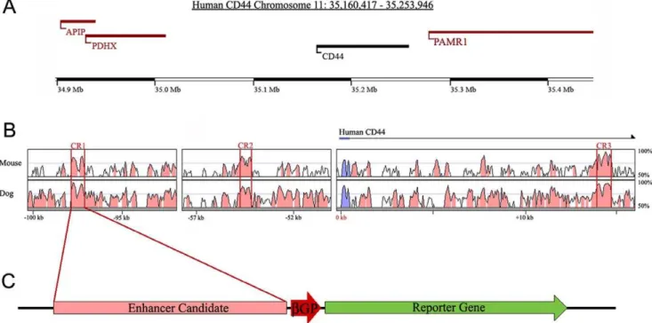
Figure 1. Prediction ofcis-regulatory elements for CD44 expression using sequence alignment analysis.(A) A genomic map of human CD44 and surrounding genes located on chromosome 11p13. (B) Multiple sequence alignment of homologous CD44 sequences using human sequence as baseline. 14 evolutionarily conserved regions were identified and predicted as potentialcis-regulatory elements for CD44 expression. Conserved regions 1–3 (CR1–3) have the highest levels of conservation. Blue regions represent CD44 coding sequence. Pink regions represent non-coding sequence. Peaks surrounded by red bars are highly conserved regions that have at least 70% conservation among species. (C) Plasmid reporter construct containing a conserved region of CD44, a minimal beta-globin-promoter (bGP), and green fluorescent protein (GFP).
from the Harvard Primer Bank (Table S2). Threshold cycles were normalized relative to GAPDH expression. Error bars represent the standard deviation of the mean.
Data Quantification
In all experiments, percentages represent the averages calculat-ed from at least three independent samples. All values are shown
as a mean6standard error of the mean. Error bars represent the standard error of the mean. In cases where results were tested for statistical significance, a student’s t-test was applied.
Immunocytochemistry
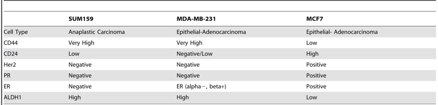
For immunocytochemistry, cells were plated on PLL treated coverslips and incubated for 24 hours and then fixed to coverslips Table 1.Expression of key factors in 3 breast cancer cell lines.
SUM159 MDA-MB-231 MCF7
Cell Type Anaplastic Carcinoma Epithelial-Adenocarcinoma Epithelial- Adenocarcinoma
CD44 Very High Very High Low
CD24 Low Negative/Low High
Her2 Negative Negative Positive
PR Negative Negative Positive
ER Negative ER (alpha2, beta+) Positive
ALDH1 High High Low
doi:10.1371/journal.pone.0050867.t001
Figure 2. CR1 directs reporter GFP expression in breast cancer cell lines.Conserved region was tested for the ability to direct reporter gene expression by transfecting breast cancer cell lines with CD44CR1-bGP-GFP construct (CD44CR1-GFP). Nuclei were stained with Hoechst 33342. (A–C) GFP expression in all three cell lines resulted from transfection of a positive control construct (CAG-GFP). (D–F) No GFP expression was detected from transfection of a negative control construct with a conserved region from NeuroD1gene. GFP expression from CR1 can be seen in MDA-MB-231 and SUM159 cells (G–H). However, no expression is seen in MCF7 cells (I).
using 4% paraformaldehyde, blocked with 10% Donkey Serum (Jackson Immunology) and then incubated with the primary antibody for 2 hours at room temperature. The following antibodies were used [CD44 (Chemicon); CD24 (Santa Cruz); NFkB-c-Rel (Chemicon); NFkB-p50 (Upstate); NFkB-p65 (Ab-cam); JUNB (Santa Cruz); FosB (Santa Cruz)]. Following incubation with primary antibody, cells were incubated with a fluorescent secondary antibody (Jackson Immunology) for 30
minutes at room temperature. Nuclei were stained with Hoechst33342.
Genomic DNA Sequencing
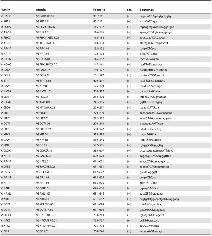
Genomic DNA was collected from the human cell lines using the Promega Genomic DNA kit as per manufacturer’s recom-mendations. Genomic DNA from each cell line was sequenced using primers specific for the conserved regions (Table S1). Table 2.Conserved transcription factor binding sites in CR1 between mouse and human.
Family Matrix From–to Str. Sequence
V$HAND V$PARAXIS.01 95–115 (+) cagaaACCAgatgtgttggtg
V$RP58 V$RP58.01 99–111 (2) aacaCATCtggtt
V$RORA V$REV-ERBA.02 115–137 (2) tagaagctgaGTCAcaggatgac
V$AP-1R V$NFE2.01 116–136 (2) agaagCTGAgtcacaggatga
V$PBXC V$PBX1_MEIS1.03 118–134 (2) aagctgagTCACaggat
V$AP-1R V$TCF11MAFG.01 118–138 (+) atcctgTGACtcagcttctat
V$AP-1F V$AP-1.01 122–132 (+) tgtgACTCagc
V$AP-1F V$AP-1.01 122–132 (2) gctgAGTCaca
V$GATA V$GATA.01 145–157 (+) tgctGATAaataa
V$HOXC V$PBX_HOXA9.01 145–161 (2) ttctTTATttatcagca
V$PAX6 V$PAX6.02 159–177 (+) gaagagtttCCAGgtatgc
V$BCL6 V$BCL6.02 161–177 (2) gcataccTGGAaactct
V$STAT V$STAT5.01 499–517 (+) tttcTTCTtcgaagttccc
V$CAAT V$NFY.03 176–190 (2) taaaCCAAacatagc
V$NKXH V$NKX31.01 203–217 (+) gacagtAAGTatacc
V$SNAP V$PSE.02 212–230 (+) tatacCCTAaagttaccaa
V$HAML V$AML3.01 241–255 (2) ggttGTGGttcagag
V$EBOX V$MYCMAX.02 259–271 (2) tcaacaCATGtga
V$IRFF V$IRF4.01 279–299 (+) aaaagaaaaaGAAAaaagaaa
V$IRFF V$IRF7.01 292–312 (+) aaaaGAAAtgaaaattggaaa
V$OCT1 V$OCT1.06 296–310 (+) gaaatgaaAATTgga
V$RBPF V$RBPJK.02 508–522 (2) cctaTGGGaacttcg
V$YBXF V$YB1.01 518–530 (2) cagatTGGCctat
V$CAAT V$NFY.01 519–533 (+) taggCCAAtctgtct
V$SP1F V$GC.01 537–551 (2) tgtggGGTGgggttg
V$CLOX V$CDPCR3.01 585–607 (2) gccctcagaaaaagatATTGctc
V$AP-1R V$BACH2.01 609–629 (2) aggcagTGAGtcagggtttac
V$AP-1R V$NFE2.01 611–631 (+) aaaccCTGActcactgcctcc
V$CREB V$TAXCREB.02 611–631 (+) aaacccTGACtcactgcctcc
V$CSEN V$DREAM.01 612–622 (2) gaGTCAgggtt
V$AP-1F V$AP-1.01 615–625 (+) cctgACTCact
V$AP-1F V$AP-1.01 615–625 (2) agtgAGTCagg
V$CARE V$CARF.01 626–636 (+) ggaagGAGGca
V$HAML V$AML1.01 631–645 (2) aactGTGGtaggaag
V$AIRE V$AIRE.01 631–657 (2) cagtgttttggaaactgTGGTaggaag
V$OCT1 V$POU2F3.01 671–695 (2) tctATGCagatctcagt
V$OCT1 V$OCT3_4.02 671–695 (+) gatctGCATagagacaa
V$FKHD V$HNF3.01 703–719 (2) tgtatgcAAACagctct
V$NFKB V$NFKAPPAB.01 725–737 (+) ctGGGAaatccct
V$NFKB V$NFKAPPAB.01 726–738 (2) aaGGGAtttccca
V$EVI1 V$EVI1.01 730–746 (2) aagacAAGAagggattt
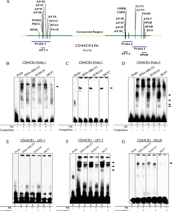
Figure 3. Specific protein factors bind with CR1.EMSAs were performed to determine thein vitrobinding activities of nuclear protein factors with CD44CR1. (A) DNA probe design using conserved mouse sequence and TFBSs within each probe. Probe 1 identified binding (indicated by arrow head) in two cell lines (MDA-MB-231 and MCF7), but not observed in SUM159. (B) Probe 2 showed strong binding present in all three cell lines (arrowheads). (C) Probe 3 showed multiple shifted bands and was successfully competed away in all three cell lines using unlabeled probes. (E) Probe AP-1-1 showed no band shift in any of the three cell lines. (F) Probe AP-1-2 resulted in a band shift in all three cell lines. All band shifts were competed away with an unlabeled probe. Arrowheads indicate bands specific to MDA-MB-231 and MCF7. (G) Probe NFkB showed a band shift that was successfully competed away in all three cell lines.
Genomic DNA was aligned using the online program ClustalW [31].
Electrophoresis Mobility Shift Assay and Supershift Single stranded DNA probes were designed from mouse CR1 and labeled with the 3’ Biotin End Labeling Kit (Thermo Scientific) as per manufacturer’s suggestions. Nuclear extracts were collected from each breast cancer cell line using NE-PER nuclear and cytoplasmic extraction reagents (Thermo Scientific). Binding reactions were performed and detected using the LightShift Chemiluminescent EMSA kit (Thermo Scientific) per manufac-turer’s recommendations. DNA-protein complexes were run on
10% non-denaturing poly-acrylamide gels and transferred onto Biodyne Plus membrane (Pall). Membranes were cross-linked in a UV imager for 15 minutes. EMSA probe sequences are inTable S3. Supershift assays were performed in a similar fashion. Antibodies were added to select reactions 15 minutes prior to addition of labeled probes.
Site Directed Mutagenesis
Site directed mutagenesis was performed as previously de-scribed [32] using primer sequences as listed inTable S4. Treated DNA was transformed into NEB5a cells (NEB) and plated onto LB-amp plates. Constructs were collected by Qiagen midi-prep Figure 4. Mutation of AP-1 and NFkB binding sites in CR1 reduces reporter GFP expression.Assays using site directed mutagenesis of
AP-1 and NFkB binding sites. (A–I) Schematic of each mutation of CR1 construct. Mutated sites are identified by a red X. (A’–I’) Transfection of each the constructs in SUM159 cells. (J) Quantification of the number of GFP-expressing cells/total number of cells counted. Control mutation at a non-conserved site (B’) showed no difference in GFP expression when compared to CR1 (A’). Single site mutations of AP-1-1, AP-1-2 and NFkB (C’-E’) showed a significant reduction of GFP expression compared to CR1. However, GFP expression was not eliminated entirely. Mutation of a combination of AP-1 and NFkB binding sites (F’-H’) did not reduce further GFP expression, however, the percentage of GFP expression was still significantly reduced compared to CR1. Mutation of all three TFBSs (I’) showed the greatest reduction of GFP expression. **p =,0.0005 ***p =,1.061025
(student’s t-test). Scale bar = 50mM.
and then sequenced to verify the resulting mutation. Mutated constructs were transfected into cells and tested for GFP expression.
Chromatin Immunoprecipitation
Chromatin immunoprecipitation (ChIP) was performed as previously described [33,34]. Sonication was performed using a Branson 450 Digital Sonicator. The chromatin extract was pre-cleared with protein A beads (NEB). NFkB-c-Rel (Chemicon); NFkB-p50 (Upstate); NFkB-p65 (Abcam); cJun(N) (Santa Cruz); cJun(D) (Santa Cruz); JUNB (Santa Cruz); FosB (Santa Cruz) antibodies were used to perform ChIP assay. Protein-DNA crosslinks were reversed with 30ml 5 M NaCl and incubating samples at 65uC for 4 hours. Proteins were digested with 0.1 mM EDTA, 20 mM Tris-HCl and 2ml Proteinase K solution (Active Motif) for 2 hours at 42uC. DNA was purified using phenol-chloroform extraction. PCR was performed to identify DNA:pro-tein interactions. PCR primers used for ChIP assays are listed in
Table S5.
shRNA-based Gene Knockdown
Short hairpin RNA (shRNA) sequence (leading strand) used for AP1-JUNB knockdown were CCTTCTACCACGACGACTCA-TACACAGCT and CACGACTACAAACTCCTGAAACC-GAGCCT. shRNA sequences for NFkB-p50 knockdown were GCAGCTCTTCTCAAAGCAGCAGGAGCAGA and GA-GAACTTTGAGCCTCTCTATGACCTGGA (OriGene Tech-nologies, Inc. , Rockville, MD). Control constructs were an empty vector and scrambled shRNA construct. Constructs were trans-fected into cell lines using Lipfectamine LTX (Life Technologies). Transfected cells were cultured for 72 hours before being fixed and stained as described above.
Results
Prediction ofcis-regulatory Elements for CD44 Expression using Sequence Alignment Analysis
To understand the molecular mechanism of CD44 expression in breast cancer cells, highly conserved regions of non-coding DNA were computationally predicted ascis-regulators of CD44 expres-sion. Multiple sequence alignment using the human CD44 genomic region as baseline revealed homologous regions in mouse, dog (Fig. 1A) and other mammalian species. A total of 14 conserved regions (CR) (.100 consecutive base pairs of sequence with.70% sequence identify) were identified. The three highest conserved regions (CR1–3, Fig. 1B) were chosen for further experimental verification, because many studies have shown that highly evolutionarily conserved noncoding DNA
sequences have a high potential to regulate gene expression [35,36]. CD44CR1 (CR1) contains 715 bp and located 95 kbp upstream of CD44 with 78% conservation. CR2 contains 611 bp with 76% conservation and is located 55 kbp upstream of CD44. CR3 contains 604 bp with 79% conservation and it is located in the first intron of the CD44 gene.
Conserved Regions have the Ability to Direct Reporter GFP Expression in Breast Cancer Cells
To test the CRs for their ability to direct gene expression, the CRs were PCR amplified from mouse genomic DNA and subcloned into an expression vector containing a b-globin minimal promoter (bGP) and green fluorescent protein (GFP) as the reporter gene (Fig. 1C). Mouse DNA was used to validate that evolutionarily conserved elements can function in different species.
The ability of the conserved regions to direct gene expression was tested using three previously characterized human breast cancer cells, MDA-MB-231, SUM159, and MCF7, each with a different CD44/CD24 expression profile (Table 1) [4,37]. Both MDA-MB-231 and SUM159 cells contain high levels of CD44 expression. In addition, SUM159 cells have been characterized with cancer stem cell like features including the ability to self-renew, reconstitute the parental cell line, survive chemotherapy, as well as form tumors with as few as 100 cells [4,37]. Thus, these cells provide different lines of validation.
First, immunofluorescence staining was performed to verify CD44 and CD24 expression level. Consistent with the genome-wide expression profiling study [4], MDA-MB-231 and SUM159 cells showed very high CD44 staining and low CD24 staining, while MCF7 showed low CD44 and high CD24 staining (Fig. S1A–C).
Then, CD44 and CD24 expression level in the three cell lines was further quantified using quantitative PCR (qPCR). Results showed that MDA-MB-231 and SUM159 cells have the high CD44 and low CD24 expression, while MCF7 cells have the opposite expression profile, i.e., a higher CD24 and lower CD44 expression (Fig. S1D).
Next, each reporter construct containing one of the top three conserved regions of CD44 was individually tested by transfection into the three cell lines. Transfection of the positive control construct, CAG-GFP, resulted in reporter GFP expression (Fig. 2A–C) and demonstrated the ability of each of the cell lines to be transfected. As negative controls, a highly conserved region in Neurod1 locus with bGP andbGP alone (data not shown), resulted in no visible GFP expression (Fig. 2D–F), indicating that not all highly conserved regions of genomic DNA norbGP alone have the ability to direct gene Figure 5. Differential AP-1 factor binding to CR1 in breast cancer cells.ChIP with AP-1 antibodies resulted in amplification of a region of CR1 with inverted repeat AP-1 binding sites. Rabbit IgG and anti-GFP antibody served as negative control. Representative results of at least two independent immunoprecipitation experiments and multiple independent PCR analyses are shown. Strong PCR amplification of CR1 region with JUNB binding was seen in SUM159 cells and with JUND binding in MCF7 cells.
expression. GFP expression was observed in MDA-MB-231 and SUM159 cell lines after transfection with CR1-GFP construct (Fig. 2G–H). More GFP-expressing cells were observed in SUM159 cells as compared to MDA-MB-231 cells, while no GFP-expressing cells were observed in MCF7 cells (Fig. 2I). Transfection of constructs containing CD44CR2 and CD44CR3 also resulted in GFP-expressing cells (data not shown, under further investigation).
Analysis of Trans-acting Factor Binding Sites on the Conserved Regions of CD44
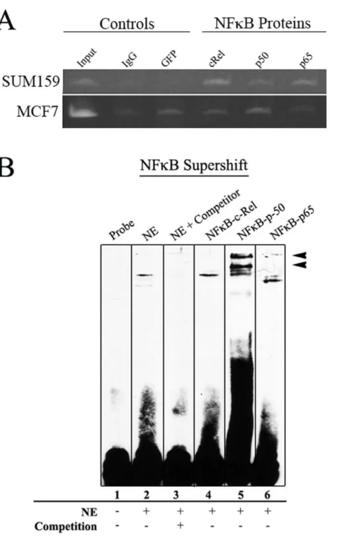
The ability of CR1 to direct different levels of reporter GFP expression among the three cell lines is most likely attributed to their interactions withtrans-acting factors. Therefore, CR1 of both mouse and human were examined fortrans-acting factor binding sites (TFBSs) and mutations in these sites. Genomic DNA of CR1 from each of the three cell lines was collected and sequenced to determine if mutations in the region that disrupt TFBSs. Figure 6. NFkB factors interact with CR1.ChIP assays were performed to identify CR1 interacting transcription factors. Rabbit IgG and anti-GFP
antibody served as negative control. (A) Strong PCR amplification of CR1 region with NFkBp50 and p65 were seen in SUM159 samples. MCF7 samples showed bands with intensities equal to the negative control. (B) Supershift with NFkB antibodies was performed with SUM159 nuclear extract. Anti NFkB-p50 and p65 antibodies were able to supershift the band, but NFkB-cRel antibody resulted in no shift.
Sequencing results show only a 5 bp span that differed between the three human cell lines in CR1 (Fig. S2). This 5 bp difference found in the SUM159 cells is located in an unconserved region of CR1 and showed no disruption of key TFBSs. This indicates that the difference in GFP expression among these cells may not be associated with the DNA sequence. Thus, we speculate that the difference in GFP expression may be the result of trans-acting factor binding in the cell lines. CR1 sequences from mouse (Table S6) and human (Table S7) both contained over 150 putative TFBSs as predicted by MatInspector [38]. These TFBSs were examined further for conservation between mouse and human sequences (Table 2). Most of these conserved TFBSs involved in breast cancer (e.g., AP-1, NFkB, and STAT5), stem cells and
embryonic development (e.g., OCT1, PAX6, GATA1), and therefore had the highest potential for regulating CD44 and for being involved in breast cancer. Our further analysis was thus focused on the activities of CR1 in regulating gene expression in breast cancer cells.
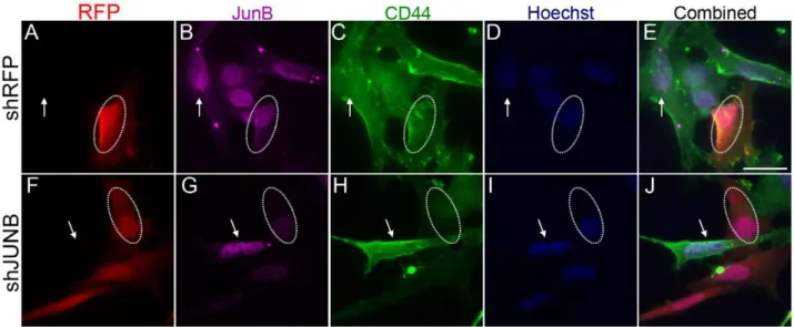
Sequence Specific Trans-acting Factor Binding with CR1 Electrophoretic mobility shift assays (EMSAs) were performed to determine if differences in GFP expression resulted from differences in trans-acting factor binding in the cells. Double-stranded, biotin labeled oligonucleotides corresponding to subre-gions of CR1 were assayed for trans-acting factor binding using nuclear extract from each of the three cell lines (Fig. 3A). The Figure 7. AP-1-JUNB knockdown decreases CD44 expression.Sum159 cells were transfected with control and JUNB shRNA constructs and then stained for JUNB and CD44 expression. Transfection with the control, scrambled DNA shRNA construct (A–E) showed no change in JUNB expression (B, circle) or CD44 expression (C, circle) when compared to un-transfected cells (arrows). Transfection with the JUNB shRNA construct (F–J) showed a reduction in JUNB expression (G, circle) and CD44 expression (H, circle) when compared to un-transfected cells (F–G, arrow).
doi:10.1371/journal.pone.0050867.g007
Figure 8. NFkB-p50 knockdown decreases CD44 expression.Sum159 cells were transfected with control and NFkB-p50 shRNA constructs and
then stained for NFkB-p50 and CD44 expression. Transfection with the control, scrambled DNA shRNA construct (A–E) showed no change in NFk B-p50 expression (B, circle) or CD44 expression (C, circle) when compared to un-transfected cells (arrows). Transfection with the NFkB-p50 shRNA construct (F–J) showed a reduction in NFkB-p50 expression (G, circle) and CD44 expression (H, circle) when compared to un-transfected cells (F–G, arrow).
shifted bands for three of the large probes spanning the length of the conserved regions in all three cell types (Fig. 3B–D) indicating protein-DNA binding activity. Probe 1 shows strong bands shifted with nuclear extracts from MDA-MB-231 and MCF7 cells only (Fig. 3B), while probe 2 has a band shifted that is equally strong with all three cell lines (Fig. 3C). Probe 3 shows a number of bands that can be competed away with an unlabeled probe (Fig. 3D). Although the bands in probe 3 are similar in all three cell lines, there was a band with SUM159 cells that is not present in the other two cell lines.
Smaller probes were then used to narrow down regions of binding and to identify specific TFBSs. A probe designed to mimic the first AP-1 site (AP-1-1) showed no band shift (Fig. 3E), while the probe for the second AP-1 site (AP-1-2) showed a number of band shifts (Fig. 3F). Although these bands were not completely competed away, there was a significant reduction in band intensity with the addition of the competition probe. A probe for the region of NFkB binding also revealed band shifts. The intensity of the band differed among cell lines, with SUM159 showing the strongest shift (Fig. 3G).
Mutation of AP-1 and NFkB Binding Sites Results in a Loss of CR1 Expression
EMSA identified regions of CR1 that were able to bind nuclear factors in each of the three cell lines. However, thesein vitroassays are not sufficient to determine if these factors have the ability to direct gene expression. To determine if the specific TFBSs are involved in the regulation of reporter GFP expression, site directed mutagenesis (SDM) was performed. The core binding sites for the two AP-1 TFBSs and NFkB binding site were deleted from the CR1 reporter construct using SDM. Mutant constructs were transfected into each of the cell lines. Wild-type CR1 and a random mutation were used as control transfections. Results show that the control transfections no significant difference in the percentage of GFP-expressing cells (Fig. 4A–B), whereas single site mutations at each AP-1 site and NFkB binding site (Fig. 4C– E) resulted in statistically significant decrease in the percentage of GFP-expressing cells in SUM159 cell line when compared to un-mutated CR1 and the control mutation (Fig. 4A–B).
Since GFP expression was not completely abolished with the deletion of a single TFBS in SUM159, we mutated a combination of TFBSs (Fig. 4F–H). Results of transfections with combinatorial mutations again showed a statistically significant decrease in the percentage of GFP-expressing cells (Fig. 4F–H). However, the percentage of GFP-expressing cells with two mutation constructs did not change significantly as compared with single-mutation constructs. To determine whether all three sites are needed for CR1 to direct GFP expression, the three binding sites were mutated (Fig. 4I). The transfection of this construct resulted in the highest decrease in the percentage of GFP-expressing cells. Interestingly, transfection of the mutant constructs into MDA-MB-231 resulted in no GFP-expressing cells (Fig. S3) suggesting regulation of CD44 in MDA-MB-231 differs from SUM159 cells. Trans-actingfactor binding assays identify components of AP-1 and NFkB binding to CR1 in SUM159 cells.
To determine whether the difference in reporter GFP expres-sion among the three breast cancer cells is due to thetrans-acting factors binding with CR1, chromatin immunoprecipitation (ChIP) assays were performed using antibodies against individual components of AP-1 and NFkB. ChIP results show that in SUM159 cells JUNB bound strongly with CR1, while in MCF7 cells only JUND bound to CR1 (Fig. 5). When ChIP assays were performed with antibodies against NFkB components (e.g., c-Rel, p50 and p65), SUM159 revealed weak binding with all three
NFkB antibodies (Fig. 6A). However, MCF7 showed no significant binding when compared to background. These results are supported by an EMSA supershift assay performed to verify specific proteins binding using antibodies against NFkB proteins c-Rel, p50 and p65 (Fig. 6B). The antibody against NFkB-p50 was able to provide a significant shift in the labeled probe. NFkB-p65 showed a weaker shift similar to NFkB-p50 as well as a band that was downshifted. Together these results support the notion that the different cell lines have different means by which they regulate CD44.
JUNB and NFkB-p50 Knockdown Represses CD44 Expression
To determine the effects of AP-1-JUNB and NFkB-p50 on CD44 expression, we performed shRNA gene knockdown experiments in SUM159 cells. Control transfections, with scram-bled control shRNA (Fig. 7A–E) or an empty vector (Fig. S3A– E), showed no change in JUNB or CD44 expression in transfected cells. Transfection of shJUNB constructs resulted in a decreased JUNB expression as shown by immunocytochemistry (Fig. 7F–J
andFig. S3F–J). Cells transfected with the shJUNB construct also showed a decrease in CD44 expression as compared to untransfected cells (Fig. 7). Similar results were seen with knockdown of NFkB-p50. Control shRNA transfection with a scrambled shRNA (Fig. 8A–E) or empty shRNA construct (Fig. S4A–E) showed no change in NFkB-p50 or CD44 expression. Knockdown of NFkB-p50 (Fig. 8F–JandFig. S4F–J) did result in a decrease in CD44 expression compared to untransfected cells. These results support the notion that JUNB and NFkBp50 interact with CR1 and regulate CD44 expression.
Discussion
In breast cancer, the up-regulation of CD44, a cell surface glycoprotein involved in cell-cell and cell-extracellular matrix adhesion, migration, differentiation and survival, is associated with cancer stem cells [39,40]. However, the mechanism for this gene up-regulation is not well understood. In this study, we identified the novelcis-element CR1, with the ability to direct reporter gene expression in a cell specific manner (Fig. 2), and the trans-acting factors AP-1 and NFkB as key factors involved in the regulation of CR1 (Fig. 3).
Genomic sequencing of CR1 from breast cancer cell lines did not reveal any major mutations that cause changes in key TFBSs (Fig. S2), which suggests that variations in reporter gene expression among these cells may be attributed to the difference intrans-acting factor binding to CR1.
Consistent with the notion that there was a difference intrans -acting factor(s) binding to CR1, mutations of TFBSs for AP-1and NFkB resulted in a significant reduction in GFP expression in two breast cancer cell lines (Fig. 4). Deletion of each site individually was able to completely eliminate reporter gene expression in MDA-MB-231 (Fig. S4). However, deletion of all three sites TFBS, individually and sequentially in SUM159 cells did not completely eliminate reporter gene expression (Fig. 4). These results indicate that factors AP-1 and NFkB are importanttrans -regulators of gene expression in breast cancer; and AP-1 and NFkB function in a cell type specific manner via various binding patterns to CR1 in different breast cancer cell lines. The inability to completely eliminate CR1 expression implies other TFs and/or co-factors may be involved in regulating CD44 expression in breast cancer stem-like SUM 159 cells.
different pattern of TF binding to CR1, i.e., JUNB in SUM159 and JUND in MCF7 (Fig. 5). ChIP results also showed that NFkB factors cRel, p50 and p65 bind to CR1 in SUM159 cells but not MCF7. This result was confirmed with an EMSA supershift with SUM159 nuclear extract, showing shifts with both NFkB-p50 and p65 (Fig. 6).
The observation that knockdown of AP-1-JUNB and NFkB-p50 reduced the expression of CD44 suggest the role of JUNB and p50 in regulating CD44 expression via their interaction with CR1. The fact that a complete loss of CD44 expression was not seen may be attributed to 1) reduced JUNB and p50 expression as opposed to a complete knockdown; 2) other factors interact with JUNB and/or p50 in the regulation of CD44 expression; and 3) other regulatory regions allowing basal expression of CD44.
Studies have shown that deletion of CD44 can lead to a reduction in recurrence of cancers [5] and metastasis [41]. By targeting the factors that result in the overexpression of CD44, we may be able to better treat breast cancer and metastatic tumors.
Previous studies have shown that AP-1 regulates CD44 expression [18,42–44]. AP-1 has an increased activity in small cell and non-small cell lung carcinomas, which lead to an increase in CD44 expression. In addition, a TRE binding element with Fra-1 in the promoter of CD44 has been identified [45,46]. These studies have established that AP-1 regulates CD44 expression via its interaction with CD44 promoter. In this study, our findings suggest that thecis-element CR1 functions via common factor AP-1 and/or NFkB and interact with the promoter to regulate CD44 expression, which provides new insight into regulatory mecha-nisms on complex CD44 expression.
Together, our findings suggest that CR1 has the potential to regulate CD44 expression in breast cancer and BCSCs via its interaction with AP-1 and NFkB factors. Further studies will focus on how CR1 interacts with the promoter to regulate CD44 expression. CD44 is known to have a complex expression patterns with ubiquitous expression and variant forms, and has been implicated in the aggressiveness and metastasis of a number of cancer types [9,11,37,47]. Therefore, the regulation of such a molecule could be equally complex. A full understanding of complex regulation of CD44 expression requires the investigation of the othercis- andtrans-regulators of CD44.
Supporting Information
Figure S1 CD44 and CD24 expression in breast cancer cell lines as detected by immunocytochemistry. Human cell lines MDA-MB-231 (a–a’’’), SUM159 (b–b’’’), and MCF7 (c– c’’’) were fixed and stained for CD44 (F10442, Millipore) and CD24 (91, Millipore). Nuclei were stained with Hoechst33342. D. Real-time PCR analysis of CD44 and CD24 mRNA levels in breast cancer cell lines. GAPDH served as endogenous control. Immunohistochemistry and Real-time PCR showed high CD44 and low CD24 expression in MDA-MB231 and SUM159 cell lines. MCF7 cells showed low CD44 and high CD24 expression. Scale bar = 100mm.
(TIF)
Figure S2 Genomic sequence alignment of conserved regions reveals no mutations in TFBSs.Genomic DNA was
obtained from the cell lines MDA-MB-231, SUM159 and MCF7. Genomic DNA was sequenced at CD44CR1 conserved region and aligned using ClustalW. Alignment of CD44CR1 sequences identified a 5 bp deletion located in SUM159 genomic DNA. However, these mutations do not change TFBSs.
(TIF)
Figure S3 JUNB knockdown decreases CD44 expres-sion. Sum159 cells were transfected with control and JUNB shRNA constructs and then stained for JUNB and CD44 expression. Transfection with the control, empty vector shRNA construct (A–E) showed no change in JUNB expression (B, circle) or CD44 expression (C, circle) when compared to un-transfected cells (arrows). Transfection with the JUNB shRNA construct (F–J) showed a reduction in JUNB expression (G, circle) and CD44 expression (H, circle) when compared to un-transfected cells (F–G, arrow).
(TIF)
Figure S4 NFkBp50 knockdown decreases CD44
expres-sion.Sum159 cells were transfected with control and NFkB-p50 shRNA constructs and then stained for NFkB-p50 and CD44 expression. Transfection with the control, empty vector shRNA construct (A–E) showed no change in NFkB-p50 expression (B, circle) or CD44 expression (C, circle) when compared to un-transfected cells (arrows). Transfection with the NFkB-p50 shRNA construct (F–J) showed a reduction in NFkB-p50 expression (G, circle) and CD44 expression (H, circle) when compared to un-transfected cells (F–G, arrow).
(TIF)
Table S1 PCR Primers for the amplification of the three conserved regions.
(DOC)
Table S2 qPCR primer sequences obtained from Har-vard Primer Bank.
(DOC)
Table S3 Probe design for EMSA.
(DOC)
Table S4 Primers used for site directed mutagenesis.
(DOC)
Table S5 Primers used for ChIP assays.
(DOC)
Table S6 Predicted transcription factor binding sites from mouse CD44CR1.
(DOC)
Table S7 Predicted transcription factor binding sites from human CD44CR1.
(DOC)
Author Contributions
Conceived and designed the experiments: LC. Performed the experiments: SMS. Analyzed the data: SMS LC. Contributed reagents/materials/ analysis tools: SMS LC. Wrote the paper: SMS LC.
References
1. Ferlay J, Shin HR, Bray F, Forman D, Mathers C, et al. (2010) Estimates of worldwide burden of cancer in 2008: GLOBOCAN 2008. Int J Cancer 127: 2893–2917.
2. Al-Hajj M, Wicha MS, Benito-Hernandez A, Morrison SJ, Clarke MF (2003) Prospective identification of tumorigenic breast cancer cells. Proc Natl Acad Sci U S A 100: 3983–3988.
3. Fillmore C, Kuperwasser C (2007) Human breast cancer stem cell markers CD44 and CD24: enriching for cells with functional properties in mice or in man? Breast Cancer Res 9: 303.
5. Jin L, Hope KJ, Zhai Q, Smadja-Joffe F, Dick JE (2006) Targeting of CD44 eradicates human acute myeloid leukemic stem cells. Nat Med 12: 1167–1174. 6. Dave B, Chang J (2009) Treatment resistance in stem cells and breast cancer.
J Mammary Gland Biol Neoplasia 14: 79–82.
7. Marotta LL, Polyak K (2009) Cancer stem cells: a model in the making. Curr Opin Genet Dev 19: 44–50.
8. Foster LC, Wiesel P, Huggins GS, Panares R, Chin MT, et al. (2000) Role of activating protein-1 and high mobility group-I(Y) protein in the induction of CD44 gene expression by interleukin-1beta in vascular smooth muscle cells. FASEB J 14: 368–378.
9. Ponta H, Sherman L, Herrlich PA (2003) CD44: from adhesion molecules to signalling regulators. Nat Rev Mol Cell Biol 4: 33–45.
10. Sheridan C, Kishimoto H, Fuchs RK, Mehrotra S, Bhat-Nakshatri P, et al. (2006) CD44+/CD24- breast cancer cells exhibit enhanced invasive properties: an early step necessary for metastasis. Breast Cancer Res 8: R59.
11. Naor D, Wallach-Dayan SB, Zahalka MA, Sionov RV (2008) Involvement of CD44, a molecule with a thousand faces, in cancer dissemination. Semin Cancer Biol 18: 260–267.
12. Hebbard L, Steffen A, Zawadzki V, Fieber C, Howells N, et al. (2000) CD44 expression and regulation during mammary gland development and function. J Cell Sci 113 ( Pt 14): 2619–2630.
13. Herrlich P, Morrison H, Sleeman J, Orian-Rousseau V, Konig H, et al. (2000) CD44 acts both as a growth- and invasiveness-promoting molecule and as a tumor-suppressing cofactor. Ann N Y Acad Sci 910: 106–118; discussion 118– 120.
14. Orian-Rousseau V (2010) CD44, a therapeutic target for metastasising tumours. Eur J Cancer 46: 1271–1277.
15. Liu C, Kelnar K, Liu B, Chen X, Calhoun-Davis T, et al. (2011) The microRNA miR-34a inhibits prostate cancer stem cells and metastasis by directly repressing CD44. Nat Med 17: 211–215.
16. Mandal CC, Ghosh-Choudhury N, Yoneda T, Choudhury GG (2011) Simvastatin prevents skeletal metastasis of breast cancer by an antagonistic interplay between p53 and CD44. J Biol Chem 286: 11314–11327. 17. Ameyar M, Wisniewska M, Weitzman JB (2003) A role for AP-1 in apoptosis:
the case for and against. Biochimie 85: 747–752.
18. Eferl R, Wagner EF (2003) AP-1: a double-edged sword in tumorigenesis. Nat Rev Cancer 3: 859–868.
19. Mao X, Orchard G, Mitchell TJ, Oyama N, Russell-Jones R, et al. (2008) A genomic and expression study of AP-1 in primary cutaneous T-cell lymphoma: evidence for dysregulated expression of JUNB and JUND in MF and SS. J Cutan Pathol 35: 899–910.
20. Damm S, Koefinger P, Stefan M, Wels C, Mehes G, et al. (2010) HGF-promoted motility in primary human melanocytes depends on CD44v6 regulated via NF-kappa B, Egr-1, and C/EBP-beta. J Invest Dermatol 130: 1893–1903.
21. Heintzman ND, Ren B (2009) Finding distal regulatory elements in the human genome. Curr Opin Genet Dev 19: 541–549.
22. Heintzman ND, Hon GC, Hawkins RD, Kheradpour P, Stark A, et al. (2009) Histone modifications at human enhancers reflect global cell-type-specific gene expression. Nature 459: 108–112.
23. Orom UA, Shiekhattar R (2011) Noncoding RNAs and enhancers: complica-tions of a long-distance relacomplica-tionship. Trends in genetics : TIG 27: 433–439. 24. Wang J, Zhuang J, Iyer S, Lin X, Whitfield TW, et al. (2012) Sequence features
and chromatin structure around the genomic regions bound by 119 human transcription factors. Genome Res 22: 1798–1812.
25. Vernot B, Stergachis AB, Maurano MT, Vierstra J, Neph S, et al. (2012) Personal and population genomics of human regulatory variation. Genome Res 22: 1689–1697.
26. Schaub MA, Boyle AP, Kundaje A, Batzoglou S, Snyder M (2012) Linking disease associations with regulatory information in the human genome. Genome Res 22: 1748–1759.
27. Cheng C, Alexander R, Min R, Leng J, Yip KY, et al. (2012) Understanding transcriptional regulation by integrative analysis of transcription factor binding data. Genome Res 22: 1658–1667.
28. Arvey A, Agius P, Noble WS, Leslie C (2012) Sequence and chromatin determinants of cell-type-specific transcription factor binding. Genome Res 22: 1723–1734.
29. Doh ST, Zhang Y, Temple MH, Cai L (2007) Non-coding sequence retrieval system for comparative genomic analysis of gene regulatory elements. BMC Bioinformatics 8: 94.
30. Brudno M, Do CB, Cooper GM, Kim MF, Davydov E, et al. (2003) LAGAN and Multi-LAGAN: efficient tools for large-scale multiple alignment of genomic DNA. Genome Res 13: 721–731.
31. Thompson JD, Higgins DG, Gibson TJ (1994) CLUSTAL W: improving the sensitivity of progressive multiple sequence alignment through sequence weighting, position-specific gap penalties and weight matrix choice. Nucleic Acids Res 22: 4673–4680.
32. Nohr J, Kristiansen K (2003) Site-directed mutagenesis. Methods Mol Biol 232: 127–131.
33. McGarvey KM, Fahrner JA, Greene E, Martens J, Jenuwein T, et al. (2006) Silenced tumor suppressor genes reactivated by DNA demethylation do not return to a fully euchromatic chromatin state. Cancer Res 66: 3541–3549. 34. Fullwood MJ, Han Y, Wei CL, Ruan X, Ruan Y (2010) Chromatin interaction
analysis using paired-end tag sequencing. Curr Protoc Mol Biol Chapter 21: Unit 21 15 21–25.
35. Visel A, Minovitsky S, Dubchak I, Pennacchio LA (2007) VISTA Enhancer Browser–a database of tissue-specific human enhancers. Nucleic Acids Res 35: D88–92.
36. Visel A, Prabhakar S, Akiyama JA, Shoukry M, Lewis KD, et al. (2008) Ultraconservation identifies a small subset of extremely constrained develop-mental enhancers. Nat Genet 40: 158–160.
37. Charafe-Jauffret E, Ginestier C, Iovino F, Wicinski J, Cervera N, et al. (2009) Breast cancer cell lines contain functional cancer stem cells with metastatic capacity and a distinct molecular signature. Cancer Res 69: 1302–1313. 38. Cartharius K, Frech K, Grote K, Klocke B, Haltmeier M, et al. (2005)
MatInspector and beyond: promoter analysis based on transcription factor binding sites. Bioinformatics 21: 2933–2942.
39. Wright MH, Calcagno AM, Salcido CD, Carlson MD, Ambudkar SV, et al. (2008) Brca1 breast tumors contain distinct CD44+/CD24- and CD133+cells with cancer stem cell characteristics. Breast Cancer Res 10: R10.
40. Zoller M (2011) CD44: can a cancer-initiating cell profit from an abundantly expressed molecule? Nat Rev Cancer 11: 254–267.
41. Wallach-Dayan SB, Rubinstein AM, Hand C, Breuer R, Naor D (2008) DNA vaccination with CD44 variant isoform reduces mammary tumor local growth and lung metastasis. Mol Cancer Ther 7: 1615–1623.
42. Young MR, Colburn NH (2006) Fra-1 a target for cancer prevention or intervention. Gene 379: 1–11.
43. Ramos-Nino ME, Blumen SR, Pass H, Mossman BT (2007) Fra-1 governs cell migration via modulation of CD44 expression in human mesotheliomas. Mol Cancer 6: 81.
44. Lamb RF, Hennigan RF, Turnbull K, Katsanakis KD, MacKenzie ED, et al. (1997) AP-1-mediated invasion requires increased expression of the hyaluronan receptor CD44. Mol Cell Biol 17: 963–976.
45. Hofmann M, Rudy W, Gunthert U, Zimmer SG, Zawadzki V, et al. (1993) A link between ras and metastatic behavior of tumor cells: ras induces CD44 promoter activity and leads to low-level expression of metastasis-specific variants of CD44 in CREF cells. Cancer Res 53: 1516–1521.
46. Risse-Hackl G, Adamkiewicz J, Wimmel A, Schuermann M (1998) Transition from SCLC to NSCLC phenotype is accompanied by an increased TRE-binding activity and recruitment of specific AP-1 proteins. Oncogene 16: 3057– 3068.