UDC 618.19-006.6:547.914 DOI:10.2298/ZMSPN1223065H
J e l e n a D. H r u b i k*, S o n j a N. K a i š a r e v i ć1, B r a n k a D. G l i š i ć1, E m i l i j a Đ. J o v i n2,
N e d a M. M i m i c a - D u k i ć2 a n d R a d m i l a Z. K o v a č e v i ć1 1 University of Novi Sad Faculty of Sciences, Department of Biology and Ecology,
Dositeja Obradovica Square 2, 21000 Novi Sad, Serbia
2 University of Novi Sad Faculty of Sciences, Department of Chemistry, Biochemistry and Environmental Protection, Dositeja Obradovica Square 3, 21000 Novi Sad, Serbia
MYRTUS COMMUNIS
AND
EUCALYPTUS CAMALDULENSIS
CYTOTOXICITY ON BREAST CANCER CELLS
ABSTRACT: In vitro cytotoxicity of methanol, ethyl acetate, n-buthanol, and water extracts of Myrtus communis L. and Eucalyptus camaldulensis Dehnh. was examined against two human breast cancer cell lines (MCF 7 and MDA-MB-231) using MTT and SRB assays. The results showed significant cytotoxic potential of examined extracts, with IC50 values ranging from 7 to 138 µg/ml for M. communis and 3-250 µg/ml for E. camald-ulensis. The two plants generally expressed similar activity, and no significant difference in cell line’s sensitivity towards extracts was observed. The results indicate to M. communis and E. camaldulensis as candidates for thorough chemical analyses for identification of ac-tive compounds, and eventually for attention in the process of discovery of new natural products in the control of cancer.
KEY WORDS: cytotoxicity, Eucalyptus camaldulensis Dehnh., MCF 7, MDA-MB-231, Myrtus communis L.
INTRODUCTION
Molecules derived from natural sources, such as plants, play a dominant role in the discovery of conventional drugs for the treatment of most human diseases, and therefore represent a basis of modern medicine (J o n e s et al., 2006). A large number of plant extracts have been screened for cytotoxic ef-fects against cancer cell lines over the last thirty years, which have resulted in some significant drugs being introduced. In addition, it was shown that some extracts are cytotoxic and selective, either between different cancer cell lines or between cancer and non-cancer cell lines, and act principally by inhibiting cell proliferation, but by different mechanisms ( d a R o c h a et al., 2001).
* Corresponding author: E-mail: [email protected]; Phone: ++381 21 485 2675
Fax: ++381 21 450 620
Myrtus communis L. (myrtle) and Eucalyptus camaldulensis Dehnh., both belonging to the Myrtaceae family, grow throughout the Mediterranean region (G r b o v i ć et al., 2010; M i m i c a - D u k i ć et al., 2010). Myrtle’s leaves and fruit are traditionally used as antiseptic, disinfectant, and hypoglyc-emic agents (E l f e l l a h et al., 1984). M. communis extracts have been con-firmed to exhibit antimicrobial (M a n s o u r i, 1999; M a n s o u r i et al., 2001; B u g a r i n, 2010) and antioxidative effects (B u g a r i n, 2010; Tu b e r o s o, 2010), while its essential oil showed considerable antioxidant and antimuta-genic effects (M i m i c a - D u k i ć et al., 2010). It is prominent that myrtle plants could be a promising source of natural antioxidants, anti-genotoxic, antimutagenic and, perhaps, chemopreventive agents (M i m i c a - D u k i ć et al., 2010). Several species of Eucalyptus are also used in traditional medicine, and are reported as agents with analgesic, anti-inflammatory, and antimicrobial properties (S i l v a et al., 2003; W i l l i a m s et al., 1998). Crude organic extract of E. camaldulensis has been proven to exhibit both antimicrobial and gastro-protective activities (A d e n i y i et al., 2006), while its essential oil showed antibacterial, antioxidant and antimutagenic effects (G r b o v i ć et al., 2010). The presence of flavonoids and tannins, which are known to have antibacterial and antifungal properties, is confirmed in organic extracts of E. globulus, which in addition revealed antimicrobial activity (E g w a i k h i d e et al., 2008). How-ever, anticancer activity of both Myrtus and Eucalyptus species has been poorly investigated, and their complete toxicity profile has yet to be determined.
In this study, the in vitro cytotoxicity of different extracts of M. communis
and E. camaldulensis was examined against MCF 7 and MDA-MB-231 human breast cancer cell lines using MTT and SRB cytotoxicity assays.
MATERIALS AND METHODS
Plant material and preparation of extracts: Plant extracts were prepared from dry leaves of the species Myrtle communis L. and Eucalyptus camaldu-lensis Dehnh., collected at the Montenegro coastline (Tivat) in August 2004. Air-dried, finely ground leaves samples were extracted by maceration with 70% methanol (8 ml per 1 g of drug) during 48h at room temperature. After filtration, solvent was evaporated in vacuum; crude residue was dissolved in hot distilled water and washed exhaustively with petrol ether to remove ballast compounds (lipids and pigments). A part of washed extract was concentrated in vacuum to dryness, and dissolved in DMSO giving final concentration of 10 mg/ml (“methanol extract”). The rest of the washed extract was partitioned with chloroform, ethyl acetate, and n-butanol, in order to obtain chloroform, ethyl acetate, n-butanol, and water soluble fractions. All of the fractions were concentrated in vacuum and dissolved in DMSO (10 mg/ml).
de-scribed (J e v t o v i ć et al., 2010; K a r a m a n et al., 2009). The cells were grown in Dulbecco’s Modified Eagle’s Medium with 4.5% of glucose (DMEM, PAA Laboratories) supplemented with 10% fetal calf serum (FCS, PAA Lab-oratories) at 37°C in a humidified atmosphere containing 5% CO2. Cells were
seeded into 96-well microtiter plates at the density 5000 cells/0.1 ml/well to ensure their exponential growth throughout the experimental period. After 24h, cells were exposed in triplicates to serial of extracts’ dilutions (50-1.56 µg/ml) dissolved in dimethyl sulfoxide (DMSO, Sigma) for 72h. The final concentra-tion of DMSO did not exceed 0.5%, and did not cause any background re-sponse in the bioassay. Control and cell-free blank wells were included in each plate. Following incubation, cytotoxicity assays were conducted.
MTT test: The principle of MTT test is based on cellular reduction of the soluble yellow MTT tetrazolium salt (3-(4,5-Dimethyl-2-thiazolyl)-2,5-diphe-nyl-2H-tetrazolium bromide) to a blue formazan product by mitochondrial dehydrogenases in viable cells. The intensity of the blue color formed by this reaction is a criterion for cell viability. Following incubation with plant extracts, the medium was removed and cells were incubated for 3h with 0.05mg/0.1ml/ well of MTT (Sigma) dissolved in serum-free DMEM. After that, the medium was removed, formazan salts were dissolved in 0.1 ml/well of 0.04 M HCl in isopropanol, and light absorbtion was measured using a plate reader (Thermo--Labsystems) on 540 nm, with reference wavelength 690 nm.
SRB test: SRB test is a colorimetric assay, based on the ability of the protein dye sulforhodamine B (SRB) to bind to protein basic amino acid resi-dues of trichloroacetic acid (TCA)-fixed cells. The incorporated dye can be solubilized for measurement, and the results are linear with cell number (Voigt, 2005). Following incubation with plant extracts, the medium was re-moved and the cells were fixed in TCA (0.025 ml of 50% w/v TCA per well) for 1 h at 4°C, washed five times with distilled water, air-dried, and stained with 0.05 ml of 0.4% SRB in 1% acetic acid for 30 min. Then, the cells were washed five times with 1% acetic acid to remove the unbound dye and air-dried. The incorporated dye was solubilized in 10 mM TRIS (pH 10.5) for 5 min, and light absorption was measured using a plate reader (ThermoLabsys-tems) on 492 nm, with reference wavelength 690 nm.
Calculation of cytotoxicity and IC50: In both tests, cell cytotoxicity was calculated as a percentage of corresponding control value (non-treated cells) obtained in a minimum of three independent experiments. The half-maximal inhibitory concentration values (IC50), defined as the concentration that in-hibits 50% of cell growth, were calculated from concentration-response curves. IC50 values were expressed as the mean of a minimum of three repeated ex-periments performed for each plant extract.
RESULTS AND DISCUSSION
categories: acute (< 1h to full toxicity), subacute (1–40h), and long term (>40h) (X i a et al., 2008). Guided by these findings, exposure time of 72h in our study has been selected to allow the expression of full cytotoxic potential of investigated plant extracts.
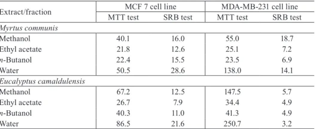
All M. communis and E. camaldulensis extracts exhibited cytotoxic ef-fects on MCF 7 and MDA-MB-231 cell lines, and the results were confirmed both by MTT and SRB assay. At the highest tested concentration (50 µg/ml), cytotoxicity of organic extracts/fractions was in the range from 40% to as high as 98%, while for water fraction it was in the range of 30-70%. The effects were dose dependent and based on dose-response curves IC50 values were determined (Table 1). In the case of M. communis, ethyl acetate and n-butanol fractions exhibited the highest antiproliferative effect on both cell lines with IC50 values ranging from 7 to 25 µg/ml. Total methanol extract and water frac-tion induced less pronounced cytotoxicity, with IC50 values ranging from 14 to 138 µg/ml. Overall pattern of the results obtained for E. camaldulensis extracts also indicate to ethyl acetate and n-butanol fractions as the most cytotoxic (IC50 5-41 µg/ml). It can also be noticed that general level and pattern of cyto-toxicity of all investigated extracts is rather similar for both tested plants.
Further commenting on obtained IC50 results and antiproliferative po-tency of tested plant extracts requires a precaution due to a noticeable differ-ence between the IC50 values obtained by the two applied cytotoxicity assays. Namely, IC50 values determined by SRB assay are in all cases lower than ones calculated from MTT assay. This dissimilarity is especially pronounced in the case of treatment of MDA-MB-231 cells by E. camaldulensis extracts, with the range of difference from 7 to 78 times. MTT and SRB assays are basically used for the assessment of cellular chemosensitivity and are related to the total cell number, therefore giving a relative measure of survival (R i s t i ć -- F i r a et al., 2008). The reason for a discrepancy in our results probably lies in a difference in the sensitivity of applied cytotoxicity assays and targets which they reflect, since they measure distinct biological parameters in living cells. The same cell line could have poor capacity to reduce MTT, and at the same time could show high values of absorbance in the SRB assay, all because SRB assay does not depend on enzymatic activity but on protein content of the cells. In contrast to MTT testing, SRB staining is less sensitive to environ-mental fluctuation such as variations in pH or depletion of glucose, and is independent of intermediary metabolism, which are all the factors that may be influenced by the test substances (L i n et al., 1999). In addition, for the study of growth kinetics, the SRB assay is more suitable because of the higher sen-sitivity and better linearity with cell number (K e e p e r s et al., 1991).
In any case, when comparing our results with the findings from the sim-ilar studies, antiproliferative potency of M. communis and E. camaldulensis on the two human breast cancer cell lines is evident. Aqueous acetone extract of
IC50 values of 17.2-17.5 µg/ml, while leaf extract had IC50 value of 19.3 µg/ml (To p ç u et al., 2011). Ethanolic extract of plants used in Thai traditional medicine showed high cytotoxic activity against MCF 7 cells (IC50 = 31-35 µg/ml), but water extract showed no cytotoxic effect (S a k p a k d e e j a r o e n and I t h a r a t, 2009). Methanolic extract of E. camaldulensis also revealed marked toxicity (IC50 = 20.7 µg/ml) on human bladder carcinoma cell line as determined by MTT test (A l - F a t i m i et al., 2005). On the other hand, leaves extract of another plant from Myrtaceae family expressed very low cytotoxic potency on MCF 7 cells (IC50 = 820±190 µg/ml), and were no cytotoxic to MDA-MB-231 cells as determined by MTT test (K a i l e h et al., 2007).
Chemical constituents responsible for the cytotoxic activities detected in our study might be speculated. Among the investigated plant extracts, total methanol extract of both M. communis and E. camaldulensis was chemically ana-lyzed. The results revealed high levels of plant phenols, especially flavonoids, flavonol-3-O-glycosides, flavonol-7-O-dyglicosides, phenol carbolic acids and their derivatives, rutin, elagic acid and their derivatives, etc. (B u g a r i n, 2010; G r b o v i ć, 2010). It is shown that some flavonoids exert cytotoxic ef-fects towards human lung embryonic fibroblasts and human umbilical vein endothelial cells by increasing level of intracellular reactive oxygen species (M a t s u o et al., 2005). In addition, structure-cytostatic activity relationship was shown after treatment of Rhesus monkey kidney and rat glial tumor cells by various flavonoids (S á n c h e z et al., 2001). All these results indicate that the compounds identified in methanol extract from our study might be respon-sible for its cytotoxicity. Other plant extracts were not chemically analyzed, but certain conclusions about type of compounds responsible for their effect might be presumed according to the results of comparable studies. In one of them, the cytotoxic activity of leaf organic extracts of E. camaldulensis was attributed to Tab. 1 – IC50 values (µg/ml) of different extracts of Myrtus communis and Eucalyptus camaldu-lensis, obtained by MTT test and SRB test on MCF 7 and MDA-MB-231 human breast cancer cell lines.
Extract/fraction MTT testMCF 7 cell lineSRB test MTT testMDA-MB-231 cell lineSRB test Myrtus communis
Methanol 40.1 16.0 55.0 18.7 Ethyl acetate 21.8 12.6 25.1 7.2 n-Butanol 22.4 15.5 23.5 6.9
Water 50.5 28.6 138.0 14.1
Eucalyptus camaldulensis
Methanol 67.2 12.5 147.5 5.7 Ethyl acetate 26.7 7.9 34.4 4.9 n-Butanol 40.3 11.0 41.3 4.9
Water 86.5 21.6 250.7 3.2
triterpenoids, mostly urosolic and oleanolic acids (To p ç u et al., 2011). Glob-ulocin A and eucaglobin isolated from leaves of E. globules had antioxidant, anti-inflammatory, and anti-melanogenesis activity (H a s e g a w a et al., 2008). Moreover, it was shown that myrtucommulone, a unique nonprenylated acylphloroglucinol contained in the leaves of M. communis, acts as a strong in-ducer of apoptosis selectively for cancer cells with lower cytotoxicity for nor-mal non-transformed cells (T r e t i a k o v a et al., 2008). In addition, all these, or structurally similar substances, could also present active compounds in ex-tracts from our study.
It is known that in vitro cytotoxicity can be cell-type specific (X i a et al., 2008; S á n c h e z et al., 2001). Regarding sensibility of the two cell lines (MCF 7 and MDA-MB-231) towards investigated plant extracts, our results did not reveal significant difference in a cellular response. Only in the case of
E. camaldulensis extracts, results of SRB test could indicate a greater sensi-tivity of MDA-MB-231 cell line. However, since this finding is not confirmed by MTT test, no conclusions on difference in sensitivity could be drawn. The same cell lines were used in testing of medicinal plant extracts in other study, and they exhibited different sensitivity towards the plant extracts (S t e e n k a m p and G o u w s, 2006). There is also an example where MCF 7 and MDA-MB-231 cell lines, depending on a tested plant extracts, had either very similar or a dif-ferent sensitivity as determined by MTT test (K a i l e h et al., 2007).
CONCLUSION
In conclusion, examined extracts of M. communis and E. camaldulensis
exhibit considerable cytotoxic potential for MCF 7 and MDA-MB-231 human breast cancer cell lines. The two plants can be recommended for thorough chemical analyses for identification of active compounds and eventually for attention in the process of discovery of new natural products in the control of cancer.
ACKNOWLEDGMENTS
This study was supported by Serbian Ministry of Education and Science Grants No. 173037 and 172058. Dr. Gordana Bogdanović from the Oncology Institute of Vojvodina, Sremska Kamenica, Serbia, is acknowledged for her valuable advices concerning cell culture experiments.
REFERENCES
A l - F a t i m i, M., F r i e d r i c h, U., J e n e t t - S i e m s, K. (2005): Cytotoxicity of plants used in traditional medicine in Yemen, Fitoterapia 76: 355–358.
B u g a r i n, D. (2010): Antioxidant, antimicrobial and antimutagenic potential of the Myrtus communis L. PhD dissertation, University of Novi Sad, Novi Sad.
d a R o c h a, A. B., L o p e s, R. M., S c h w a r t s m a n n, G. (2001): Natural products in anti-cancer therapy, Curr. Opin. Pharmacol. 1: 364–369.
E g w a i k h i d e, P. A., O k e n i y i, S. O., A k p o r h o n o r, E. E., E m u a, S. O. (2008): Studies on bioactive metabolites constituents and antimicrobial evaluation of leaf extracts of Eucalyptus globulus, Agricultural Journal 3: 42–45.
E l f e l l a h, M. S., A k h t e r, M. H., K h a n, M. T. (1984): Anti-hyperglycemic effect of an ex-tract of Myrtus communis in streptozotocin-induced diabetes in mice, J. Ethnopharma-col. 11: 275–281.
G r b o v i ć, S. (2010): Chemical features and biological activities of the E. camaldulensis Dehnh. and E. gunnii Hook. from Montenegro. PhD dissertation, University of Novi Sad, Novi Sad. G r b o v i ć, S., O r č i ć, D., C o u l a d i s, M., J o v i n, E., B u g a r i n, D., B a l o g, K., M i m i -c a - D u k i ć, N. (2010): Variation of essential oil -composition of Eu-calyptus -camaldulensis (Myrtaceae) from Montenegro coastline, APTEFF 41: 151–158.
H a s e g a w a, T., T a k a n o, F., T a k a t a, T., N i i y a m a, M., O h t a, T. (2008): Bioactive mono terpene glycosides conjugated with gallic acid from leaves of Eucalyptus globulus, Phytochemistry 69: 747–753.
J e v t o v i ć, V. S., P e l o s i, G., I a n e l l i, S., K o v a č e v i ć, R. Z., K a i š a r e v i ć, S. (2010): Synthesis, structural studies and biological activity of a dioxovanadium(V) complex with pyridoxal semicarbazone, Acta. Chim. Slov. 57: 363–369.
J o n e s, W. P., C h i n, Y. W., K i n g h o r n, A. D. (2006): The role of pharmacognosy in mod-ern medicine and pharmacy, Curr. Drug Targets 7: 247–264.
K a i l e h, M., B e r g h e, W. V., B o o n e, E., E s s a w i, T., H a e g e m a n, G. (2007): Screening of indigenous Palestinian medicinal plants for potential anti-inflamatory and cytotoxic activity, J. Ethnopharmacol. 113, 510–516.
K a r a m a n, M., K a i š a r e v i ć, S., S o m b o r s k i, J., K e b e r t, M., M a t a v u l j, M. (2009): Biological activities of the lignicolous fungus Meripilus giganteus (Pers.: Pers.) Karst, Arch. Biol. Sci. 61: 853–861.
K e e p e r s, Y. P., P i z a o, P. E., P e t e r s, G. J., V a n A r k - O t t e, J., W i n o g r a d, B., P i n e d o , H. M. (1991): Comparison of the sulforhodamine B protein and tetrazolium (MTT) assays for in vitro chemosensitivity testing, Eur. J.Cancer 7: 897–900.
L i n, Z. X., H o u l t, J. R. S., R a m a n, A. (1999): Sulphorhodamine B assay for measuring proliferation of a pigmented melanocyte cell line and its application to the evaluation of crude drugs used in the treatment of vitiligo, J. Ethnopharmacol. 2: 141–150.
M a n s o u r i, S. (1999): Inhibition of Staphylococcus aureus mediated by extracts from Ira-nian plants, Pharm. Biol. 5: 375–377.
M a n s o u r i, S., F o r o u m a d i, A., G h a n e i e, T., N a j a r, A. G. (2001): Antibacterial activ-ity of the crude extracts and fractionated constituents of Myrtus communis, Pharm. Biol. 39: 399–401.
M i m i c a - D u k i ć, N., B u g a r i n, D., G r b o v i ć, S., M i t i ć - Ć u l a f i ć, D., V u k o v i ć - - G a c i ć, B., O r č i ć, D., J o v i n, E., C o u l a d i s, M. (2010): Essential oil of Myrtus communis L. as a potential antioxidant and antimutagenic agents, Molecules 15: 2759– 2770.
R i s t i ć F i r a, A. M., P e t r o v i ć, I. M., K o r i ć a n a c, L. B., V a l a s t r o, L. M., P r i v i t -e r a, G., C u t t o n -e, G. (2008): Ass-essm-ent of th-e inhibitory -eff-ects of diff-er-ent radiation qualities of chemotherapeutic agents on a human melanoma cell line, Phys. Medica 24: 187–195.
S a k p a k d e e j a r o e n, I., I t h a r a t, A. (2009): Cytotoxic compounds against breast adeno-carcinoma cells (MCF-7) from Pikutbenjakul, J. Health Res. 23: 71–76.
S á n c h e z, I., C a l e n d r ó n, J., R u i z, B., Te l l e z, J., C a l d a z a, L., T a b o a d a, J. (2001): In vitro cytotoxicity of flavonoids against MK2 and C6 tumour cells, Phytother. Res. 15: 290–293.
S i l v a, J., A b e b e, W., S o u s a, S. M., D u a r t e, V. G., M a c h a d o, M. I. L., M a t o s, F. J. A. (2003): Analgesic and anti-inflammatory effects of essential oils of Eucalyptus, J. Ethnopharmacol. 89: 277–283.
S i n g a b, A. N., A l - S a y e d, E., M a r t i s k a i n e n, O., S i n k k o n e n, J., P i h l a j a, K. (2011): Phenolic constituents of Eucalyptus camaldulensis Dehnh., with potential anti-oxidant and cytotoxic activities, Rec. Nat. Prod. 5: 271–280.
S t e e n k a m p, V., G o u w s, M. C. (2006): Cytotoxicity of six South African medicinal plant extracts used in the treatment of cancer, S. Afr. J. Bot. 72: 630–633.
To p ç u, G., Ya p a r, G., T ü r k m e n, Z., G ö r e n, A. C., Ö k s ü z, S., S c h i l l i n g, J. K., K i n g s t o n, D. G. I. (2011): Ovarian antiproliferative activity directed isolation of trit-erpenoids from fruits of Eucalyptus camaldulensis Dehnh., Phytochem. Lett. 4: 421–425. T r e t i a k o v a, I., B l a e s i u s, D., M a x i a, L., W e s s e l b o r g, S., S c h u l z e - O s t h o f f,
K., C i n a t l, Jr J., M i c h a e l i s, M., W e r z, O. (2008): Myrtucommulone from Myrtus communis induces apoptosis in cancer cells via mitochondrial pathway involving cas-pase-9, Apoptosis 13: 119–131.
Tu b e r o s o, C. I. G., R o s a, A., B i f u l c o, E., M e l i s, M. P., A t z e r i, A., P i r i s i, F. M., D e s s e, M. A. (2010): Chemical composition and antioxidant activities of Myrtus com-munis L. berries extracts, Food Chem. 123: 1242–1251.
Vo i g t, W. (2005): Sulforhodamine B assay and chemosensitivity, Methods Mol. Med. 110: 39–48.
W i l l i a m s, L. R., S t o c k l e y, J. K., Ya n, W., H o m e, V. N. (1998): Essential oils with high antimicrobial activity for therapeutic use, Int. J. Aromather. 4: 30–40.
ЦИТОТОКСИЧНОСТ MYRTUS COMMUNIS И EUCALYPTUS
CAMALDULENSIS НА ћЕЛИЈАМА КАНЦЕРА ДОЈКЕ
Јелена Д. хрубик1, Соња Н. Каишаревић1, Бранка Д. Глишић1, Емилија Ђ. Јовин2,
Неда М. Мимица-Дукић2 и Радмила З. Ковачевић1
1 Универзитет у Новом Саду, Природно-математички факултет – Департман за
биологију и екологију, Трг Доситеја Обрадовића 2, 21000 Нови Сад, Србија
2 Универзитет у Новом Саду, Природно-математички факултет – Департман за
хемију, биохемију и заштиту животне средине, Трг Доситеја Обрадовића 3, 21000 Нови Сад, Србија
Резиме
Испитивана је in vitro цитотоксичност метанолског, етил-ацетатног, n
-бута-нолског и воденог екстракта Myrtus communis and Eucalyptus camaldulensis на две
хумане ћелијске линије канцера дојке (MCF 7 и MDA-MB-231) помоћу два теста цитотоксичности (МТТ и SRB). Резултати су показали значајан цитотоксичан по-тенцијал испитиваних екстраката, са IC50 вредношћу у опсегу од 7 до 138 µg/ml
за екстракте M. communis и 3-250 µg/ml за екстракт E. camaldulensis. Обе биљке
су показале сличну активност, a није показана ни значајна разлика у сензитив-ности две ћелијске линије на испитане биљне екстракте. На основу добијених
резултата, M. communis and E. camaldulensis се могу препоручити за темељну
хемијску анализу у циљу идентификације активних једињења, а такође и као биљке које би могле имати значај у изучавању нових природних продуката у контроли канцера.
КЉУЧНЕ РЕЧИ: Eucalyptus camaldulensis Dehnh., MCF 7, MDA-MB-231,