TCDD-Induced Activation of Aryl
Hydrocarbon Receptor Inhibits Th17
Polarization and Regulates Non-Eosinophilic
Airway Inflammation in Asthma
Xiao-ming Li☯, Juan Peng*☯, Wen Gu, Xue-jun Guo*
Department of Respiratory Medicine, Xinhua Hospital, Shanghai Jiao Tong, University School of Medicine, Shanghai 200092, China
☯These authors contributed equally to this work.
*[email protected](JP);[email protected](XJG)
Abstract
The aryl hydrocarbon receptor (AhR), a transcription factor of the bHLH/PAS family, has recently been demonstrated to regulate T cell differentiation. Whether AhR activation partici-pates in allergic airway inflammation remains unknown. In the current study, using a non-eosinophilic asthma model, we demonstrate that 2, 3, 7, 8-tetrachlorodibenzo-P-dioxin (TCDD), a potent AhR ligand, reduced the airway infiltration of neutrophils, airway hyperre-sponsiveness and Th17 cytokine expression. Furthermore, stimulation with TCDD promoted Treg differentiation and inhibited Th17 differentiation. However, the maturation of dendritic cells may not be inhibited by AhR activation. This study thus indicates a critical role of TCDD-induced AhR activation in the regulation of non-eosinophilic airway inflammation.
Introduction
Asthma, a complex respiratory disease, is characterized by airway inflammation, bronchial hyperresponsiveness and airway structural remodeling [1,2]. It is driven by Th2 and Th17 cell differentiation and activation, which leads to eosinophilic and neutrophilic airway infiltration, respectively. Recent studies have demonstrated that neutrophilic airway inflammation primar-ily occurs in more severe asthma [3,4,5]. Dendritic cells play a key role in T cell differentiation and result in various Th subsets [6]. The functional impairment of regulatory T lymphocytes (Tregs) boosted the activation of effector T lymphocytes via the production of proinflamma-tory dendritic cells [7,8]. Therefore, the effective regulation of T cell differentiation has impor-tant value in controlling airway inflammation in asthma.
The aryl hydrocarbon receptor (AhR) is a ligand-activated transcription factor that belongs to the bHLH-PAS protein family [9,10]. All major cell types, including bronchial epithelial cells, express AhR [11]. AhR activation leads to receptor translocation from the cytosol to the nucleus and subsequent binding to its dimerization partner, the aryl hydrocarbon receptor
OPEN ACCESS
Citation:Li X-m, Peng J, Gu W, Guo X-j (2016) TCDD-Induced Activation of Aryl Hydrocarbon Receptor Inhibits Th17 Polarization and Regulates Non-Eosinophilic Airway Inflammation in Asthma. PLoS ONE 11(3): e0150551. doi:10.1371/journal. pone.0150551
Editor:Carolyn J. Baglole, McGill University, CANADA
Received:July 17, 2015
Accepted:February 15, 2016
Published:March 3, 2016
Copyright:© 2016 Li et al. This is an open access article distributed under the terms of theCreative Commons Attribution License, which permits unrestricted use, distribution, and reproduction in any medium, provided the original author and source are credited.
Data Availability Statement:All relevant data are within the paper.
nuclear translocator (ARNT). The AhR-ARNT complex binds to an enhancer sequence of drug-metabolizing enzymes, such as cytochrome P450 1A1 (CYP1A1), which is important for detoxification [12]. AhR activation has recently been demonstrated to play an important role in immune system regulation [13,14]. Moreover, several studies have indicated that AhR acti-vation regulates the differentiation of both Th17 and Tregs [15,16]. Quintanaet alreported that AhR activation by its ligand 2, 3, 7, 8-tetrachlorodibenzo-p-dioxin (TCDD) induced func-tional Tregs and suppressed experimental autoimmune encephalomyelitis. Moreover, AhR activation by 6-formylindolo [3, 2-b] carbazole (FICZ) enhanced Th17 differentiation and aggravated experimental autoimmune encephalomyelitis in mice [17]. Thus, we hypothesized that TCDD-induced activation of AhR may play an immunoregulatory role in airway inflam-mation in asthma.
The current findings demonstrated that TCDD alleviated airway inflammation by decreas-ing neutrophil recruitment and Th17 cytokine expression. Moreover, stimulation with TCDD promoted Treg differentiation and inhibited Th17 differentiation. Furthermore, TCDD-induced activation of AhR enhanced FOXP3 expression but reduced RORγexpression. How-ever, the maturation of dendritic cells may not be inhibited by AhR activation. Taken together, our findings indicate a critical role of TCDD-induced AhR activation in the regulation of non-eosinophilic airway inflammation.
Materials and Methods
Mice
Six week-old female BALB/c mice were purchased from Shanghai SLAC Co. (Shanghai, China). All mice were maintained under pathogen-free conditions at the animal center of Xin-hua Hospital. The health of the mice was monitored every other day. All mice were divided into 3–4 groups, and each group comprised 5 mice. All animal care and handling protocols were approved by the Institutional Animal Care and Use Committee at Xinhua Hospital (Shanghai, China). All surgery was performed under sodium pentobarbital anesthesia, and all efforts were made to minimize suffering.
Induction of non-eosinophilic asthma and TCDD treatment
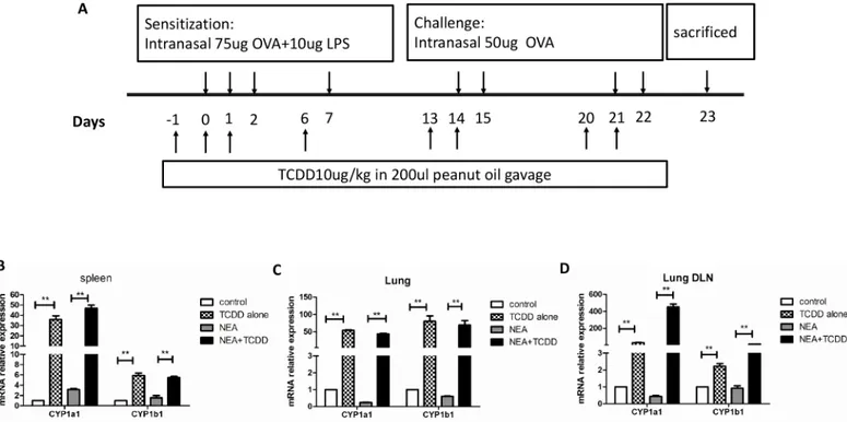
We generated a non-eosinophilic asthma(NEA)model, which indicates that the inflammatory cells in the airway are not mainly eosinophils, as described by Kim and his colleagues [18]. In brief, 6-wk-old mice were intranasally sensitized with OVA (75μg) and LPS (10μg) (Sigma-Aldrich) on days 0, 1, 2, and 7 and then challenged with OVA (50μg) alone on days 14, 15, 21, and 22. The control group was simultaneously administered PBS. To determine the effect of TCDD on airway inflammation, the mice were gavaged (10μg/kg) one day prior to being sensi-tized and challenged, which reflects a total of eight times (Fig 1A). TCDD (purity 99.1%, 50μg/ ml in toluene) was purchased from AccuStandard, Inc. and diluted in peanut oil. The control groups were simultaneously gavaged with peanut oil. Twenty-four h after the last challenge, the mice were euthanized, and the sera, cBALFs, lungs, spleens and lung-draining mediastinal lymph nodes were harvested and analyzed.
Measurement of methacholine AHR
Twenty-four hours after the last challenge, measurements of the airway hyper responsiveness (AHR) were performed using a Buxco’s modular and invasive system (Buxco Electronics Inc., NY, USA). Changes in the airway resistance (RL) and lung dynamic compliance (Cdyn) were measured as described by Amdur and Mead [19]. Briefly, each anesthetized mouse was
tracheostomized and intubated with an appropriate cannula and subsequently laid supine inside the body plethysmograph chamber connected to the ventilator. After a stable baseline airway pressure (<5% variation over 2.5 min) was achieved, the mice were administered aero-solized PBS or various concentrations of methacholine (3.125, 6.25, 12.5, or 25 mg/ml) via a jet nebulizer into the head chamber. The minimum values for the RL and Cdyn were calculated in response to increasing concentrations of inhaled methacholine; the values were expressed as a percent change from the baseline value as previously reported [20].
Lung histology
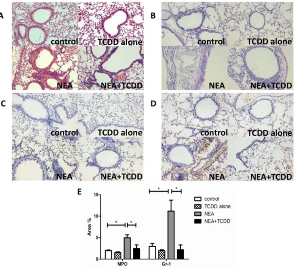
The right lung was removed and immediately fixed in 4% buffered paraformaldehyde for 24 h at room temperature. Paraffin-embedded sections were created and stained with hematoxylin and eosin (H&E) or periodic acid-Schiff (PAS). The airway inflammation and mucus produc-tion were estimated using light microscopy under ×200 magnificaproduc-tions. Samples of the secproduc-tions were incubated overnight at 4°C with rabbit-anti-MPO (Abcam) and rabbit-anti-Ly-6G (Gr-1) (Biolegend) antibody to identify the infiltration of neutrophils. The immunostained sections were quantitatively characterized via digital image analysis using Image Pro-Plus 6 software. The percentage of the positive staining area of the airway was counted from 10 randomly selected fields per section.
Analysis of the sera and BALF
Blood was harvested via cardiac puncture. The serum was collected after 2–3 hours of coagula-tion and 15 minutes of centrifugacoagula-tion at the speed of 500 g. The tracheas of the mice were Fig 1. AhR activation by TCDD promoted the expression of CYP1a1 and CYP1b1 genes.Mice were intranasally sensitized with OVA and LPS, and TCDD was gavaged one day prior to being sensitized and challenged. (A) Timeline of the OVA/LPS immunization/challenge and the TCDD treatment protocol. (B) mRNA expression of CYP1a1 and CYP1b1 in spleens was determined via real-time PCR. (C) mRNA expression of CYP1a1 and CYP1b1 in lungs was determined via time PCR. (D) mRNA expression of CYP1a1 and CYP1b1 in lung-draining mediastinal lymph nodes was determined via real-time PCR. N = 5 per group and*P<0.05,**P<0.01.
lavaged three times via a catheter with 0.8 ml sterile PBS. The BALF was centrifuged for 5 min-utes at 300 g, and the supernatant was harvested. The precipitated cells were resuspended in 20μl PBS for cellular composition using May-Gruenwald Giemsa staining. The supernatants from the sera and BALF were stored at -80°C until cytokine level analysis.
Cell isolation and culture
The spleens and lung-draining mediastinal lymph nodes were removed from the mice. The tis-sues were grind in Dhanks balance buffer on a mesh. The suspension was collected and centri-fuged at a speed of 500 g for 10 minutes. Erythrocytes were lysed using a red blood cell lysis buffer. The cells were resuspended in RPMI 1640 medium with 10% bovine serum and further cultured with OVA (0, 10, 30 and 100μg/ml) for 3 days; the supernatants were analyzed for cytokine expression by ELISA (eBioscience).
Cell staining and Flow cytometry
For intracellular cytokine analysis, the cells from the spleen and lung-draining mediastinal lymph node were restimulated with leukocyte activation cocktail (BD Bioscience) 2μl/ml for 5 hours. The cells were permeabilized and stained with a Cytofix/Cytoperm kit (eBioscience). CD4-FITC, CD25-PE, and IL-17-PE-Eflour-610 (eBioscience) were used to label Treg and Th17 cells. The analysis was performed on a BD FACSCalibur (BD Bioscience).
Real time PCR
Total RNA was extracted using TRIzol reagent (Invitrogen), and cDNA was synthesized using a PrimeScript TM RT reagent kit (Takara). The gene expression was examined with an ABI 7500 real time PCR system using a SYBR Premix Ex Taq TM II kit. The data were normal-ized to a reference, Actb. The following primer pairs were used: CYP1a1 forward,
TTCCTGTCCTCCGTTACCTG, and reverse,GCCCTTCTCAAATGTCCTGT. CYP1b1 forward, GAGAGAGTGCCATC CACCAG, and reverse,GTAGTGACCGAACGCCAGAC. FOXP3 forward, GCCCATCCAATAAACTGTGG, and reverse,GTATCCGCTTTCTCCTGCTG. RORγforward,
CCTCCTGCCACCTTGAGTAT, and reverse,TCTGGACCCTGTTCTGGTT.
Measurement of DC maturation in Lung mononuclear cells
Lung tissue was chopped and incubated in 37°C with 300μg/ml collagenase (Sigma) and 42μg/ml of DNase (Sigma). Following digestion for 90 min, lung mononuclear cells were attained using lymphocyte separation medium (DAKEWE). DCs were isolated from the lung mononuclear cells using CD11c magnetic beads (Miltenyi Biotec). The cells from each group of mice were mixed and analyzed. Flow cytometry analyses were performed to identify the purity of the dendritic cells. CD11c-PE (eBioscience) was used to label the DCs. CD80-FITC, CD86-PE, and CD83-FITC (eBioscience) were used to measure DC maturation.
Statistical analysis
Results
AhR activation by TCDD promotes the expression of CYP1a1 and
CYP1b1 genes
Activated AhR binds to its dimerization partner, which enhances the expression of the target genes, such as cytochrome P450 1a1 (CYP1a1) and CYP1b1 [12]. To determine whether AhR was activated by TCDD, we first assessed the CYP1a1 and CYP1b1 gene expression. As shown
inFig 1B–1D, the expressions of CYP1a1 and CYP1b1 genes were dramatically increased in
the spleens, lungs and lung-draining mediastinal lymph nodes from the TCDD-treatment mice compared with the non-treatment mice. These findings indicate that AhR was clearly activated by TCDD treatment.
TCDD-induced AhR activation alleviates non-eosinophilic airway
inflammation and airway hyperresponsiveness
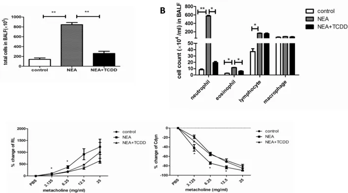
The AhR is expressed in lung tissue [21,22], and AhR activation plays a key role in the inflam-matory reaction [17]. Thus, we investigated the regulatory function of TCDD in non-eosino-philic airway inflammation. Six-wk-old mice were intranasally sensitized with OVA and LPS, and TCDD was gavaged one day prior to being sensitized and challenged. The lung histology was analyzed via HE or PAS staining. As shown inFig 2A and 2B, the inflammatory cells and mucus production were not significantly different in the lungs between the control and TCDD alone groups. However, these variables were substantially decreased in the lungs from the TCDD-treatment OVA/LPS sensitized mice compared with the non-treatment mice. Further-more, we analyzed the neutrophil infiltration in the lungs by MPO and Gr-1 staining. As shown inFig 2C and 2D, the neutrophil infiltration was also significantly decreased in the lungs from the TCDD-treatment OVA/LPS sensitized mice compared with the non-treatment mice. A quantitative analysis indicated the same results (Fig 2E). Through an analysis of the cellular composition in BALF, we determined that eosinophils and neutrophils were both sig-nificantly decreased in the TCDD-treatment group compared with the non-treatment group
(Fig 3A and 3B). When challenged with various concentrations of methacholine, the RL was
significantly decreased, whereas the Cdyn was significantly increased in the TCDD-treatment group compared with the non-treatment group (Fig 3C). Taken together, these findings suggest that TCDD-induced AhR activation prevents the development of allergen-induced non-eosin-ophilic airway inflammation and airway hyperresponsiveness.
TCDD-induced AhR activation regulates non-eosinophilic airway inflammation, potentially through the inhibition of pro-inflammatory cytokine synthesis.
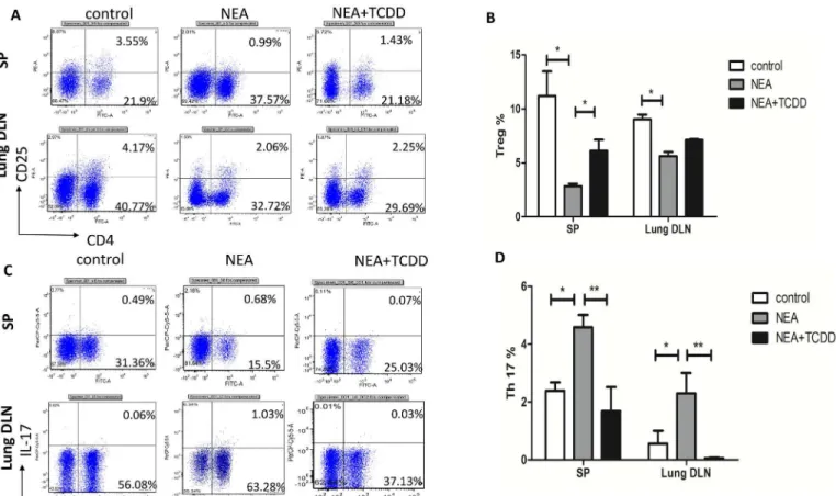
TCDD-induced AhR activation regulates Th17 and Treg differentiation
AhR activation plays an important role in the regulation of the immune system [13,14]; thus, we subsequently investigated whether TCDD-induced AhR activation was involved in T cell differentiation in non-eosinophilic airway inflammation. Cells from the spleens and lung-draining mediastinal lymph nodes were restimulated with leukocyte activation cocktail 2μl/ml for 5 hours, and the intracellular cytokine expression patterns were assessed. The lymphocytes from the OVA/LPS sensitized mice exhibited induced IL-17-producing cells and reduced CD4 +CD25+Tregs. Following TCDD treatment, the IL-17-producing cells were substantially decreased and the CD4+CD25+Treg cells were significantly increased (Fig 5A–5D), which is consistent with a previous report by Singhet al[25]. Furthermore, we assessed the gene expres-sion in the lymphocytes from the spleens that were restimulated with leukocyte activation Fig 2. TCDD-induced AhR activation reduced non-eosinophilic airway inflammation.(A) Lung histology was analyzed via HE staining. (B) Lung histology was analyzed via PAS staining. (C) Lung histology was analyzed via neutrophilic marker-MPO staining. (D) Lung histology was analyzed via neutrophilic marker-Gr-1 staining. (E) Quantitative analysis of neutrophil infiltration in the lungs. The percentage of the positive staining area of the airway was counted from 10 randomly selected fields per section.*P<0.05.cocktail for 5 hours. The RORγmRNA expression was significantly decreased and FOXP3 was significantly increased in the lymphocytes from the mice treated with TCDD compared with the non-treatment mice (Fig 6A and 6B). Thus, TCDD-induced AhR activation may inhibit Th17 differentiation but enhance Treg differentiation.
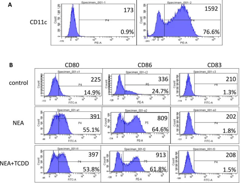
TCDD-induced AhR activation may not inhibit dendritic cell maturation
Dendritic cells (DCs) play an important role in the induction of tolerance or allergic sensitiza-tion to allergens [6]; thus, we investigated the maturation of dendritic cells in non-eosinophilic airway inflammation. DCs were isolated from lung mononuclear cells using magnetic activated cell sorting (MACS). Flow cytometry analyses confirmed that the proportion of CD11c+ cells was greater than 70% (Fig 7A). Furthermore, the flow cytometry analysis indicated that the expression CD80, CD86 and CD83 on dendritic cells was significantly induced in the OVA/ LPS sensitized mice. However, the expression levels were not substantially different following TCDD treatment (Fig 7B). Therefore, dendritic cell maturation may not be inhibited by AhR activation.Discussion
Recently, AhR activation has been demonstrated to regulate T cell differentiation [13,14]. In the present study, we demonstrate that TCDD-induced AhR activation reduced non-eosino-philic airway inflammation via the inhibition of Th17 differentiation and IL-17 expression and the promotion of Treg differentiation and IL-10 production. Furthermore, our findings indi-cate that AhR activation may not inhibit dendritic cell maturation in a non-eosinophilic asthma model.
Fig 3. TCDD-induced AhR activation reduced non-eosinophilic airway inflammation and airway hyperresponsiveness.(A-B) Total cells and cellular composition in BALF were assessed using cytospin with May-Gruenwald Giemsa staining. (C) Airway responsiveness to aerosolized methacholine was evaluated using a Buxco’s modular and invasive system. Changes in RL and Changes in Cdyn. N = 5 per group and*P<0.05,**P<0.01.
Non-eosinophilic asthma, which is mainly characterized by neutrophilic airway inflamma-tion, is an important inflammatory subtype of asthma. It is associated with steroid-resistance [26]. Following an antigen challenge, a substantial number of mucus production and inflam-matory cells, including both eosinophils and neutrophils, infiltrated into the lung tissue in the OVA/LPS sensitized mice. However, TCDD administration reversed allergen-induced non-eosinophilic airway inflammation. In addition, the airway hyperresponsiveness was substan-tially decreased by treatment with TCDD. These findings collectively suggest that TCDD-induced AhR activation alleviates the severity of non-eosinophilic asthma.
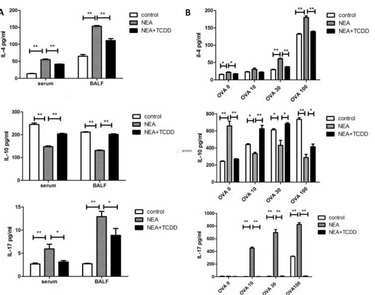
Allergic asthma has traditionally been associated with the imbalance of Th1 and Th2 cell differentiation, which results in eosinophilic airway inflammation. However, recent experi-mental and clinical evidence indicates that severe asthma is more prone to neutrophilic airway inflammation, which is related to Th17 cell responses [23]. Eosinophilic airway inflammation, which is driven by type-2 immune responses, is characterized by the production of IL-4, IL-5, and IL-13, whereas neutrophilic airway inflammation, which is driven by type-17 immune Fig 4. TCDD-induced AhR activation enhanced IL-10 expression and inhibited IL-17 and IL-4 expression.(A) Cytokine expression in the serum and BALF was determined via ELISA. (B) Cytokine expression from the OVA-specific T cells was determined via ELISA. N = 5 per group and*P<0.05,**P<0.01.
responses, is characterized by the production of IL-17. Consistent with the cellular profiles in BALF, the production of the Th2 and Th17 cytokines was substantially decreased in the serum and BALF from the TCDD-treatment mice. The same results were demonstrated via the ex vivo OVA restimulation of lung-draining mediastinal lymph node cells and splenocytes. Thus, Fig 5. TCDD-induced AhR activation promoted Treg differentiation and inhibited Th17 differentiation.Cells from the spleens and lung-draining mediastinal lymph nodes were restimulated with leukocyte activation cocktail for 5 hours. (A-B) Five hours later, CD4+CD25+ cells were analyzed via cell surface staining. (C-D) IL-17-producing cells were analyzed via intracellular staining. The numbers within the quadrants indicate the percentage of positive cells in the total cells; the percentage of positive cells in the CD4+ cells (Treg% and Th17%) was subsequently calculated. N = 5 per group and*P<0.05, **P<0.01.
doi:10.1371/journal.pone.0150551.g005
Fig 6. TCDD-induced AhR activation promoted FOXP3 expression and inhibited RORγexpression.Cells from the spleens were restimulated with
leukocyte activation cocktail for 5 hours. (A) mRNA expression of FOXP3 in lymphocytes was determined via real-time PCR. (B) mRNA expression of RORγ
in lymphocytes was determined via real-time PCR. N = 5 per group and*P<0.05,**P<0.01.
TCDD-induced AhR activation alleviates non-eosinophilic airway inflammation potentially via the inhibition of both Th2-type and Th17-type immune responses. Furthermore, using flow cytometry, we demonstrated that IL-17-producing cells were substantially decreased and CD4+CD25+Tregs were significantly increased following TCDD treatment. Moreover, we have tentatively identified these cells as Tregs in the absence of FoxP3 staining. The RT-PCR results indicated that the mRNA expression of RORγwas significantly decreased and FOXP3 was significantly increased in the lymphocytes restimulated via a leukocyte activation cocktail from the mice treated with TCDD compared with the non-treatment mice. Therefore, TCDD-induced AhR activation substantially affects the immunoregulatory function of non-eosino-philic asthma. These findings are consistent with a recent report that TCDD ameliorated colitis via the up-regulation of Tregs and down-regulation of Th17 cells [25].
Fig 7. Effect of TCDD-induced AhR activation on the maturation of dendritic cells.Lung mononuclear cells were harvested using lymphocyte separation medium. DCs were isolated from lung mononuclear cells using CD11c magnetic beads (Miltenyi Biotec). (A) Flow cytometry analyses were performed to determine the purity of the dendritic cells. (B) Frequencies of CD80+, CD86+, and CD83+ cells on dendritic cells were determined via flow cytometry.
Dendritic cells (DCs) are important for the initiation of T cell-dependent immunity, and AhR activation has been demonstrated to affect DC functions [27]. Finally, we investigated the effect of TCDD-induced AhR activation on DC maturation in non-eosinophilic airway inflam-mation. DCs were isolated from lung mononuclear cells using magnetic activated cell sorting (MACS). However, the expressions of CD80, CD86 and CD83 on dendritic cells were not sig-nificantly changed by TCDD-induced AhR activation, which suggests that DC maturation may not be inhibited by AhR activation. Previous studies have demonstrated that the chemotactic migration of DCs also plays an important role in the immune pathogenesis of asthma [28]; thus, whether AhR activation disturbs the chemotactic migration of DCs requires further investigation.
In conclusion, our study demonstrates that TCDD-induced AhR activation inhibits the development of non-eosinophilic airway inflammation by regulating the differentiation of Th17 and Tregs. These findings may provide a new therapeutic approach for non-eosinophilic asthma.
Acknowledgments
We thank the staff in the Center for Medical Research of Xinhua Hospital.
Author Contributions
Conceived and designed the experiments: JP XJG. Performed the experiments: XML JP. Ana-lyzed the data: XML WG. Contributed reagents/materials/analysis tools: JP XJG. Wrote the paper: JP XML XJG.
References
1. Anderson GP. Endotyping asthma: new insights into key pathogenic mechanisms in a complex, hetero-geneous disease. Lancet.2008; 372:1107–1119. doi:10.1016/S0140-6736(08)61452-XPMID: 18805339
2. Holgate ST, Arshad HS, Roberts GC, Howarth PH, Thurner P, Davies DE. A new look at the pathogen-esis of asthma. Clin Sci (Lond).2010; 118:439–450.
3. McGrath KW, Icitovic N, Boushey HA, Lazarus SC, Sutherland ER, Chinchilli VM, et al. A large sub-group of mild-to-moderate asthma is persistently noneosinophilic. Am J Respir Crit Care Med.2012; 185:612–619. doi:10.1164/rccm.201109-1640OCPMID:22268133
4. Aujla SJ, Alcorn JF. T(H)17 cells in asthma and inflammation. Biochim Biophys Acta. 2011; 1810:1066–1079. doi:10.1016/j.bbagen.2011.02.002PMID:21315804
5. Trejo Bittar HE, Yousem SA, Wenzel SE. Pathobiology of severe asthma. Annu Rev Pathol.2015; 10:511–545. doi:10.1146/annurev-pathol-012414-040343PMID:25423350
6. Vroman H, van den Blink B, Kool M. Mode of dendritic cell activation: the decisive hand in Th2/Th17 cell differentiation. Implications in asthma severity? Immunobiology.2015; 220:254–261. doi:10.1016/ j.imbio.2014.09.016PMID:25245013
7. Matsumoto K, Inoue H, Fukuyama S, Kan-O K, Eguchi-Tsuda M, Matsumoto T, et al. Frequency of Foxp3+CD4+CD25+ T cells is associated with the phenotypes of allergic asthma. Respirology.2009; 14:187–194. doi:10.1111/j.1440-1843.2008.01472.xPMID:19192224
8. Palomares O, Martín-Fontecha M, Lauener R, Traidl-Hoffmann C, Cavkaytar O, Akdis M, et al. Regula-tory T cells and immune regulation of allergic diseases: roles of IL-10 and TGF-β. Genes Immun.2014; 15:511–520. doi:10.1038/gene.2014.45PMID:25056447
9. Tian Y. Ah receptor and NF-kappaB interplay on the stage of epigenome. Biochem Pharmacol.2009; 77: 670–680. doi:10.1016/j.bcp.2008.10.023PMID:19014911
10. Hahn ME. Aryl hydrocarbon receptors: diversity and evolution. Chem Biol Interact.2002; 141: 131–
160. PMID:12213389
12. Mimura J, Fujii-Kuriyama Y. Functional role of AhR in the expression of toxic effects by TCDD. Biochim Biophys Acta. 2003; 1619:263–268. PMID:12573486
13. Esser C, Rannug A, Stockinger B. The aryl hydrocarbon receptor in immunity. Trends Immunol.2009; 30: 447–454. doi:10.1016/j.it.2009.06.005PMID:19699679
14. Stevens EA, Mezrich JD, Bradfield CA. The aryl hydrocarbon receptor: a perspective on potential roles in the immune system. Immunology.2009; 127: 299–311. doi:10.1111/j.1365-2567.2009.03054.x PMID:19538249
15. Veldhoen M, Hirota K, Westendorf AM, Buer J, Dumoutier L, Renauld JC, et al. The aryl hydrocarbon receptor links TH17-cell-mediated autoimmunity to environmental toxins. Nature.2008; 453: 106–109. doi:10.1038/nature06881PMID:18362914
16. Veldhoen M, Hirota K, Christensen J, O’Garra A, Stockinger B. Natural agonists for aryl hydrocarbon receptor in culture medium are essential for optimal differentiation of Th17 T cells. J Exp Med.2009; 206: 43–49. doi:10.1084/jem.20081438PMID:19114668
17. Quintana FJ, Basso AS, Iglesias AH, Korn T, Farez MF, Bettelli E, et al. Control of T(reg) and T(H)17 cell differentiation by the aryl hydrocarbon receptor. Nature.2008; 453:65–71. doi:10.1038/ nature06880PMID:18362915
18. Kim YK, Oh SY, Jeon SG, Park HW, Lee SY, Chun EY, et al. Airway exposure levels of lipopolysaccha-ride determine type 1 versus type 2 experimental asthma. J Immunol. 2007; 178: 5375–5382. PMID: 17404323
19. Pichavant M, Goya S, Hamelmann E, Gelfand EW, Umetsu DT. Animal models of airway sensitization. Curr Protoc Immunol.2007; Chapter 15:Unit 15.18.
20. Glaab T, Daser A, Braun A, Neuhaus-Steinmetz U, Fabel H, Alarie Y, et al. Tidal midexpiratory flow as a measure of airway hyperresponsiveness in allergic mice. Am J Physiol Lung Cell Mol Physiol.2001; 280:L565–573. PMID:11159041
21. Dolwick KM, Schmidt JV, Carver LA, Swanson HI, Bradfield CA. Cloning and expression of a human Ah receptor cDNA. Mol Pharmacol.1993; 44:911–917. PMID:8246913
22. Li W, Donat S, Dohr O, Unfried K, Abel J. Ah receptor in different tissues of C57BL/6J and DBA/2J mice: use of competitive polymerase chain reaction to measure Ah-receptor mRNA expression. Arch Biochem Biophys.1994; 315:279–284. PMID:7986069
23. McKinley L, Alcorn JF, Peterson A, Dupont RB, Kapadia S, Logar A, et al. TH17 cells mediate steroid-resistant airway inflammation and airway hyperresponsiveness in mice. J Immunol.2008; 181: 4089–
4097. PMID:18768865
24. Borish L, Aarons A, Rumbyrt J, Cvietusa P, Negri J, Wenzel S. Interleukin-10 regulation in normal sub-jects and patients with asthma. J Allergy Clin Immunol. 1996; 97:1288–1296. PMID:8648025 25. Singh NP, Singh UP, Singh B, Price RL, Nagarkatti M, Nagarkatti PS. Activation of aryl hydrocarbon
receptor (AhR) leads to reciprocal epigenetic regulation of FoxP3 and IL-17expression and ameliora-tion of experimental colitis. PLoS One. 2011; 6:e23522. doi:10.1371/journal.pone.0023522PMID: 21858153
26. McGrath KW, Icitovic N, Boushey HA, Lazarus SC, Sutherland ER, Chinchilli VM, et al. A large sub-group of mild-to-moderate asthma is persistently noneosinophilic. Am J Respir Crit Care Med.2012; 185:612–619. doi:10.1164/rccm.201109-1640OCPMID:22268133
27. Schulz VJ, van Roest M, Bol-Schoenmakers M, van Duursen MB, van den Berg M, Pieters RH, et al. Aryl hydrocarbon receptor activation affects the dendritic cell phenotype and function during allergic sensitization. Immunobiology.2013; 218:1055–1062. doi:10.1016/j.imbio.2013.01.004PMID: 23433705