Adiponectin inhibits oxidization-induced differentiation
of T helper cells through inhibiting costimulatory
CD40 and CD80
Y. Xiao, T. Deng, Z. Shang and D. Wang
Department of Hematology, Tongji Hospital, Tongji Medical College, Huazhong University of Science and Technology, Wuhan, China
Abstract
Adiponectin is a multifunctional adipokine that has several oligomeric forms in the blood stream, which broadly regulates innate and acquired immunity. Therefore, in this study, we aimed to observe the differentiation of T helper (Th) cells and expression of costimulatory signaling molecules affected by adiponectin. The mRNA and protein expression levels of adiponectin and its receptors in oxidized low density lipoprotein cholesterol-treated endothelial cells were assayed by real time PCR and immuno-fluorescence. The endothelial cells were then treated with adiponectin with or without adipoR1 or adipoR2 siRNA and co-cultured with T lymphocytes. The distribution of Th1, Th2 and Th17 subsets were assayed byflow cytometry. The effects of adiponectin on costimulatory signaling molecules HLA-DR, CD80, CD86 and CD 40 was also assayed byflow cytometry. The results showed that endothelial cells expressed adiponectin and its receptor adipoR1 and adipoR2, but not T-cadherin. Adiponectin suppressed Th1 and Th17 differentiation through adipoR1 receptor, contributed to the inhibition of CD80 and CD40, and inhibited differentiation of Th1 and Th17 by inhibiting antigen presenting action.
Key words: Adiponectin; T helper cells; HLA-DR; CD80; CD40
Introduction
Adipose tissues can secrete a variety of adipokines such as leptin, resistin, adiponectin, plasminogen activator inhibitor-1, which participate in the regulation of a number of chronic diseases such as diabetes, cardiovascular diseases and cancers (1–4). Adiponectin is recognized as an
impor-tant protector to the above diseases and it is usually mark-edly reduced in many kinds of metabolic diseases (5). It is a protein hormone produced by adipose tissues and released into the blood stream at a concentration of approximately 0.5–30mg/mL, which is 1,000-fold higher than most other
hor-mones (6). The effects of adiponectin in the regulation of insulin sensitivity have been documented since 1996 (7,8). Recently, adiponectin has also been shown to play a role in the protection and inhibition of cardiovascular diseases (9,10). Growing evidence shows that atherosclerosis (AS) is a chronic inflammatory disease in which autoimmunity may play an important role. T lymphocytes are important immune cells participating in AS. An increase in the total number of T lymphocytes and activation of T lymphocytes in peripheral blood can be detected in hyperlipidemia and unstable angina patients (11). Also, a large number of T lymphocytes, especially CD4+T lymphocytes, are found
in atherosclerotic plaque in humans and rat models (12).
Again, T lymphocytes can react with related antigen of artery atheromatous plaque, such as oxidized low density lipoprotein cholesterol (ox-LDL) (13), heat shock protein (14), etc., so as to promote the formation of AS. In addition, the inhibition of Th1 or Th17 pathways also reduces athero-sclerotic lesions (15).
The activation of naive CD4+T lymphocytes requires
two signals delivered by antigen presenting cells:first, the interaction of specific major histocompatibility complex-peptide with the T-cell receptor, and second, a costimu-latory signal. Among the costimucostimu-latory molecules, CD80 and CD86 are the best characterized. The CD40/CD40L system is also a crucial costimulatory molecule involved in differentiation of Th1 and Th17 (16,17). Some reports show that shutting down the CD40/CD40L system is very effective in dampening inflammation inin vitrocellular systems as well asin vivoanimal models of experimental AS (18,19). Many signalings were coupled to CD40; for example, P38 MAPK/TNFaand PI3K/Akt /NF-kappab have been shown to enlarge systemic inflammation and promote differentiation of Th1 and Th17 (20).
Here, we aimed to observe the effect of adiponectin on the differentiation of T helper (Th) cells and expression of
costimulatory signaling molecules in T lymphocytes treated with ox-LDL.
Material and Methods
Ethics statement
Peripheral blood was drawn from healthy adult volun-teers. Before the experiments, we obtained approval for our study from the Ethics Committee of Tongji Hospital, Tongji Medical College, Huazhong University of Science and Technology and written informed consent from all study participants.
Separation and culture of human umbilical vein endothelial cells (HUVEC)
Newborn umbilical cords were obtained under aseptic condition and washed in cold PBS. Then, the umbilical veins were injected with 10 mL collagenase (1 mg/mL) for digestion of endothelial cells at room temperature for 30 minutes. Digested cells were collected into a 50 mL sterile centrifuge tube by washing twice with sterile PBS at 1000gat room temperature for 10 min and then cultured in DMEM medium supplemented with 15% fetal bovine serum at 37°C in 5% CO2. The culture media was replaced
by fresh media every 2 days.
Real-time PCR
The total RNA was isolated from HUVEC cells by using TRIzols Reagent (Thermofisher Scientific, USA). Then, RNA was reverse transcribed to cDNA using PrimeScriptt
RT reagent Kit (Takara, China) and analyzed using a Mini8 Plus Detection System (Coyote, USA). Real-time PCR was performed in a total of 20mL reaction volume containing 1mL of cDNA, 0.6mL each of forward- and reverse-specific primers (from 25 pmol/mL primer stock) and Sybr green I. All qRT-PCR reactions were performed using the following reaction conditions: initial denaturation at 94°C for 4 min followed by 35 cycles at 94°C for 30 s, 60°C for 30 s, and then 72°C extension for 10 min. The primers of adipo-nectin were 50-TATTGGTCCTAAGGGAGACATCGC-30and
50-TTTTGGTGATACTACCGAGGTGAC-30; for adipoR1 50
-CTGACTGGCTAAAGGACAACGACTA-30and 50-TCGTAG
AAGGCGTAAGTATGTCTT-30; for adipoR2 50-CGAAACC
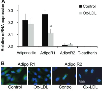
Figure 1.mRNA expression of adiponectin and its receptors in endothelial cells (n=4) assayed by real-time PCR (A). The mRNA expression level of adipoR1 decreased significantly at 8 h after treat-ment of oxidized low density lipoprotein choles-terol (ox-LDL).B, Adiponectin receptors identified by immunofluorescence (400). **Po0.01 com-pared to control (pairedt-test).
CGTCATTCAGCAGT-30 and 50-TTACCTTCCGACCGAA
CCTAC-30; for T-cadherin 50-TGTGGGTTAGTATTGGTGT
ATGTATGAGT-30and 50-TTTGATTCTGTGGACTTGGGA
GGTC-30. Target genes were normalized to b-actin and
quantified using 2-DDCtmethod (21).
Immunofluorescence
The HUVEC cells were fixed, permeabilized and subsequently combined with rabbit adipoR1, anti-adipoR2 or anti-T-cadherin antibodies from Abcam (UK). Then, the cells were incubated with FITC-conjugated anti-IgG antibodies (Sigma, USA) in the dark. The cells were mounted and observed on a fluorescence microscope (Olympus, USA).
Separation of peripheral lymphocytes
Heparinized whole blood was collected from healthy adult volunteers. Peripheral blood mononuclear cells (PBMCs) were isolated by Ficoll-Paque plus (Absin, China) density gradient centrifugation. Then, PBMCs were incubated in complete medium at 37°C in 5% CO2 for 2 h until the
monocytes adhered to the bottom of cultureflasks. Then the lymphocytes suspended in medium were isolated.
After, PBMCs were incubated in complete medium at 37°C in 5% CO2for 2 h until the monocytes adhered to
the bottom of culture flasks. The lymphocytes suspended in medium were isolated using a CD4+T Cell
Isola-tion Kit (Miltenyi Biotec, Germany) by positive selecIsola-tion of CD45RA+CD4+ T cells and negative selection of
CD45RO+CD4+T cells. Cells were cultivated at 37°C in
10% human AB plasma-containing RPMI 1640 medium, supplemented with penicillin-streptomycin. A total of 5
105CD45RA+CD4+T cells were cultured in 96-well plates
along with 1105beads coated with 5 mg/mL anti-CD3, anti-CD28 according to the manufacturer’s instructions.
Co-culture of HUVECs and lymphocytes
In co-cultured experiments, HUVECs (2105cells/well) were treated with or without 10 mg/mL ox-LDL (Yuanyuan Biotechnology, China) for 4 h after pretreatment with 5mg/mL adiponectin (Glycosilated Polypeptide, Prospec, Israel) for 2 h. Then, the medium was discarded and lympho-cytes were added to the HUVECs at a ratio of 1:1 so that HUVECs and lymphocytes were cultured in the same well. After incubation at 37°C in 5% CO2for 48 h, the
lympho-cytes were collected.
Flow cytometric analysis of Th subtypes
Lymphocytes were fixed with 4% paraformaldehyde for 10 min at room temperature and permeabilized in
permeabilizing solution (eBioscience, USA). After blocking with 5% BSA for 20 min, cells were stained with appro-priate anti-IL-4, IL-17 and IFN-gamma antibodies (Abcam, UK) on ice for 1 h. Isotype-matched antibodies were used as controls. The expression levels of antigens are reported as a percentage of positive cells in total cells.
Flow cytometric analysis of costimulatory molecules
To assess the effect of adiponectin on costimulatory molecules, HUVECs were incubated with 10mg/mL ox-LDL for 4 h after pretreatment with adiponectin for 2 h. Then they were washed twice with fresh media and treated with the monoclonal anti-human HLA, CD80, CD86 and CD40 (Bioleg-end, CA, USA). For each analysis, 105cells were incubated with 10mL of monoclonal antibody at room temperature for 20 min. The cells were then washed twice and re-suspended in PBS containing 1% FBS and 0.1% NaN3(Sigma) and
immediately analyzed with FACS (Becton Dickinson, USA). Isotype-matched antibodies were used as controls. The levels of antigen expression are reported as a per-centage of positive cells in the total cells.
RNA interference
siRNAs Lentivector against AdipoR1 and AdipoR2, or control siRNA were obtained from Abm, Inc. (Canada). Endothelial cells were infected with control siRNA lenti-virus, AdipoR1 siRNA or AdipoR2 siRNA with a multiplicity of infection of 1:10. After 48 h, transfected endothelial cells were incubated with 10 mg/mL ox-LDL for 4 h after pre-treatment with adiponectin for 2 h. Control experiments have revealed that AdipoR1 and AdipoR2 mRNA levels are reduced by480%.
Statistical analysis
Data are reported as means±SE. The two groups
were compared using the paired t-test. A two-way ANOVA was used to analyze Th subset distribution. A P value of o0.05 was considered to be statistically
significant. Statistics were calculated using SPSS/Win-dows version 15.0.
Results
Endothelin cells expressed adiponectin and its receptors
By using real-time PCR, we found that endothelial cells express the mRNA of adiponectin, adipoR1 and adipoR2, but not T-cadherin. The expression of adipoR1 was greater than that of adipoR2. After stimulation of 10mg/mL ox-LDL, mRNA expression of adipoR1 decreased significantly at 8 h after treatment, while adipoR2 was not affected (Figure 1A). Immunofluorescence staining showed that adipoR1 and adipoR2 proteins were expressed in endothelial cells and adipoR1 decreased significantly at 8 h after oxidation stress (Figure 1B).
Adiponectin rescued the decrease of adipoR1 induced by ox-LDL.
To investigate whether adiponectin acted through reg-ulating its receptor adipoR1 expression, we stimulated the endothelial cells with 10 mg/mL ox-LDL for 4 h and assessed adipoR1 at 0, 4, 8, and 12 h later. The expres-sion of adipoR1 significantly decreased after ox-LDL treat-ment, particularly at 8 and 12 h. With 5mg/mL adiponectin co-incubation, the decrease of adipoR1 was rescued from 8 h after ox-LDL treatment (Figure 2).
Adiponectin inhibited the differentiation of Th1 and Th17
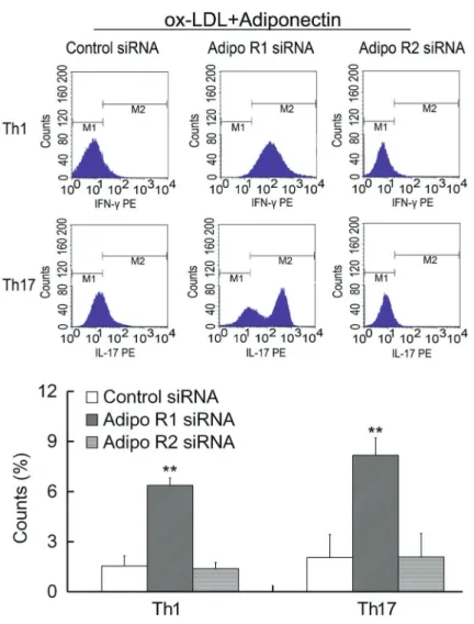
Th1 cells mainly secrete IFN-gamma, Th2 cells mainly secrete IL-4 and Th17 cells mainly secrete IL-17. Monensin (20 mL, 10 mg/mL) was used to inhibit the secretion of newly produced cytokines in Golgi body of lymphocytes. After intracellular staining, the distribution of Th1, Th2 and Th17 subsets were examined with flow cytometry. The results suggested that Th1 and Th17 differentiation induced by oxidation stress were suppressed with adipo-nectin treatment (Figure 3). Moreover, the inhibitory effects
were abrogated by the treatment of adipoR1 siRNA (Figure 4), indicating that the inhibitory effects of adiponectin were mediated by adipoR1.
Adiponectin inhibited the expression of CD80 and CD40
Constitutive expression of HLA, CD80, CD86 and CD40 molecules was observed on endothelial cells. Among them, HLA, CD80 and CD40 were up-regulated in the pres-ence of ox-LDL, and adiponectin inhibited the expression of CD80 and CD40 induced by ox-LDL (Figure 5A). More-over, the inhibitory effects were abrogated by the treat-ment of adipoR1 siRNA (Figure 5B).
Discussion
Several studies have shown that adiponectin can reduce the area of lipid plaque, and inhibit neointimal thickening, smooth muscle cell proliferation and migration, and the expression of adhesion molecules on endothelial cells (22). It is reported that adiponectin suppresses TNF
alpha-induced expression of adhesion molecules and mitogen activated protein (MAP) pathway-induced smooth muscle cell proliferation. In macrophages, adiponectin inhibits the expression of scavenger receptor and the formation of foam cells (23). Therefore, accumulation of adiponectin at the site of the lesions of endothelial cells plays a protective effect against AS. Currently, there are many reports about the relationship between adiponectin and AS. However, the specific roles of adiponectin in the regulation of immune responses remain unknown.
Using oxidized-injured endothelial cells, we aimed to determine whether and how adiponectin is involved in the immunological response. Therefore, we first tested the mRNA expression of adiponectin and its receptors in endothelial cells. We verified that endothelial cells express adiponectin and its receptor adipoR1 and adipoR2, but not T-cadherin. Ox-LDL can inhibit the expression of adipoR1, which can be rescued by additional administration of adiponectin. Next, the endothelial cells were treated with adiponectin with or without adipoR1 or adipoR2 siRNA, and then co-cultured with T lymphocytes. The distribution
of Th1, Th2 and Th17 subsets were observed. The results showed that ox-LDL induced the differentiation of Th1 and Th17, and adiponectin suppressed Th1 and Th17 differ-entiation through adipoR1 receptor.
Among the accessory molecules involved in the acti-vation of naïve CD4+T lymphocytes , the up-regulation of
CD40 is of particular interest in AS because the CD40 pathway is intimately involved in exaggerated infl amma-tion (24). Indeed, stimulaamma-tion of CD40-bearing cells trig-gers multiple inflammatory signals, resulting in leukocyte recruitment and amplification of tissue injury.
In conclusion, we found that endothelial cells were mediated in the abnormal differentiation of Th subsets, and adiponectin inhibited the differentiation of Th1 and Th17 subsets induced by ox-LDL through inhibiting CD80 and CD40 molecules.
Acknowledgments
This work was supported by Natural Science Founda-tion of Hubei Province (No. 2016CFB545).
References
1. Morley TS, Xia JY, Scherer PE. Selective enhancement of insulin sensitivity in the mature adipocyte is sufficient for systemic metabolic improvements. Nat Commun2015; 6: 7906, doi: 10.1038/ncomms8906.
2. Jensen MK, Bertoia ML, Cahill LE, Agarwal I, Rimm EB, Mukamal KJ. Novel metabolic biomarkers of cardiovascular disease. Nat Rev Endocrinol 2014; 10: 659–672, doi: 10.1038/nrendo.2014.155.
3. Park J, Morley TS, Kim M, Clegg DJ, Scherer PE. Obesity and cancer - mechanisms underlying tumour progression and recurrence. Nat Rev Endocrinol2014; 10: 455–465, doi: 10.1038/nrendo.2014.94.
4. Khandekar MJ, Cohen P, Spiegelman BM. Molecular mech-anisms of cancer development in obesity.Nat Rev Cancer 2011; 11: 886–895, doi: 10.1038/nrc3174.
5. Matsuda M, Shimomura I. Roles of adiponectin and oxida-tive stress in obesity-associated metabolic and cardiovas-cular diseases.Rev Endocr Metab Disord2014; 15: 1–10, doi: 10.1007/s11154-013-9271-7.
6. Lisowska A, Tycinska A, Knapp M, Sawicki R, Lisowski P, Musial WJ, et al. Adiponectin - an independent marker of coronary artery disease occurrence rather than a degree of its advancement in comparison to the IMT values in per-ipheral arteries.Clin Chim Acta 2012; 413: 749–752, doi: 10.1016/j.cca.2012.01.006.
7. Paniagua JA. Nutrition, insulin resistance and dysfunctional adipose tissue determine the different components of meta-bolic syndrome.World J Diabetes2016; 7: 483–514. 8. Weyer C, Funahashi T, Tanaka S, Hotta K, Matsuzawa Y,
Pratley RE, et al. Hypoadiponectinemia in obesity and type 2 diabetes: close association with insulin resistanceand hyper-insulinemia.J Clin Endocrinol Metab2001; 86: 1930–1935, doi: 10.1210/jcem.86.5.7463.
9. Sawicka M, Janowska J, Chudek J. Potential beneficial effect of some adipokines positively correlated with the adi-pose tissue content on the cardiovascular system.Int J Cardiol 2016; 222: 581–589, doi: 10.1016/j.ijcard.2016. 07.054.
10. Ehsan M, Singh KK, Lovren F, Pan Y, Quan A, Mantella LE, et al. Adiponectin limits monocytic microparticle-induced endothelial activation by modulation of the AMPK, Akt and NFkB signaling pathways.Atherosclerosis2016; 245: 1–11, doi: 10.1016/j.atherosclerosis.2015.11.024.
11. Steppich BA, Moog P, Matissek C, Wisniowski N, Kühle J, Joghetaei N, et al. Cytokine profiles and T cell function in acute coronary syndromes.Atherosclerosis2007; 190: 443– 451, doi: 10.1016/j.atherosclerosis.2006.02.034.
12. Spitz C, Winkels H, Bürger C, Weber C, Lutgens E, Hansson GK, et al. Regulatory T cells in atherosclerosis: critical immune regulatory function and therapeutic potential.Cell Mol Life Sci2016; 73: 901–922, doi: 10.1007/s00018-015-2080-2.
13. Frodermann V, van Puijvelde GH, Wierts L, Lagraauw HM, Foks AC, van Santbrink PJ, et al. Oxidized low-density lipoprotein-induced apoptotic dendritic cells as a novel therapy for atherosclerosis. J Immunol 2015; 194: 2208– 2218, doi: 10.4049/jimmunol.1401843.
14. Ketelhuth DF, Hansson GK. Adaptive Response of T and B Cells in Atherosclerosis.Circ Res2016; 118: 668–678, doi: 10.1161/CIRCRESAHA.115.306427.
16. Pilon C, Levast B, Meurens F, Le Vern Y, Kerboeuf D, Salmon H, et al. CD40 engagement strongly induces CD25 expression on porcine dendritic cells and polarizes the T cell immune response toward Th1.Mol Immunol2009; 46: 437– 447, doi: 10.1016/j.molimm.2008.10.014.
17. Iezzi G, Sonderegger I, Ampenberger F, Schmitz N, Mars-land BJ, Kopf M. CD40-CD40L cross-talk integrates strong antigenic signals and microbial stimuli to induce develop-ment of IL-17-producing CD4+T cells.Proc Natl Acad Sci U S A2009; 106: 876–881, doi: 10.1073/pnas.0810769106. 18. Tiniakou I, Drakos E, Sinatkas V, Van Eck M, Zannis VI, Boumpas D, et al. High-density lipoprotein attenuates Th1 and th17 autoimmune responses by modulating dendritic cell maturation and function.J Immunol2015; 194: 4676– 4687, doi: 10.4049/jimmunol.1402870.
19. Zhao Z, Wu Y, Cheng M, Ji Y, Yang X, Liu P, et al. Activation of Th17/Th1 and Th1, but not Th17, is associated with the acute cardiac event in patients with acute coronary syn-drome.Atherosclerosis2011; 217: 518–524, doi: 10.1016/ j.atherosclerosis.2011.03.043.
20. Gao S, Zeng L, Zhang X, Wu Y, Cui J, Song Z, et al. Attenuated Streptococcus pneumoniae vaccine candidate
SPY1 promotes dendritic cell activation and drives a Th1/ Th17 response.Immunol Lett2016; 179: 47–55, doi: 10.1016/ j.imlet.2016.08.008.
21. Li L, Li Y, Dai Z, Liu M, Wang B, Liu S, et al. Lipid Metabolism in vascular smooth muscle cells infuenced by HCMV infec-tion.Cell Physiol Biochem2016; 39: 1804–1812, doi: 10.1159/ 000447880.
22. Ouchi N, Kihara S, Funahashi T, Matsuzawa Y, Walsh K. Obesity, adiponectin and vascular inflammatory disease. Curr Opin Lipidol 2003;14: 561–566, doi: 10.1097/0004 1433-200312000-00003.
23. Ouchi N, Kihara S, Arita Y, Nishida M, Matsuyama A, Okamoto Y, et al. Adipocyte-derived plasma protein, adipo-nectin, suppresses lipid accumulation and class Ascavenger receptor expression in human monocyte-derived macro-phages.Circulation2001; 103: 1057–1063, doi: 10.1161/01. CIR.103.8.1057.