Iranian Journal of Basic Medical Sciences
ijbms.mums.ac.ir
Electrophysiologic and clinico‐pathologic characteristics of
statin‐induced muscle injury
Mohammed Abdulrazaq , Farqad (amdan *, Waseem Al‐Tameemi
Department of Physiology, College of Medicine, Al‐Nahrain †niversity, Baghdad, )raqDepartment of Medicine, College of Medicine, Al‐Nahrain †niversity, Baghdad, )raq
A R T ) C L E ) N F O A B S T R A C T
Article type:
Original article Objective(scharacteristics of statin‐induced muscle injury as well as clinical features of patients who develop this ): )n this study, we aimed at evaluation of electrophysiological and histopathalogical condition in terms of frequency and pattern of evolution.
Materials and Methods: Forty patients age ‐ years including subjects with type diabetes mellitus, with cardiovascular diseases and with hyperlipidemia, who were receiving atrovastatin mg/day for variable period, were studied. Thirty three healthy subjects age ‐ years served as control group. Creatine phosphokinease level, thyroid function, motor unit potential parameters and muscle fiber conduction velocity of biceps brachii and tibialis anterior muscles were measured. Results: Creatine phosphokinase level was elevated in statin users, particularly in those with diabetes mellitus. Less than % of statin users experienced symptoms related to muscle injury. Muscle fiber conduction velocity of the biceps brachii muscle was significantly reduced. Statin users with diabetes mellitus showed significant changes in electrophysiological parameters as compared to those with cardiovascular diseases and hyperlipidemia. Muscle biopsies showed muscle fiber variation in size, fibrosis and mild inflammatory cell infiltration. )mmunohistochemical evaluation of muscle biopsies showed positive expression of Bcl‐ and one patient showed positive P immunohistochemical expression with elevated level of creatine phosphokinase.
Conclusion: Atorvastatin increased average creatine kinase, statins produce mild muscle injury even in asymptomatic subjects. Diabetic statin users were more prone to develop muscle injury than others. Muscle fiber conduction velocity evaluation is recommended as a simple and reliable test to diagnose statin‐induced myopathy instead of invasive muscle biopsy.
Article history: Received: Nov , Accepted: May , Keywords: Statin EMG MFC‡ (istopathology )mmune histochemistry
Muscle injury
►Please cite this article as:
Abdulrazaq M, (amdan F, Al‐Tameemi W. Electrophysiologic and clinico‐pathologic characteristics of statin‐induced muscle injury. )ran J
Basic Med Sci ; : ‐ .
Introduction
Statins are considered to be safe, well‐tolerated efficient drugs for the treatment of hypercholes‐ terolemia; Therefore, they are frequently prescribed
, . The most severe adverse effects that are seen following statins administration are various forms of myotoxicity including myopathy, myalgia, myositis and rhabdomyolysis . Symptoms usually develop within four weeks but can be delayed up to four years after starting statins therapy , .
According to the †S National Lipid Association Statin Safety Assessment Task Force, myopathy, defined as muscle symptoms and creatine phosphor‐ kinase CPK levels times normal, occurs in five
patients per , person‐years , .
Myalgia with or without CPK elevation affects ‐ % of patients, and asymptomatic CPK elevation up to times normal is noted in ‐ % of patients , whereas normal CPK values do not exclude statin‐
induced myopathy .
Statin‐induced muscle weakness is dose and time‐ related and can be accompanied by myopathic electromyographic EMG changes. )n this regard, usually muscle necrosis is observed in muscle biopsy , . Myopathic changes are usually seen in the proximal muscles and a normal EMG does not exclude statin‐induced myopathy, because it primarily affects
type )) muscle fibers .
)n certain circumstances, when statins trigger toxic myopathy, they cause clinical weakness, fibrillations, positive sharp waves, and even myotonic discharges with early recruitment of short‐duration motor unit action potentials M†APs become apparent in weak
muscles , .
The objective of our study was to assess electro‐ physiological and histopathalogical characteristics and clinical features of statin‐induced muscle injury in patients, in term of frequency and pattern of evolution.
*Corresponding author: Farqad (amdan. Department of Physiology, College of Medicine, Al‐Naharin †niversity, Baghdad, )raq. email:
Materials
and
Methods
Forty patients aging . ± . years old –
years old were recruited from those attending the outpatient medical clinic or those who were hospitalized in Al‐)mamian Al‐Kadhimiyian Medical
City, Baghdad, )raq between January and May .
The study was approved by the )nstitute Review Board of the College of Medicine, Al‐Nahrain †niversity. The protocol was carefully explained to each subject before entering the study and their written consent was obtained.
Twenty‐five . % patients had type diabetes
mellitus DM and took statin according to ADA guidelines above years with multiple risk factors for cardiovascular disease C‡D or low density lipoprotein
LDL > mg/dl , . % patients had C‡D
acute coronary syndrome and history of ischemic heart
disease and % patients had unclassified
hyperlipidemia hypercholesterolemia .
All patients received atorvastatin mg/day for a
period of one to seven months. % patients
received the drug for two months, . % for
three or four months and only . % patient for seven months.
Patients with documented history of muscle disorder, renal impairment, liver diseases, thyroid dysfunction, and those who had long‐term steroids use, alcohol consumption and concurrent treatment with other lipid‐lowering agents fibrates, bile acid sequestrates and niacin were excluded. Patients who were treated with amiodarone and proton pump inhibitors, or those who had electrolytes abnormalities, surgery or trauma in the past months or recent intramuscular injection were also excluded. Exclusion criteria were chosen based on the fact that the aforementioned conditions or diseases are considered as risk factors to increase
the tendency for myopathies .
Participants were asked if they had pain. Those who answered yes were asked to specify the severity and location of their pain. We assessed them
using functional activity score and the patients
were scored as follows:
A. No limitation: the patient’s activity is not limited by pain.
B. Mild limitation: the patient’s activity is mild to moderately limited by pain.
C. Severe limitation: the patient ability to do the activity is severely limited by pain.
For practical issues and because of difficult interpretation by some illiterate patients, some modifications were applied as mild for grade A , moderate for grade B , severe for grade C and not specified.
Thirty three volunteers females and males
aging to years old mean±SD = . ± . were
enrolled in the current study as the control group.
Biochemicaltests
Serum CPK was measured using commercially available kits purchased from Abbot Laboratories
Kit No. D ‐ , ARC()TECT plus‐c , †SA . CPK
was considered abnormal if it exceeded the range of
‐ )†/l in male patients and ‐ )†/l in
female patients.
Thyroid function test T , T and TS( was done by antigen‐antibody reaction using Mini ‡)DAS equipment
SN. ivd , France . ‡)DAS ‐ SN.
kit was used for T , ‡)DAS ‐ SN.
for T , and ‡)DAS Code no. ‐ for TS(.
The expected normal values were . ‐ . nmol/l for
T , ‐ nmol/l for T , and . ‐ . μ)†/ml for TS(.
Electrophysiologicalexamination
This was done using Micromed, ‐channel
electromyography SN. G( ( NW B, model
, code G( ESSM/EDC, )taly .
The biceps brachii and tibialis anterior muscles of the left side were chosen for all studies. The subjects were comfortably lying prone with both the arms and the legs extended and held fixed in an abducted position. The skin over the muscle was thoroughly treated with antiseptic solution. The examination room temperature was roughly maintained at
‐ °C.
Electromyographic activity was analyzed using monopolar needle EMG electrodes Micromed code
D)N . Another electrode which served as
ground Micromed code was fitted across the
muscle belly. Twenty M†Ps were analyzed for duration, amplitude and polyphasia during minimal volitional effort. The setup used in this test was as follows: gain
at microvolt/cm, sweep speed msec/cm, and
filter setting (z to K(z.
The method of Troni et al was adopted for
measurement of muscle fiber conduction velocity MFC‡ using the same monopolar needle electrode as an active recording electrode and surface silver cup‐shaped disc electrode Micromed Code tpco as reference recoding electrode. The muscle stimulated at distal position from the recording electrode by means of the same monopolar needle electrode. The latency, amplitude, and C‡ were estimated. The setup of the test was as follows: stimulation insured
to be supramaximal, band pass filter (z‐ k(z
and the time base varied between to msec per division.
Musclebiopsy
†nder local or general anesthesia, muscle biopsies were taken from the tibialis anterior muscle of six patients and examined using clamp fixation method. Four out of six samples were symptomatic with elevated CPK and the other two patients were asymptomatic. Each sample had two sections namely
Table1.Demographic features of the statin users vs control subjects
Parameters Control subjectsN= Statin users N=
Gender Females Males . % . % % %
Thyroid function test T T
TS(
. ± .
. ± .
. ± .
. ± .
. ± .
. ± . Age years
Weight Kg CPK range †/l
. ± .
. ± .
‐
. ± . . ± .
. ‐ *
* =P< . Mann‐Whitney † test , CPK= creatine phosphokinase, T = Triiodothyronine, T = Triiodothyronine, TS(= Thyroid stimulating
hormone
specimen was put in % formalin solution for
(ematoxylin and Eosin test and further immune‐ histochemical studies. The immune‐histochemical
staining was done using Bcl‐ Dako, code NO ,
Denmark and p Dako Cytomation )(C kit LSAB
System‐(RP, code K , Denmark . All samples
were examined by histopathology consultant.
Statisticalanalysis
All statistical analyses were done using SPSS
software version and Microsoft office Excel .
The values were presented as mean±SD , median and
range. The comparison between patient group and control group was made using t‐test for parametric
variables whereas Chi square and Mann‐Whitney † test were employed for non‐parametric variables. Comparison between more than two groups was made by using ANO‡A and Kruskal‐Wallis tests. P‐value less
than . was considered significant.
Results
Demographicdata
No difference was observed between control subjects and statin users concerning their age, gender, weight, and thyroid function indices, whereas CPK level was significantly higher among statin users Table .
Age and weight were not different between the control subjects and statin users with different underlying pathologies while CPK level was significantly higher P< . in diabetic statin users as
compared to the control subjects Table . ANO‡A showed that age, weight and the duration of statin
treatment . ± . months; . ± . months;
and . ± . months, respectively were not different among statin users with various underlying pathologies. Similarly, Kruskal‐Wallis test showed that
CPK was not different among statin users with various underlying pathologies.
Clinicaldata
Seventeen statin users . % showed
symptoms related to myopathy while the rest of
subjects . % were symptoms free. None of the
patients in the present study presented signs and symptoms of rhabdomyolysis. Symptomatic statin users had different degrees of muscle pain severity as shown in Figure A. Different parts of the body had muscle pain as demonstrated in Figure B.
Neurophysiologicdata
(ere, M†Ps were analyzed in the patient's
group and in the control group. )n the biceps brachii muscle, the amplitude and duration of the M†Ps were significantly reduced in patients as compared to the control subjects P< . and P< . , respectively
but this was not seen in the tibialis anterior muscle Table . Moreover, no difference was noticed between symptomatic and non‐symptomatic statin users regar‐ ding M†Ps parameters in the muscles tested.
Table illustrates the number of M†Ps phases polyphasia in statin users and control subjects. Starin users had significantly increased polyphasia more than phases as compared to the control group in the biceps brachii and tibialis anterior
muscles P< . . Furthermore, no difference was
noticed between symptomatic and non‐ symptomatic statin users concerning the number of M†Ps phases in examined muscles.
The M†P amplitude recorded from biceps brachii muscle was significantly reduced P< . in statin
users with DM and C‡D as compared to the control subject.
Table2. Comparison of age and weight among control subjects and statin users with different pathologies
Parameter Control SubjectsN=
Statin users N= DM
N= C‡DN= N= (L
Age Year
Weight Kg . + . . ± . . + .. + . . + .. + . . + .. + .
CPK †/l . ± . ‐ . ± .‐ * . ± .‐ . ± .‐
*= P< . Mann‐Whitney † test , CPK= creatine phosphokinase, DM= diabetes mellitus, C‡D= cardiovascular disease,
Figure1.Percentage of statin users showing the severity A and location B of muscle pain Table3. M†P parameters in statin users vscontrol subjects
M†P parameter Control subjectsN= Statin usersN=
Biceps brachii Amplitude m‡ Duration ms . ± .. ± . . ± . * . ± . **
Tibialis anterior Amplitude m‡ Duration ms . ± .. ± . . ± . . ± .
* P < . , ** P < . , student t test , M†P=Motor unit potential
Similarly,M†Psduration recorded from biceps
brachii and tibialis anterior muscles was significantly different P< . in statin users with DM as compared
to the control subjects Table . †sing ANO‡A test, the M†P amplitude and duration recorded from the two muscle samples were not different among statin users with various underlying pathologies.
The latency of evoked muscle fiber response was not different between statin users and control subjects whether it was measured in the biceps brachii or tibialis anterior muscles. On the other hand, the recorded amplitude of the biceps brachii and tibialis anterior muscles showed significant decrement in patients in comparison with the control subjects
P< . . )n addition, C‡ of the biceps brachii muscle
was significantly reduced in statin users as compared to
the control subjects P< . , whereas it showed non‐
significant reduction in the tibialis anterior muscle Table . The amplitude
recorded from tibialis anterior muscle was the only parameter that was significantly reduced P< . in
the symptomatic vs asymptomatic statin users.
The amplitude of muscle fiber response evoked
from biceps brachii was significantly reduced
P< . in statin users with DM and C‡D as
compared to the control subjects. Likewise, the latency and C‡ were also significantly reduced
P< . in statin users with DM in comparison with
the control subjects. The amplitude of muscle fiber response evoked from tibialis anterior muscle was significantly reduced P< . in statin users with
DM when compared to the control subjects Table . Furthermore, using ANO‡A test, neither the latency nor the amplitude or C‡ was different among statin users with different underlying pathologies.
A significant inverse correlation was demonstra‐ ted between CPK level and MFC‡ of the tibialis
anterior muscle r= ‐ . and P= . .
Table4. M†Ps phases in statin users vscontrol subjects
Group Biceps brachii M†Ps phases
Control group Frequency % . . . . . . .
Statin users Frequency % . . . . . * . .
Tibialis anterior M†Ps phases
Control group Frequency % . . . . . . ‐
Statin users Frequency % . . . . * . * . ‐
* P=< . Chi square test , M†P=Motor unit potential
Table5. M†P parameters of biceps brachii and tibialis anterior muscles among control subjects and statin users with different underlying
pathologies
Parameter Control subjects N=
Statin users N= DM
N= C‡DN= N= (L
Biseps
Brachii Amplitude m‡ Duration ms . ± .. ± . . ± .. ± . * . ± . . ± . . ± .. ± . Tibialis
Anterior Amplitude m‡ Duration ms . ± .. ± . . ± .. ± . ** . ± . . ± . . ± .. ± .
*= P< . , **= P< . , = P< . , = P< . ANO‡A test , statin users vs control subjects , M†Ps= Motor unit potentials, DM=
Table6. Muscle fiber parameters in statin users vscontrol subjects
Parameters Control subjectsN= Statin users N=
Biceps brachii Amplitude mv Latency msec
C‡ m/s
. ± . . ± . . ± .
. ± . . ± . **
. ± . *
Tibialis anterior Amplitude mv Latency msec C‡ m/s
. ± . . ± . . ± .
. ± . . ± . **
. ± . * = P< . , ** = P< . , student t test , C‡= conduction velocity
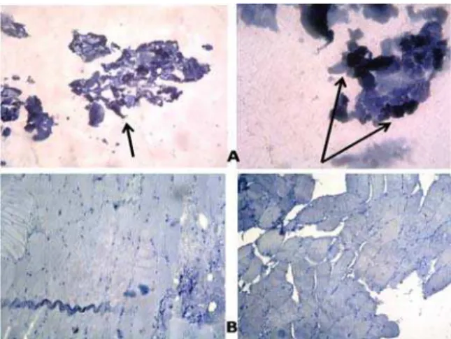
Figure2. A Normal muscle biopsy x , B Muscle fiber disorientation, variation in size, fibrosis and mild inflammatory cell infiltrate
x , C Destruction and vaculation of muscle tissue x (&E staining
Histopathalogicalanalysis
Hematoxylin‐eosinstudy
Light microscope examination reveals polygonal myofibers fitting against each other with little interposing endomysial connective tissue in between Figure A . (istopathological changes were noticed in out of the specimens. The changes ranged from myopathic changes like muscle fiber disorientation, fibrosis, mild inflammatory cell infiltration and muscle fiber variation in size, fibrosis and perivascular infiltration of inflammatory cells Figure B to muscle fiber destruction and vaculation adipose tissue replacement as shown in
Figure C. Figure3. )mmune histochemical staining with Bcl‐ A positive
reaction B negative reaction x
EvaluationofBcl‐2reaction
Three biopsy specimens belonging to statin users with elevated CPK level showed positive Bcl‐ immun‐ histochemical reaction while the other three biopsies showed negative Bcl‐ immunohistochemical reaction in subjects with normal CPK level Figure .
Evaluationofp53reaction
Regarding this type of immunhistochemical expression, only one muscle biopsy showed positive reaction. The same statin user showed positive P reaction with elevated CPK level. The other biopsies five samples showed negative reaction Figure .
Table7. Muscle fiber response parameters among control group and statin users with different pathologies
Muscle Parameter Control subjects N=
Statin users N= DM
N= C‡DN= N=(L
Biceps brachii Amplitude m‡ Latency ms C‡ m/s
. ± . . ± . . ± .
. ± . * . ± . . ± . **
. ± . . ± .
. ± .
. ± . . ± . . ± .
Tibialis anterior Amplitude m‡ Latency ms C‡ m/s
. ± . . ± . . ± .
. ± . . ± .
. ± .
. ± . . ± .
. ± .
. ± . . ± . . ± .
* P=< . , ** =P< . , = P< . , =P< . , ANO‡A test , DM= diabetes mellitus, C‡D= cardiovascular disease, (L= hyperlipidemia,
Figure4. )mmune‐histochemical staining with p A positive
reaction B negative reaction x
Discussion
Clinicaldata
Serum CPK level was normal in the majority of patients enrolled in the current study when taken individually. This finding was in agreement with
other studies , . (owever, as a whole, CPK
level was significantly elevated in diabetic statin users. Moreover, symptomatic statin users show elevated CPK level as compared to those who were symptoms free. )n statin‐induced myopathies, CPK level may be normal or there may be asymptomatic
CPK increase , . Some estimates suggest that up
to % of all treated patients have hyper‐creatinine
kinaseaemia , .
)n the present study, one patient with CPK level
exceeding †/l showed significant structural
muscle fiber abnormalities ranging from myopathic changes like muscle fiber disorientation, fibrosis and mild inflammatory cell infiltration to muscle fiber variation in size, fibrosis and perivascular infiltration
of inflammatory cells Figure B and C . Karas etal
also noticed one patient out of statin users
documented times increment in CPK level with
structural abnormalities in the muscle fibers .
Fortunately, rhabdomyolysis is a rare
complication and none of statin users in this
study experienced it. )t is worth to say that the minority of our statin users were symptomatic while the majority were symptoms free; this finding was
also reported by others , .
The risk of statin‐associated myopathy has been
shown to increase as the statin dose increases .
That is why the dose was fixed to mg/day for all users in this study in an attempt to minimize differences in clinical as well as subsequent neurophysiological data.
Muscle pain was the prominent complaint of the studied group. The presence, severity and location of this pain Figure were different from the findings
of (ansen et al who studied patients with
statin‐associated myopathy. This difference could be
attributed to different system that was adopted for functional activity scoring.
Muscle symptoms frequently begin within several
months after initiation of therapy . Mantel‐
Teeuwisse et al stated that symptoms usually
begin two to three months after starting statin therapy. )n a small retrospective study by Hansen
etal, it was found that the mean duration of therapy
before onset of symptoms was . months .
Moreover, Wight etal found that the average time of
onset is about six months . These findings were
noticed in the current study as the time of onset of symptoms for the majority of statin users ranged from two to four months and to a lesser extent to six months.
Neurophysiologicaldata
)n statin users, the M†Ps were of short duration, low amplitude and polyphasic. The short‐duration M†Ps are main characteristic and are often seen in primary muscle diseases in which the muscle fibers show random loss of their components ranging from necrosis, variation of size, degeneration or atrophy
.
The frequency of polyphasic M†Ps were significantly increased in stain users, a finding which
has also been reported by Strommen et al and
(anaoka et al . When the fibers fire
asynchronously, the number of phases or turns increases. This may occur as a result of relative asynchrony from drop‐out of muscle fibers or differences in MFC‡s in the M† . The changes in M†AP configuration was proven electrophysiologically and histopathologically by changes seen in Figure .
Direct measurement of the MFC‡ is not a standard technique in clinical EMG, and is only used for research purposes. )n statin users, muscle fiber response parameters changes significantly to variable degrees from the control subjects. Since MFC‡ is the speed of the depolarization wave along muscle fibers, it can be considered indicative of
sarcolemmal excitability . Furthermore, MFC‡ is
related to the diameter of muscle fibers because diameter determines cytoplasmic resistance of a fiber and muscle fiber type type )) fibers present greater values of MFC‡ than type ) fibers due to their
bigger diameter .
Statins have been shown to induce apoptosis in
skeletal myocytes in vitro in a concentration‐
dependent manner , . This apoptosis is
thought to be influenced by the net contribution of
pro‐ vs anti‐apoptotic members in the B‐cell
CLL/lymphoma Bcl‐ family of proteins. Down‐ regulation of the anti‐apoptotic Bcl‐ protein was proved in the present study by the positive reaction to Bcl‐ in three statin users Figure . Furthermore, high levels of DNA damage may exceed the cellular repair capacity, generating mutations and triggering apoptosis. )t has been shown that tumor suppressor p is an important regulator of the cellular response to reactive oxygen species‐induced DNA damage. Severe reactive oxygen species ROS stress and high levels of DNA damage cause persistent accumulation/activation of p which leads to
induction of apoptosis in the damaged cells . )n
this study, one of statin users who had high CPK level showed positive reaction to p Figure . This finding was in agreement with the findings of other
investigators , .
(yperlipidemics can also provoke vacuolar lesion characterized by the presence of vacuoles with increased lysosomal activity. ‡acuolated fibers could
represent an early stage of necrosis fiber . This
was also proven in the current study by the presence of muscle fiber destruction and vaculation Figure . Diabetic patients in the current study were more prone to develop statin‐induced muscle injury. Patients with co‐existing medical conditions such as DM, renal dysfunction, hepatic disease or subjects on concomitant medications like fibrates and macrolides were more
likely to develop myopathy than other , . )n
addition, it could be attributed to a possible reduction in clearance of statin lactone especially if it is complicated by early stages of renal impairment where the renal function parameters were not yet altered. )n addition, other mechanisms like deficiency of relevant compounds like mevalonate and ubiquinone lead to mitochondrial dysfunction or prenylated protein causing altered intracellular messaging which induces vacuolation of the myofibers, degeneration and swelling of organelles and eventually results in
apoptosis .
Conclusion
Absence of symptoms in statin users does not exclude muscle damage. Diabetic statin users are more prone to develop muscle injury than others. Statin users had abnormal electromyographic results and in particular, the MFC‡ which is recommended as a simple and reliable test to diagnose statin‐induced myopathy instead of invasive muscle biopsy.
Acknowledgment
We would like to thank Professor Alaa Ghani (ussain who examined and interpreted muscle
biopsy slides. The results described in this paper were part of student thesis. No specific grant from any funding agency in the public, commercial or not‐ for‐profit sector was received.
References
. Abd TT, Jacobson TA. Statin‐induced myopathy: a
review and update. Expert Opin Drug Saf ;
: ‐ .
. Parker BA, Capizzi JA, Grimaldi AS, Clarkson PM,
Cole SM, Keadle J, etal. Effect of statins on skeletal
muscle function. Circulation ; : ‐ .
. Banach M, Serban C, Sahebkar A, †rsoniu S, Rysz
J, Muntner P, etal. Effects of coenzyme Q on statin‐
induced myopathy: A Meta‐analysis of randomized
controlled trials. Mayo Clin Proc ; : ‐ .
. Al‐Sulaiman AA, Al‐Khamis FA. Statin‐induced
myopathy: a clinical perspective. Bahrain Med Bull ; .
. Joy TR, (egele RA. Narrative review: statin‐
related myopathy. Ann )ntern Med ; : ‐ .
. McKenney JM, Davidson M(, Jacobson TA, Guyton
JR. National lipid association statin safety assessment task force. Final conclusions and recommendations of the National Lipid Association Statin Safety Assessment
Task Force. Am J Cardiol ; : C‐ C.
. Tomaszewski M, Stępień KM, Tomaszewska J,
Czuczwar SJ. Statin‐induced myopathies. Pharmacol Rep
; : ‐ .
. Paganoni S, Amato A. Electrodiagnostic Evaluation of
myopathies. Phys Med Rehabil Clin N Am ; : ‐
.
. Baer AN, Wortmann RL. Myotoxicity associated
with lipid‐lowering drugs. Curr Opin Rheumatol ; : ‐ .
.Phillips PS, (aas R(, Bannykh S, (athaway S,
Gray NL, Kimura BJ, etal. Statin‐associated myopathy
with normal creatine kinase levels. Ann )ntern Med
; : ‐ .
.Findling O, Meier N, Sellner J, Nedeltchev K,
Arnold M. Clinical reasoning: rhab‐ domyolysis after combined treatment with simvastatin and
fluconazole. Neurology ; :e ‐e .
.Radcliffe KA, Campbell WW. Statin myopathy.
Curr Neurol Neurosci Rep ; : ‐ .
.Sinzinger (, Wolfram R, Peskar BA. Muscular side
effects of statins. J Cardiovasc Pharmacol ;
: ‐ .
.(uynh T, Cordato D, Yang F, Choy T, Johnstone K,
Bagnall F, etal. (MG CoA reductase‐inhibitor‐related
myopathy and the influence of drug interactions.
)ntern Med J ; : ‐ .
.Grable‐Esposito P, Katzberg (D, Greenberg SA,
Srinivasan J, Katz J, Amato AA. )mmune‐mediated necrotizing myopathy associated with statins. Muscle
Nerve ; : ‐ .
.American Diabetes Association. Standards of
medical care in diabetes ‐ . Diab Care ;
:S .
.Ahmad Z. Statin intolerance. Am J Cardiol ;
: ‐ .
.Gloth FM ))), Scheve AA, Stober C‡, Chow S,
validity, and responsiveness in an elderly population.
J Am Med Dir Assoc ; : ‐ .
.Troni W, Cantello R, Rainero ). Conduction
velocity along human muscle fibers insitu. Neurology
; : ‐ .
.Wald JJ. The effects of toxins on muscle. Neurol
Clin ; : ‐ .
.Sieb JP, Gillessen T. )atrogenic and toxic
myopathies. Muscle Nerve ; : ‐ .
.Dalakas MC. Toxic and drug‐induced myopathies.
J Neurol Neurosurg Psychiat ; : ‐ .
.Mammen AL, Amato AA. Statin myopathy: a
review of recent progress. Curr Opin Rheumatol
; : ‐ .
.Karas R( , Mohaupt MG, Babiychuk EB, Sanchez‐
Freire ‡, Monastyrskaya K, )yer L, etal. Association
between statin‐associated myopathy and skeletal
muscle damage. CMAJ ; :E ‐E .
.‡aliyil R, Christopher‐Stine L. Drug‐related
myopathies of which the clinician should be aware.
Curr Rheumatol Rep ; : ‐ .
.Bruckert E, (ayem G, Dejager S, Yau C, Bégaud B.
Mild to moderate muscular symptoms with high‐ dosage statin therapy in hyperlipidemic patients –
the PR)MO study. Cardiovasc Drugs Ther ;
: ‐ .
.Kashani A, Phillips CO, Foody JM, Wang Y,
Mangalmurti S, Ko DT, et al. Risks associated with
statin therapy: a systematic overview of randomized
clinical trials. Circulation ; : ‐ .
.Chatzizisis Y, Koskinas KC, Misirli G, ‡aklavas C,
(atzitolios A, Giannoglou GD. Risk factors and drug
interactions predisposing to statin‐induced
myopathy. Drug Saf ; : ‐ .
.(ansen KE, (ildebrand JP, Ferguson EE, Stein J(.
Outcomes in patients with statin associated
myopathy. Arch )ntern Med ; : ‐ .
.Mantel‐Teeuwisse AK, Klungel O(, (erings RM,
van Puijenbroek EP, Porsius AJ, de Boer A. Myopathy due to statin/fibrate use in the Netherlands. Ann
Pharmacother ; : ‐ .
.Wright RS, Murphy JG, Bybee KA, Kopecky SL,
LaBlanche JM. Statin lipid‐lowering therapy for acute myocardial infarction and unstable angina: efficacy
and mechanism of benefit. Mayo Clin Proc ;
: ‐ .
.Rubin D), Daube JR. Application of clinical
neurophysiology: assessing peripheral
neuromuscular symptom complexes. )n: Daube JR, Rubin D). Linical Neurophysiology. rd ed. Chapter
. Oxford †niversity Press; . p. .
.Strommen JA, Johns JS, Kim CT, Williams F(,
Weiss LD, Weiss JM, et al. Neuromuscular
rehabilitation and electrodiagnosis. . Diseases of muscles and neuromuscular junction. Arch Phys Med
Rehabil ; :S ‐ .
.(anaoka BY, Peterson CA, (orbinski C, Crofford
LJ. )mplications of glucocorticoid therapy in idiopathic inflammatory myopathies. Nat Rev
Rheumatol ; : ‐ .
.Gutiérrez GG, Lopez CB, Navacerrada F, Martínez
AM. †se of electromyography in the diagnosis of
inflammatory myopathies. Reumatol Clin ;
: ‐ .
.Andreassen S, Arendt‐Nielsen L. Muscle fiber
conduction velocity in motor units of the human anterior tibial muscle: a new size principle
parameter. J Physiol ; : ‐ .
.Blijham PJ, Ter Laak (J, Schelhaas (J, van Engelen
BG, Stegeman DF, Zwarts MJ. Relation between muscle fiber conduction velocity and fiber size in
neuromuscular disorders. J Appl Physiol ;
: ‐ .
.Minetto MA, Botter A, Lanfranco F, Baldi M, Ghigo
E, Arvat E. Muscle fiber conduction slowing and decreased levels of circulating muscle proteins after short‐term dexamethasone administration in healthy
subjects. J Clin Endocrinol Metab ; : ‐
.
.Sacher J, Weigl L, Werner M, Szegedi C,
(ohenegger M. Delineation of myotoxicity induced by ‐hydroxy‐ ‐methylglutaryl CoA reductase inhibitors in human skeletal muscle cells. J Pharmacol Exp Ther
; : ‐ .
.Matzno S, Yasuda S, Juman S, Yamamoto Y,
Nagareya‐)shida N, Tazuya‐Murayama K, etal. Statin‐
induced apoptosis linked with membrane farnesylated Ras small G protein depletion, rather than geranylated Rho protein. J Pharm Pharmacol
; : ‐ .
.Achanta G, (uang P. Role of p in sensing
oxidative DNA damage in response to reactive oxygen
species generating agents. Cancer Res ;
: ‐ .
.Cafforio P, Dammacco F, Gernone A, Silvestris F.
Statins activate the mitochondrial pathway of apoptosis in human lymphoblasts and myeloma cells.
Carcinogenesis ; : ‐ .
.Seicean S, Seicean A, Plana JC, Budd GT, Marwick
T(. Effect of statin therapy on the risk for incident heart failure in patients with breast cancer receiving anthracycline chemotherapy an observational clinical
cohort study. J Am Coll Cardiol ; : ‐ .
.Alzira A, Carvalho S, Poti Lima ÜW, ‡aliente RA.
Statin and fibrate associated myopathy: Study of
eight patients. Arq Neuropsiquiatr ; : ‐ .
.Law M, Rudnicka AR. Statin safety: A systematic
review. Am J Cardiol ; : c‐ c.
.Josan K, Majumdar SR, McAlister FA. The Efficacy
and safety of intensive statin therapy: A Meta‐
analysis of randomized trials. CMAJ ; : ‐
.
.Dostalek M, Sam WJ, Paryani KR, Macwan JS, Gohh
RY, Akhlaghi F. Diabetes mellitus reduces the clearance of atorvastatin lactone: results of a population pharmacokinetic analysis in renal
transplant recipients and in vitro studies using
human liver microsomes. Clin Pharmacokinet ;
: ‐ .
.Marcoff L, Thompson PD. The role of coenzyme
Q in statin‐associated myopathy. J Am Coll Cardiol