Please cite this paper as:
Kunjiappan S, Bhattacharjee Ch, Chowdhury R. Hepatoprotective and antioxidant effects of Azolla microphylla based gold nanoparticles against acetaminophen induced toxicity in a fresh water common carp fish (Cyprinus
Received: Oct. 19, 2014; Accepted: Feb. 5, 2015 Vol. 2, No. 2, Spring 2015, page 88-110
Online ISSN 2322-5904 http://nmj.mums.ac.ir
Original Research
Hepatoprotective and antioxidant effects of
Azolla microphylla
based gold
nanoparticles against acetaminophen induced toxicity in a fresh water
common carp fish (
Cyprinus carpio
L
.
)
Selvaraj Kunjiappan1, Chiranjib Bhattacharjee1*, Ranjana Chowdhury1
1
Department of Chemical Engineering, Jadavpur University, India
Abstract
Objective(s): Our present study sought to evaluate hepatoprotective and antioxidant effects of methanol extract of Azolla microphylla phytochemically synthesized gold nanoparticles (GNaP) in acetaminophen (APAP) - induced hepatotoxicity of fresh water common carp fish.
Materials and Methods: GNaP were prepared by green synthesis method using methanol extract of Azolla microphylla. Twenty four fishes weighing 146 ± 2.5 g were used in this experiment and these were divided into four experimental groups, each comprising 6 fishes. Group 1 served as control. Group 2 fishes were exposed to APAP (500 mg/kg) for 24 h. Groups 3 and 4 fishes were exposed to APAP (500 mg/kg) + GNaP (2.5 mg/kg) and GNaP (2.5 mg/kg) for 24 h, respectively. The hepatoprotective and antioxidant potentials were assessed by measuring liver damage, biochemical parameters, ions status, and histological alterations.
Results: APAP exposed fish showed significant elevated levels of metabolic enzymes (LDH, G6PDH and MDH), hepatotoxic markers (GPT, GOT and ALP), reduced hepatic glycogen, lipids, protein, albumin, globulin, increased levels of bilirubin, creatinine, and oxidative stress markers (TBRAS, LHP and protein carbonyl), altered the tissue enzymes (SOD, CAT, GSH-Px and GST) non-enzyme (GSH), cellular sulfhydryl (T-SH, P-SH and NP-SH) levels, reduced hepatic ions (Ca2+, Na+ and K+), and abnormal liver histology. It was observe that GNaP has reversal effects on the levels of above mentioned parameters in APAP hepatotoxicity.
Conclusion: Azolla microphylla phytochemically synthesized GNaP protects liver against oxidative damage and tissue damaging enzyme activities and could be used as an effective protector against acetaminophen-induced hepatic damage in fresh water common carp fish.
Keywords: Azolla microphylla, Acetaminophen, Antioxidant Cyprinus carpio L.,
Hepatotoxicity, Gold nanoparticles
*Corresponding author: Chiranjib Bhattacharjee,Professor, Department of Chemical Engineering, Jadavpur
University, Kolkata-700 032, India.
Nanomed J, Vol. 2, No. 1, Spring 2015 89
Introduction
The liver is one of the prime target organs, which regulates many biological functions such as drug metabolism, amino acid metabolism, lipid metabolism and glycolysis [1].
Liver injury or liver dysfunction is the most serious ailment and are mainly caused by excess consumption of alcohol, high doses of acetaminophen, chemoth-erapeutic agents, hepatic viral infection, dantrolene sodium, valporic acid, per-oxidised oil and isonicotinic acid hydrazide, etc [2].
Acute and chronic liver diseases constitute a global concern and medical treatments for these diseases are often difficult to handle and have limited efficiency [3]. Therefore, there has been considerable interest in role of complementary and alternative medicines for the treatment of liver diseases. Developing therapeutically effective agents from natural products may reduce the risk of toxicity when the drug is used clinically.
It has been reported that, among a variety of drugs, acetaminophen (APAP or N-acetyl-p-aminophenol or paracetamol or 4-hydroxyacetanilide) is the most common cause of drug-induced liver injury [4]. APAP is one of the best-selling and widely used over-the-counter analgesic and antipyretic drug. Although considered safe at therapeutic doses, at higher doses (3h following 500 mg/kg body weight), acetaminophen produce acute liver failure (ALF) [5-7].
When taken in therapeutic doses, greater than 90% of acetaminophen is metabolized to phenolic glucuronide and sulfate in the liver by glucuronyltransferases and sulfo-transferases and subsequently excreted in the urine [8].
Of the remaining acetaminophen, about 2% is excreted in the urine unchanged; approximately 5 to 10% is metabolized by cytochrome P450, mainly the enzyme CYP2E1 [9], to N-acetyl-p -benzoquinon-eimine (NAPQI) (10), a highly reactive, electrophilic molecule that causes harm by formation of covalent bonds with other
intracellular proteins [11]. This toxic metabolites neutralized by glutathione (GSH), which is an important cellular free radical scavenging non-enzymes for neutralization of toxic metabolites produced by drugs and foreign chemicals [12]. If glutathione is not replenished, NAPQI will begin to accumulate in the hepatocytes [13].
NAPQI can form covalent bonds with cysteinyl thiol groups of cellular proteins to form protein-(cystein-S-yl)-APAP adducts [14], which may modify the structure and functions of the proteins [15]. This cellular functional disturbance leads to a decrease in calcium ATPase activities and increase in levels of cytosolic calcium [16, 17].
Abnormal cellular calcium homeostasis can alter the permeability of the cell, causing the formation of blebs in the cell membrane and loss of membrane integrity [18]. Medicinal plants have been gaining importance of effective source of both traditional and modern medicines to treat or prevent human and animal diseases. World health organization has recomme-nded traditional plant derivatives as safe remedies for ailments for microbial and non-microbial origin [19].
The medicinal effects of these plants lies in some chemical substances such as flavor-noids, phenolic compounds, alkaloids, anthroquinones, carbohydrates, proteins and vitamins that produces definite therapeutic action on the human and animal body [20]. Plant secondary metabolites (Flavonoids and phenolic compounds) are of great importance for the bioactivities, related to their antioxidant activities [21] and many enzymatic reactions, resulting in a decrease of platelet activation and aggr-egation, against cardiovascular diseases, cancer chemoprevention, acute liver failure [22] and anti-inflammatory activity [23-25].
regions of the world. Azolla microphylla is one of the species from the genus Azolla; it is a pteridophyte plantae belonging to the Salvinacea family [26].
The phytochemical investigation on the
Azolla microphylla shows that tannins,
polyphenols, sugar, anthroquinone glycos-ides and steroids are present [27]. Becerra et al. [28], Lumpkin and Plucknett [29], Van Hove and López [30] concluded that, Azolla is the most promising aquatic plant for livestock feed due to its ease of cultivation, productivity and nutritive value. Recently this research group has isolated and purified rutin and quercetin from Azolla microphylla for the first time [31]. Therefore Azolla microphylla is one of the important sources for various antioxidants and hepatoprotective agents. Researchers in the field of nanotechnology are gaining new insights into its versatile applications in the treatment of various diseases, including hepatotoxicity. Research into the preparation and biological applicability of noble metal nanoparticles with a nearly
mono-dispersed size distribution and arbitrarily variable size and geometry has attracted
considerable research interest.
It is reported that metal nanoparticles, especially gold nanoparticles have drawn more attention of scientists because of their unique nature of stability, oxidation resistance and biocompatibility [32]. Gold nanoparticles widely applied in the field of medicine such as chemical bio-sensing, imaging, drug delivery and therapeutic labeling [33].
Gold nanoparticles have a growing role in medical biotechnology.
Production of nanoparticles can be achieved mainly through chemical, physical, and biological methods.
Biological methods for nanoparticles synthesis using microorganisms, enzymes and plants or plant extracts have been suggested as possible ecofriendly alterna-tives to chemical and physical methods. However, major drawback of using microbes such as bacteria, fungi and yeast are the requirement of manipulation of
reaction parameters such as pH, temperature and incubation time for the synthesis of nanoparticles [34].
On the other hand, due to high rate of generation of nanoparticles, plant based synthesis is comparatively simpler and more cost-effective[35].
Plant phytochemicals such as polyphenols, flavonoids, triterpenes, tannins, glycosides, vitamins, proteins and steroids are capable of reducing the gold ions (Au3+ or Au1+) to neutral gold nanoparticles (Au0) and ensures good control over size distribution and crystallinity of the nanoparticles (36). The phytochemicals present in Azolla microphylla serve a dual role as effective reducing agents to reduce gold and also as stabilizers to provide a robust coating on the gold nanoparticles in a single step. However, no reports have been found on the protective effect of Azolla
microphylla phytochemically synthesized
gold nanoparticles on APAP-induced hepatotoxicity.
Thus, we hypothesized that phytochemicals rich in Azolla microphylla mediated synthesized gold nanoparticles would prevent APAP-linked abnormalities.
The present study examined the hepatoprotective and antioxidant effects of
Azolla microphylla methanol extract
mediated green synthesized gold nanoparticles in acetaminophen-induced hepatotoxicity in common carp fish
(Cyprinus carpio L.) in terms of
hepatotoxic markers, oxidative stress, antioxidant, hepatic ions status, glycogen, cholesterol, protein, bilirubin, lipid levels and H & E staining.
Materials and Methods Materials
Acetaminophen was received as a gift from Pharma-Fabricon pharmaceuticals Ltd, Madurai, India.
All other chemicals and solvents were of analytical grade and were obtained from Hi-media laboratories Pvt. Ltd. Mumbai, India, and Merck, Mumbai, India.
Nanomed J, Vol. 2, No. 1, Spring 2015 91 Ltd. Surat, India and Qualigen Diagnostics
Ltd. Mumbai, India.
Plant collection and preparation of Azolla microphylla extract
Azolla microphylla fern were received
from Vivekananda Kendra-NARDEP (Natural Resources Development Project), Vivekanandapuram, Kanyakumari, India. The whole parts of the fern were washed with tap water, rinsed with distilled water and air-dried under shade with good ventilation at room temperature (≈37˚C) for a week.
The fine powder (≈60 mesh size) was
obtained from dried plant material by using kitchen blender (Bajaj electronics Ltd, India).
About 500g of Azolla microphylla powder was weighed and macerated with 1000 mL of 99.9% methanol in a 2000 mL conical
flask and kept at room temperature for 72 h.
After 72 h, the methanol extract of Azolla
microphylla was filtered with Whatman
No: 1 filter paper.
The filtered extract was centrifuged at 10000 rpm for 10 min at 4˚C. We discarded the precipitate that contains debris and collected the supernatant in a brown bottle and stored in refrigerator at 4°C until further studies.
Green chemistry approach for the synthesis of gold nanoparticles
1 mM solution of hydrogen tetrarch-oloroaureate (III) hydrate (Himedia laboratories Pvt. Ltd. Mumbai) was prepared using de-ionized water. 5 mL (concentration 5%, v/v) of the methanolic extract of Azolla microphylla was mixed with 25 mL aqueous solution of HAuCl4.3H2O (1 mM).
The mixture solution was left on constant magnetic stirring at room temperature
(≈35°C) for 30minutes and observed for
change in color.
The bio-reduction of gold ions into gold nanoparticles in the solution was monitored by periodic sampling of aliquots (1 mL) and subsequently measuring
UV-vis spectra of the solution. UV-UV-visible spectrophotometry analysis was carried out by a computer controlled UV–vis spectrophotometer (Varian Cary 50 UV-Spec) between 400 and 800 nm possessing a scanning speed of 400 nm/min.
Characterization of nanoparticles
Characterization of the biosynthesized gold nanoparticles was carried out before starting the experiment for their potent hepatoprotective and anti-oxidative effects in common carp fish.
Hydrogen tetracholoroaureate (III) hydrate (aqueous) treated methanol extract of
Azolla microphylla solution was
centri-fuged at 10000 rpm for 15 min at 4˚C and the resulted supernatant solution was maintained at -80˚C for 24 h and then freeze dried in a lyophilizer (Christ Gefriertrocknungsanlagen GmbH Model 1–4) for 48 h.
Molecular shape and size of the lyophilized gold nanoparticles was characterized using FESEM and HRTEM analyses.
The dose for the experimental study of the lyophilized gold nanoparticles was calculated as 2.5 mg/kg body weight from previous studies [37].
Total antioxidant assay of gold nanoparticles
The total antioxidant capacity of the methanol extract of Azolla microphylla
mediated synthesized gold nanoparticles was evaluated by the method of Prieto et al. [38].
The assay is based on the reduction of Mo (VI) to Mo (V) by various concentrations of green gold nanoparticles (1, 2, 3, 4 and 5 mg/mL).
De-ionized water (0.3 mL) in place of gold nanoparticles was used as the blank. The total antioxidant activity is expressed as the number of gram equivalents of ascorbic acid.
Fish husbandry and maintenance
Fresh water common carp (Cyprinus
carpio L.) was obtained from commercial
hatcheries, Jadavpur, Kolkata, India. 48 healthy fish of uniform size of length (12 ± 2 cm) and weight (145.46 ± 1.24 g) were segregated from the stock and acclimatized to laboratory conditions for 7 days in an aquarium (60 × 30 × 40 cm) with 50 L of dechlorinated tap water (6-8 individuals /aquarium).
The total mortality of fish was less than 1%. During experiment, water temperature was 25 ± 1˚C, pH was 7.1 ± 0.25, dissolved oxygen was 8.015 ± 0.5 mg/L, hardness was 425 ± 3.5 mg/L CaCo3,
turbidity was 2.5NTU and total solids were 14.05 ± 0.1 mg/L.
Fishes were reared in a re-circulation system with 12 h light-dark photoperiod and fed with commercial diets (Tokyu baby pellet, Floating type, Japan) twice a day.
Feeding was withheld 1day prior to exposure, with an average weight of 146 ± 2.5 g at the start of the experiment.
Toxicity studies
In this study, the fishes were randomly divided into two groups consist of 6 fish in each.
Group 1 – Control, Group 2 - GNaP treated, Group 2 Fish were treated with
Azolla microphylla mediated synthesized
gold nanoparticles (GNaP) (2.5 mg/kg body wt) for 48 h in static tank containing 5L of water. The control group fish were fed with commercial baby pellet feed. At the end of the 48 h treatment, the fish were sacrificed by decapitation.
Blood was collected and allowed to clot at room temperature, centrifuged at 3000 rpm
at 4˚C for 20 min to obtain serum in which
analysis of total protein, cholesterol and triglycerides were performed. Liver was dissected immediately and fixed with 10% buffered formalin for histological analysis.
Experimental design
The fishes were dived into four groups consist of 6 fish in each. Fishes were treated as follows: Group I Control fish (CON) were fed with commercial baby pellet feed (crude protein-46%, crude fat-6%, crude fibre-5%, crude ash- 12% and spirulina nitrogen free extract-20%) throughout the experimental period. Group II Fish were treated with acetaminophen (APAP) for 24 h in static tank containing 5 L of water. Acetaminophen; 500 mg/kg was dissolved in 100 mL water at 70˚C and cooled to room temperature and added to the static tank. Group III Fish were treated with acetaminophen + Azolla
microphylla mediated synthesized gold
nanoparticles (APAP + GNaP) for 24 h in static tank containing 5 L of water. Acetaminophen; 500 mg/kg was dissolved in 100 mL water at 70˚C and cooled to room temperature and combined with methanol extract of Azolla microphylla
mediated synthesized gold nanoparticles (2.5 mg/kg body wt) and added to the static tank. Group IV Fish were treated with Azolla microphylla mediated synthesized gold nanoparticles (GNaP) (2.5 mg/kg body wt) alone for 24h in static tank containing 5 L of water. At the end of the experimental period, the fishes were sacrificed by decapitation. Blood was collected and allowed to clot at room temperature and then centrifuged at 3000
rpm at 4˚C for 20 min to obtain serum in
Nanomed J, Vol. 2, No. 1, Spring 2015 93
Assessment of carbohydrate metabolic enzymes
The enzyme assays of Lactate dehydrogenase (LDH, E.C.1.1.1.27), Glucose-6-phosphate dehydrogenase (G6PDH, E.C.1.1.1.49) and Malate dehydrogenase (MDH, E.C.1.1.1.37) were determined spectrophotometrically using commercial kits (Span Diagnostics, Ltd. Surat, India).
Biochemical assessment of hepatotoxicity.
Glutamate pyruvate transaminase (GPT, E.C.2.6.1.2), Glutamate oxalate transaminase (GOT, E.C.2.6.1.1) and Alkaline phosphatase (ALP, E.C.3.1.3.1) were assayed spectrophotometrically with
clinical test kits according to manufacturer’s
protocol (Span Diagnostics Ltd. Surat, India). Glycogen content was estimated by the method of Morales et al. [39].
Cholesterol was estimated by the method of Natio [40]. Triglycerides were estim-ated by the method of Buccolo [41]. Protein content was estimated by the method of Lowry et al. [42]. Albumin content was estimated by the method of Gendler [43]. Globulin content was obtained by subtracting albumin content from total protein. Bilirubin content was estimated by the method of Jendrassik [44]. Creatinine was measured by the method of Spencer [45]. Levels of ions (Ca2+, Na+ and K+) in the liver tissue homogenates were estimated using Atomic Absorption Spectrophotometer (Perkin Elmer, Analyst spectra 200 Atomic absorption spectroscopy) with certified standards by AOAC method [46].
The detection limits for the assay of Ca2+, Na+ and K+ tend to range between 0.06, 0.01, 0.03 and 100 ± 10 mg/L, respectively.
Assessment of enzymes involved in free radical scavenging activities
Superoxide dismutase (SOD, E.C.1.15.1.1) was assayed by the method of Marklund and Marklund [47]. Catalase (CAT, E.C.1.11.1.6) was assayed by the method of Chance and Maehly [48]. Glutathione peroxidase (GSH-Px, E.C.1.11.1.9) activities were assayed by the method of
Mohandas et al. [49]. Glutathione-S-transferase (GST, E.C.2.5.1.14) was measured by the method of Rajasekar and Anuradha [50]. Briefly, SOD activity was assayed by the inhibition of nicotinamide adenine dinucleotide (reduced) phenazine methosulphate nitrobluetetrazolium reaction system as adapted and the results are expressed as units (U) of SOD activity/mg protein. CAT and GSH-Px activities were assayed by measuring the amount of substrate consumed (hydrogen peroxide and glutathione, respectively) after carrying out the reactions for a specified period of time and the results have been expressed as units (U) of activity/mg protein. Activity of glutathione-s-transferase (GST) was expressed as 1, 2-dichloro-4-nitrobenzene µmol/mg protein/min.
Non-enzymatic glutathione (GSH) was estimated in the liver homogenates using dithionitrobenzoic acid (DTNB) by the method of Ellman [51]. The absorbance was read at 412nm and the results were expressed as µmol/mg protein/min.
Assessment of oxidative stress markers and –SH
Lipid peroxidation of liver homogenate was estimated by measuring the thiobarb-ituric acid reactive substances (TBARS) and was expressed in terms of MDA content (nanomol/mg protein), according to the method of Uchiyama and Mihara [52]. Lipid hydroperoxide (LHP) was measured by the method of Ohkawa et al. [53] and expressed as µmol/mg protein. The level of protein carbonyl was measured by the method of Levine et al. [54]. Total (T-SH), non-protein (NP-SH) and protein bound (P-SH) sulfhydryl groups were determined by the method of Sedlak and Lindsay [55].
Histopathological studies
concentrations of alcohol in ascending order and finally in absolute alcohol (10 min each). The tissue was then embedded in paraffin wax. Sections of 5µm thickness were made using a microtome.
The tissues were de-paraffinized with xylene and treated with 100, 90 and 70% alcohol for removing undesirable pigments and other elements. The sections were then stained with hematoxylin-eosin (H & E) and observed under microscope. Photographs of each slide were taken at 10× and 40× magnifications.
Statistical analysis
Values are expressed as mean ± standard deviation. Data within the groups are analyzed using one-way analysis of variance (ANOVA) followed by Dunnett multiple comparisons test.
Values were considered statistically significant when p<0.05.
Statistical analyses were carried out using SPSS statistics version 20 software.
Results
Characterization of the green synthesized gold nanoparticles
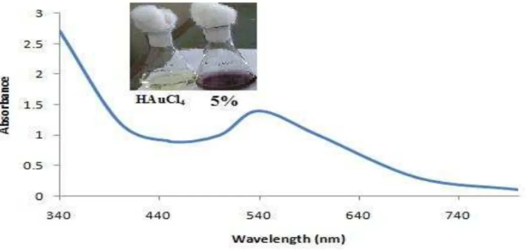
The formation and stability of the gold nanoparticles were confirmed by colour change from original yellow to pink to dark pink which is measured by UV-Vis spectrophotometry. The dark pink colour observed is characteristic for unique surface plasmon resonance (SPR) of different sizes of gold nanoparticles. Figure 1 showed the UV-Vis absorption spectra of biosynthesized gold nanoparticles.
The gold surface plasmon resonance (SPR)
band (λmax 540 nm), indicates the
presence of spherical nanoparticles in the reaction mixture.
According to the Mie theory, the small gold nanoparticles exhibited only one surface plasmon resonance absorption
band (56). Kinetics of the formation of gold nanoparticles, 5% v/v methanol
extract of Azolla microphylla in 1mM HAuCl4 solution are shown in Figure2.
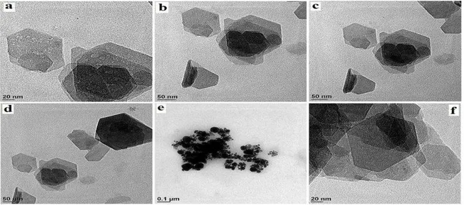
Figure 3 depicts the field emission scanning electron microscope images of the biosynthesized gold nanoparticles. The overall morphological shapes of the gold nanoparticles are spherical and rectangular at higher magnification. In Figure 4, the HRTEM images clearly proved the size and shape of the gold nanoparticles as a function of concentration of the phenolic compounds present in the plant extract.
The shapes of the gold nanoparticles were spherical, triangular, hexagonal and rod shaped. Gold nanoparticles corresponding to HRTEM images exhibited the variation in the particle size ranging from 3 to 20nm with average of 8.3 nm.
Total antioxidant capacity of gold nanoparticles
The methanol extract of Azolla
microphylla mediated biosynthesized gold
nanoparticles were subjected to screening for their total antioxidant capacity.
The antioxidant capacity of the various concentrations of green synthesized gold nanoparticles was summarized in Table 1. All the concentrations of green synthesized gold nanoparticles were significantly showed antioxidant activity resulted in ranges from 48.0 ± 0.05 to 82.04 ± 0.03 mg/g ascorbic acid equivalent, while 3 mg/mL gold nanoparticles showed efficient antioxidant capacity.
Toxicity studies
In vivo toxicity studies of the synthesized nanoparticles were performed by exam-ining the changes of blood serum and histological analysis. The fish exposed with GNaP (2.5 mg/kg body wt) for 48 h and examined for any changes in the morphology, behavior and mortality.
Nanomed J, Vol. 2, No. 1, Spring 2015 95
Figure 1. UV-vis-absorption spectra of 5% methanol extract phytochemically synthesized gold nanoparticles.
The inset image shows the 1mM HAuCl4 solution and green synthesized gold nanoparticles solution.
Figure 2. Kinetics of the formation of gold nanoparticles.
Figure 4. Transmission electron microscope images of green synthesized gold nanoparticles.
Table 1. Total antioxidant capacity of methanol extract of Azolla microphylla mediated synthesis of gold
nanoparticles.
Concentration of gold nanoparticles
(mg/mL)
Total antioxidant capacity mg/g ascorbic acid equivalent
1 48.0±0.05
2 68.2±0.02
3 82.4±0.03
4 66.1±0.02
5 64.2±0.03
Values are listed as mean± SD
The fish did not showed any symptoms of toxicity such as fatigue, change in orientation of color, weight loss, etc. Comparative analysis of blood serum in the gold treated and control fish, clearly showed that there was no significant alteration (Table 2).
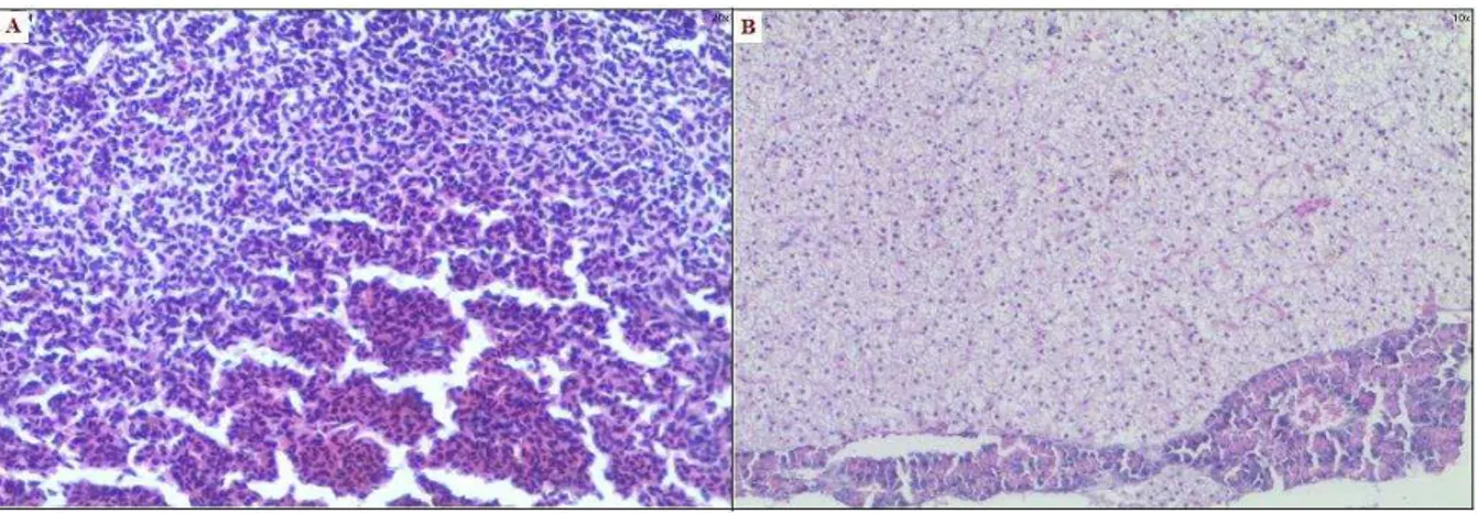
Thus the histopatho-logical effect of the control and gold nanoparticles treatment was observed using light microscope. The histological finding of the liver of the non-toxic effect of gold nanoparticles was showed in Figure 5.
The liver histological studies showed the control liver with normal hepatic portal triad and central vein (Figure 5a) and the
treatment of gold nanoparticles at a dosage of 2.5 mg/kg body wt for 48 h did not lead to any disruptions in the histology showed normal hepatocytes in comparison with control (Figure 5b).
Effect of gold nanoparticles on tissue gross morphology
Nanomed J, Vol. 2, No. 1, Spring 2015 97
Figure 5. Toxicity studies of green synthesized gold nanoparticles in carp fish liver. Histopathological studies
of fish liver collected from 48h treatment of gold nanoparticles, stained with hematoxylin and eosin (H and E) showed normal morphology. (a) Control fish liver section showed normal morphology and cells arranged around the central vein (b) Gold nanoparticles treated fish liver sections also showed normal architecture with clear central vein.
Table 2. Blood plasma analysis revealing the nontoxic effects of green synthesized gold nanoparticles in Carp fish.
Parameters CON GNaP
Protein (mg/dL) 4.435±0.32 4.442±0.19
Cholesterol (mg/dL) 11.959±0.97 12.018±0.12
Triglyceride (mg/dL) 5.44±0.40 5.628±0.42
Values are listed as mean± SD (n=6 in each group) CON control, GNaP green synthesized gold nanoparticles
Figure 6. Hepatoprotective effects of gold nanoparticles over Acetaminophen-induced liver damage. H and E
GNaP treated and control fish showed smooth and shiny, with normal appearance of liver as that of control fish.
Effect of gold nanoparticles on liver tissue histopathology
In the histopathological studies, liver sections (H&E staining) of control fish showed normal hepatic architecture (Fig.6A), whereas that APAP treated fish showed total loss of hepatic architecture with intense peripheral central vein necrosis (A*), fatty changes (B*), congestion of sinusoid, hepatic cell hyperplasia, crowding of the central vein, apoptosis (C*) (Fig.6B), in case of fish treated with APAP+GNaP (Figure 6C), these changes were reduced (i.e. mild vacuolar degeneration, reduced hepatocellular damage and absence of congestion).
The above abnormalities were effectively controlled in the GNaP (Fig. 6D) administered fish tissues.
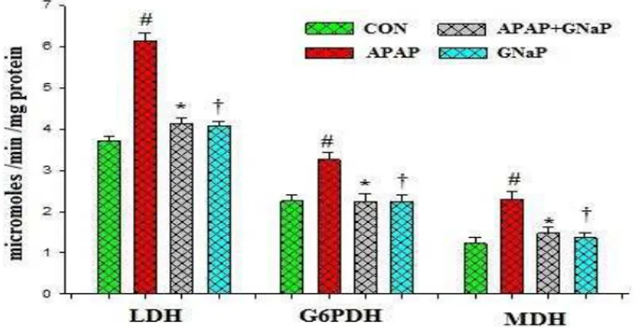
Effect of gold nanoparticles on carbohydrate metabolic enzymes
The levels of carbohydrate metabolic enzymes such as LDH, G6PDH and MDH were significantly (p<0.05) higher in APAP-exposed fish when compared with the control (Figure 7), which indicates that the effect of carbohydrate metabolic enzymes was seen in the severity of liver tissue damage of exposed fish rather than the control. The elevated levels of these enzymes were reduced by treatment of APAP+GNaP. No significant effect on these metabolic enzymes with GNaP-treated fish was observed.
Figure 7. Effect of green synthesized gold nanoparticles on carbohydrate metabolic enzymes of control and
experimental fish. Values are mean± SD (n=6 in each group). #p<0.05 as compared to CON. *p<0.05 as compared to APAP. †p<0.05 as compared to APAP and CON. CON control, APAP acetaminophen, GNaP green synthesized gold nanoparticles.
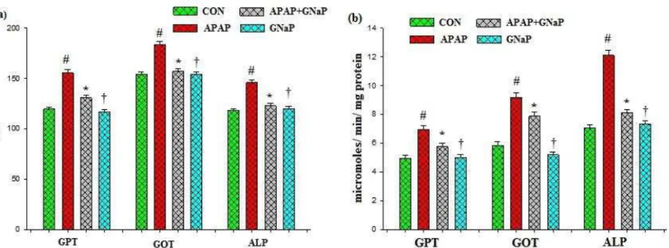
Effect of gold nanoparticles on hepato-toxic markers
The effects of GNaP on hepatotoxic marker enzymes such as GPT, GOT and ALP in blood and liver tissue of control and experimental fish are summarized in Figure 8 a,b, respectively. Significant elevation in the marker enzyme activities
Figure 8. Effect of green synthesized gold nanoparticles on hepatotoxic markers in blood (a) and liver (b) of control and experimental fish. Values are mean± SD (n=6 in each group). #p<0.05 as compared to CON. *p<0.05 as compared to APAP. †p<0.05 as compared to APAP and CON. CON control, APAP acetaminophen, GNaP green synthesized gold nanoparticles.
Effect of gold nanoparticles on oxidative stress markers
The effects of GNaP on oxidative stress parameters are summarized in Table 3. The lipid peroxidation markers such as LHP, TBARS and protein carbonyl, were significantly elevated (p<0.05) in the APAP-treated fish when compared with the
control. This indicates the severity of hepatic damage under APAP exposure, whereas in APAP+GNaP treated fish, the levels of lipid peroxidation (LPO) markers were significantly depleted (p<0.05) to values near to those in control fish. GNaP-treated fish did not showed any changes in the levels of LPO markers.
Table 3. Effect of green synthesized gold nanoparticles on oxidative stress markers in liver of control and experimental
fish.
Parameters CON APAP APAP+GNaP CON+GNaP
LHPd 0.4341±0.05 0.07483±0.01a 0.2687±0.18b 0.4209±0.04c TBARSe 1.318±0.08 3.389±0.22a 1.919±0.04b 1.879±0.06c Protein Carbonyld 2.902±0.27 4.908±0.66a 3.140±0.50b 3.005±0.46c
Values are listed as mean± SD (n=6 in each group)
CON control, APAP acetaminophen, GNaP green synthesized gold nanoparticles
a (p < 0.05) as compared to CON b
(p < 0.05) as compared to APAP
c
(p < 0.001) as compared to CON and APAP+GNaP. Data within the groups were analyzed using one-way ANOVA followed by
Dunnett multiple comparisons test. d µmoles/mg protein, e nanomoles/mg protein
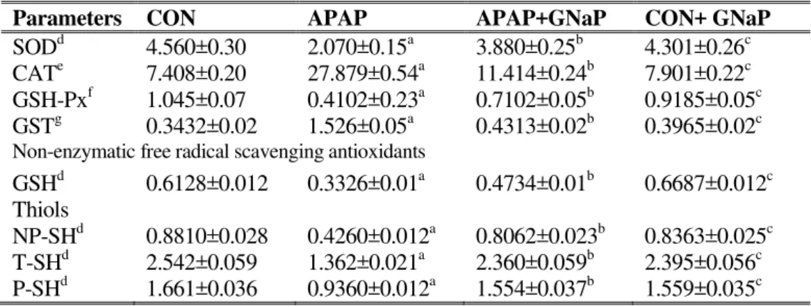
Effect of gold nanoparticles on enzymatic and non-enzymatic antioxidants and thiols
The effects of GNaP on enzymatic (SOD, CAT, GSH-Px and GST) and non-enzymatic (GSH) antioxidants and cellular thiols levels (T-SH, NP-SH and P-SH) in the liver homogenates are summarized in Table APAP-treated fish showed a decrease in the levels of SOD, GSH-Px, GSH, T-SH, NP-SH and P-NP-SH and an increase in the levels of
CAT and GST, when compared with the control fish. However, the levels of the above enzymatic and non-enzymatic antioxidants significantly (p<0.05) altered upon treatment with APAP+GNaP treated fish.
Table 4. Effect of green synthesized gold nanoparticles on enzymatic and non-enzymatic free radical scavenging antioxidants and thiols in liver of control and experimental fish.
Parameters CON APAP APAP+GNaP CON+ GNaP
SODd 4.560±0.30 2.070±0.15a 3.880±0.25b 4.301±0.26c CATe 7.408±0.20 27.879±0.54a 11.414±0.24b 7.901±0.22c GSH-Pxf 1.045±0.07 0.4102±0.23a 0.7102±0.05b 0.9185±0.05c GSTg 0.3432±0.02 1.526±0.05a 0.4313±0.02b 0.3965±0.02c Non-enzymatic free radical scavenging antioxidants
GSHd 0.6128±0.012 0.3326±0.01a 0.4734±0.01b 0.6687±0.012c Thiols
NP-SHd 0.8810±0.028 0.4260±0.012a 0.8062±0.023b 0.8363±0.025c T-SHd 2.542±0.059 1.362±0.021a 2.360±0.059b 2.395±0.056c P-SHd 1.661±0.036 0.9360±0.012a 1.554±0.037b 1.559±0.035c
Values are listed as mean± SD (n=6 in each group)
CON control, APAP acetaminophen, GNaP green synthesized gold nanoparticles
a (p < 0.05) as compared to CON b
(p < 0.05) as compared to APAP
c
(p < 0.05) as compared to CON and APAP+GNaP. Data within the groups were analyzed using one-way ANOVA followed by
Dunnett multiple comparisons test.
d µmoles/min/mg protein
e
µmoles of H2O2 consumed/min/mg protein
Effect of gold nanoparticles on blood plasma and hepatic proteins, bilirubin, glycogen and lipids
Table 5 gives the details of the serum protein, lipids (triglyceride, cholesterol) of control and experimental fish. The levels of protein, triglyceride and cholesterol were significantly (p<0.05) reduced in the APAP-treated fish compared to those of the control fish. Whereas, the metabolic alterations were restored to near-control
(p<0.05) in the APAP+GNaP treated fish.
GNaP treated control fish did not showed any alterations in the protein and lipid levels. Table 6 showed the activities of the liver homogenate in protein, albumin,
globulins, bilirubin, glycogen, triglyceride, cholesterol and creatinine of control and experimental fish.
The levels of protein, albumin, globulins, glycogen, triglyceride and cholesterol were significantly (p<0.05) reduced and increased the value of bilirubin and creatinine in the APAP-treated fish compared to those of the control fish. Whereas, the metabolic alterations of these parameters were significantly restored to near-control (p<0.05) in the APAP+GNaP treated fish.
GNaP treated control fish did not showed any alterations in the proteins, creatinine and lipid levels.
Table 5. Effect of green synthesized gold nanoparticles on blood plasma protein and lipid levels of control and
experimental fish.
Parameters CON APAP APAP+GNaP CON+GNaP
Proteind 4.357±0.33 1.507±0.12a 2.846±0.17b 4.404±0.18c Cholesterold 12.363±0.97 8.983±0.28a 10.539±0.60b 12.312±0.61c Triglycerided 5.144±0.40 2.499±0.12a 3.543±0.36b 4.968±0.21c
Values are listed as mean± SD (n=6 in each group)
CON control, APAP acetaminophen, GNaP green synthesized gold nanoparticles
a
(p < 0.05) as compared to CON
b
(p < 0.05) as compared to APAP
c
(p < 0.05) as compared to CON and APAP+GNaP. Data within the groups were analyzed using one-way ANOVA followed
by Dunnett multiple comparisons test d
Table 6. Effect of green synthesized gold nanoparticles on hepatic proteins, glycogen, bilirubin and lipids in liver of control and experimental fish.
Parameters CON APAP APAP+GNaP CON+GNaP
Proteind 1.858±0.018 1.443±0.013a 1.585±0.014b 1.760±0.018c Albumind 0.08027±0.001 0.02496±0.009a 0.03524±0.009b 0.08370±0.009c Globulind 1.778±0.018 1.419±0.013a 1.550±0.014b 1.617±0.018c Glycogend 1.664±0.023 0.8699±0.012a 1.373±0.018b 1.552±0.021c Bilirubind 0.03748±0.003 0.06695±0.008a 0.05828±0.002b 0.04070±0.001c Cholesterold 9.039±0.43 7.103±0.27a 8.401±0.35b 9.073±0.34c Triglycerided 4.531±0.29 0.2774±0.08a 1.635±0.14b 4.110±0.22c Creatinined 0.435±0.08 0.983±0.11a 0.548±0.07b 0.431±0.078c
Values are listed as mean± SD (n=6 in each group)
CON control, APAP acetaminophen, GNaP green synthesized gold nanoparticles
a
(p < 0.05) as compared to CON
b
(p < 0.05) as compared to APAP
c
(p < 0.05) as compared to CON and APAP+GNaP. Data within the groups were analyzed using one-way ANOVA followed
by Dunnett multiple comparisons test d
mg/g tissue
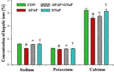
Effect of gold nanoparticles and Azolla microphylla on hepatic ions
The effects of GNaP on hepatic ions such as Na+, K+ and Ca2+ in the liver homogenate of control and experimental fish are summarized in Figure 9. The levels of hepatic ions were significantly
(p<0.05) reduced in the APAP-treated fish
compared to those of the control fish. Whereas, the altered hepatic ions were significantly elevated to near-control
(p<0.05) in the APAP+GNaP treated fish.
GNaP treated control fish did not showed any alterations in the hepatic ions.
Figure 9. Effect of green synthesized gold nanoparticles on the hepatic ions status of control and experimental
fish. Values are mean± SD (n=6 in each group). #p<0.05 as compared to CON. *p<0.05 as compared to APAP.
†p<0.05 as compared to APAP and CON. CON control, APAP acetaminophen, GNaP green synthesized gold
Discussion
Nanoparticles have drawn more attention in biology and medicine because they can be used as carriers for delivering small molecules such as drugs, proteins and genes [57]. Most commonly studied metal nanoparticles include gold, silver, titanium oxide and iron nanoparticles [58]. Among these, gold being inert and relatively less cytotoxic is extensively used for various applications including drug and gene delivery [59-61]. Biosynthesis of gold nanoparticles with the help of medicinal plants has come into the limelight in nanobiotechnology due to the growing need to develop environmental friendly benign technologies [62]. No evidence indicates that green synthesized gold nanoparticles causes toxicity at the clinical, histological, cellular and molecular levels [63].
In this study, we have reported total antioxidant capacity of various concentrations of green synthesized gold nanoparticles. From the analysis, it was evident that antioxidant capacity increases with increase in concentration up to certain level (3 mg/mL) and beyond this concentration saturation is observed. The results are in agreement with the content of flavonoids found in the surface of the gold nanoparticles.
Previous studies have demonstrated that,
Cassia fistula aqueous extract and
phytochemically synthesized gold nano-particles as progression of hypoglycemic treatment for diabetic mellitus in mice [62]. A Bacillus licheniformis biomass mediated synthesized gold nanoparticles has also been reported for its antioxidant as well as antidiabetic effect against streptozocin-induced diabetes in mice [37]. However, studies on possible beneficial effects of plant phytochemicals mediated synthesized gold nanoparticles against drug-induced toxicity.
The present study evaluated the effect of green synthesized gold nanoparticles on APAP-induced hepatotoxicity and liver alterations in a fresh water fish (Cyprinus
carpio L.). In the current work, no
mortality, alteration in behavior of experimental fish, changes in swimming behavior, breathing and orientation or color pattern were not observed during the experimental period. The appearance of control fish liver showed smooth and shiny, with normal, whereas APAP-treated fish liver appeared in dark black colour, which is confirmed the liver toxicity of APAP.
GNaP treated fish showed normal appearance of liver as that of control fish. Examination of pathological changes in the livers of both control and experimental fish revealed the highest hepatoprotective activity of GNaP. Our finding was similar to those of Wonkchalee et al. [64], who treated hepatotoxic Syrian hamster with
Thunbergia laurifolia Linn.
The observed hepatoprotective activity might be due to its several flavonoids on the surface of the gold nanoparticles and
Azolla microphylla extract.
Acetaminophen-induced hepatic failure is the second leading cause of liver transplantation and accounts for considerable levels of morbidity and mortality [65].
Hepatotoxicity of acetaminophen has been attributed to the formation of reactive metabolite (N-acetyl-p -benzoquinone-imine) by the actions of hepatic CYP450 enzymes [66]. However, an overdose of APAP causes depletion of cellular glutathione level in the liver due to toxic NAPQI, directly react with glutathione [67].
Depletion of glutathione may have two adverse effects. First, it reduces the inactivation of the reactive metabolite and tends to increase its covalent binding to proteins [68, 69].
Second, it may therefore aggravate the toxic effects of the reactive metabolite [70, 71].
revealed that increased permeability of the hepatocytes and cellular leakage. An increase in the levels of such enzymes reflects the liver damage and related oxidative stress [72, 73]. Lactate dehydrogenase catalyzes the reversible oxidation of pyruvate to lactate in the terminal step of glycolysis. It is also involved in gluconeogenesis in tissues in which lactate is converted to glycogen through gluconeogenesis [74]. An increased LDH activity in fresh water fish by Arsenite treatment was reported. G6PDH activity was moderately increased in paranchymal and inflammatory cells. Inflammatory and epithelial cells are responsible for ROS (reactive oxygen species) generation. Moreover G6PDH is responsible for increased metabolic activity of liver by glucose oxidation (oxidation of G-6-P) via hexose monophosphate (HMP) shunt which is essential for synthesis of fat. HMP shunt pathway is the major source of NADPH, which maintains the reductive environment for all biosynthetic processes using NADPH as a co-factor [75]. Increased level of MDH in APAP induced fish indicated the change in the metabolic pathways of energy generation and glucose synthesis by gluconeogenesis [76].
Generally liver damage reflects disturbances of cellular metabolism, which lead to characteristic changes in cellular and liver enzymes [77, 78]. Acetaminophen-induced hepatic damage was accompanied by significant elevation of both blood and hepatic tissue GPT, GOT and ALP about 60%. The increased levels of GPT, GOT and ALP, soluble enzymes in cytoplasm of liver cells may be a result of liver injury lead to increased permeability of cell membranes [74]. GPT and GOT are two mitochondrial enzymes, which plays an important role in the conversion of amino acids to keto acids. These results are in agreement with those reported earlier by Senthilkumar et al. [79]. Treatment with green synthesized gold nanoparticles, 2.5 mg/kg body
weight, significantly restored elevated levels of both blood plasma and liver tissue GPT, GOT and ALP.
Lipid peroxidation is an important parameter of oxidative stress and usually was reflected by increased levels of MDA and TBARS, a lipid peroxidation end product of lipid hydroperoxide (LHP) [50]. The increased levels of TBARS, LHP in the liver tissue of the fresh water fish indicated the tissue lipid peroxidation and oxidative stress exerted by APAP. This could be due to APAP-mediated gene-ration of ROS and increased peroxidation. On the other hand, excess of ROS generation initiates protein-oxidation, forming protein carbonyls and advanced oxidation protein product (AOPP), which have been described as reliable markers for protein damage [80, 81]. Our result is supported by other researchers, who have observed that oral exposure of APAP (500 mg/kg) in a fresh water fish (Pangasius
sutchi) for 24h duration induces liver
damage and alters the antioxidants. Abraham [82] suggests that, paracetamol-induced renal damage was accompanied by an increase in lipid peroxidation on rats. Vengerovskii et al. [83] reported, paracetamol induced LPO processes in the liver, along with calcium ions and pro-inflammatory cytokines, damage the barrier and matrix functions of hepatocyte membranes. Lipid peroxidation in liver is induced mainly by hepatotoxic chemicals including paracetamol and alcohol either directly or indirectly [84].
A novel finding in the present study is that, green synthesized gold nanoparticles (2.5 mg/kg) when administered in a fresh water fish helps in the prevention of hepatic damage (completely) caused by paracet-amol.
the levels of bilirubin and creatinine. Since there is a close relationship between the rate of protein synthesis in the liver tissue and total protein concentrations in the plasma. The depleted level of total protein (composed of albumin and globulin) in plasma reflects the decrease of protein synthesis in liver tissue. Administration of green synthesized gold nanoparticles and
Azolla microphylla methanol extract
showed significant effect on both blood and liver tissue protein, albumin and globulin levels. The rise of globulin level in the experimental fish may improve the immune system mediated immune responses [85].
Experimentally determined elevated levels of liver bilirubin and creatinine indicate the severe hepatic damage and renal impairment. It also confirms the hepat-otoxic nature of APAP. Bilirubin is considered as an index for the assessment of hepatic function and abnormality indicates hepatobiliary disease and severe disturbance of hepatocellular architecture [86]. An increase in creatinine indicates the pathological changes in hepaticbiliary flow [87].
The main function of the liver is to store energy in the form of glycogen or lipids (cholesterol or triglycerides). High dose of APAP reduces the levels of glycogen, cholesterol and triglycerides in the carp fish, indicates liver injury. In our study, green synthesized gold nanoparticles +APAP were restored the depleted levels of glycogen and lipids near-control fish. Simultaneously Azolla microphylla
phytochemically synthesized gold nano-particles were also maintained the levels of glycogen and lipids significantly higher than the APAP exposed fish.
In the present study, we found that the activities of antioxidant enzymes such as SOD, CAT, GSH-Px and GST and non-enzymatic GSH were significantly altered by higher doses of APAP. The altered levels of these enzymes were maintained
by Azolla microphylla phytochemically
synthesized gold nanoparticles. Similarly,
APAP+GNaP treated fish restored the levels near to control. SOD, CAT, GSH-Px are known to be inactivated by H2O2, O2*
-and *OH respectively. SOD -and CAT are the major antioxidant defense components [88]. CAT primarily catalyzes the conversion of H2O2 toH2O. Increased CAT
activity in the liver of APAP treated fish revealed the free radical induced damage of hepatic cells. Excessive generation of free radicals may result in alterations in the biological activity of macromolecules. It has been proved that, CAT is so efficient that it cannot be saturated by H2O2 at any
concentrations [89]. SOD has been reported as one of the most important enzymes in the enzymatic defense system. It scavenges the superoxide anion to form H2O2, thus diminishing the toxic effect
caused by this radical. SOD is necessary because superoxide reacts with sensitive and critical cellular targets. It reacts the NO radical and makes toxic peroxynitrite (ONOO-) and also SOD catalyze the dismutation of superoxide (O2*-) radicals
to oxygen and H2O2 [90]. In Azolla
microphylla phytochemically synthesized
gold nanoparticles, a significant decline in hepatic SOD activity was observed and thus reduces the reactive free-radical induced oxidative damage in liver.
H2O2 is normally detoxified in cells by
either CAT or GSH-Px. In GSH-Px catalyzes the reduction of hydrogen and organic H2O2, utilizing reduced glutathione
Addit-ionally, APAP-mediated GSH depletion could also be one of the reasons for the down regulated activity of GSH-Px in the liver tissue of APAP-treated fish.
Glutathione-S-transferases (GST) are an inducible phase II detoxification enzymes that catalyze the conjugation of glutathione with reactive metabolites formed during phase I of metabolism [93]. Induction of GST synthesis is a protective mechanism that occurs in response to APAP exposure. Our finding is supported by Sun et al. (94) who explained the decline in GSH level at lower concentrations and inductions of GST activity were observed in Carassius
auratus, exposed to pyrene. Glutathione
(GSH) provides a first line of defense and scavenges free radical oxygen species. GSH is a tripeptide (Cys-Gly-Glu), non-enzymatic biological antioxidant, present in liver. GSH plays an important role in protecting the liver against APAP-induced hepatotoxicity, because NAPQI is detoxified by conjugation with GSH.
During overdose of APAP, protein sulfhydryls groups (P-SH, T-SH, and NP-SH) in liver tissue are significantly reduced. Protein sulfhydryls groups are important targets of oxidation stress, where protein oxidation can acts as a cellular redox switch to modulate protein function, particularly those involving cell death [95]. The depletion of non-protein thiols and protein thiols in the liver, about 50-85% of which is APAP toxic byproduct NAPQI, as well as in enhanced lipid peroxidation. Lipid peroxidation resulting from oxidative stress contributes to the initiation and progress of liver damage. ROS can cause the S-hydroxylation of protein sulfhydryls to slightly oxidized state, sulfenic acid, which is reversible. Sulfenic acid can be further oxidized to sulfinic acid and sulfonic acid, which are irreversible modifications. The changes or loss of protein sulfhydryl groups may cause the mitochondrial dysfunction that is observed at high dose of APAP.
The ability of the Azolla microphylla
phyto-chemically synthesized gold
nanoparticles reduces the toxic byproduct NAPQI, by regeneration of glutathione and also maintains the protein sulfhydryls groups in the liver.
The APAP –treated fish liver tissue showed reduced Na+, K+ and Ca2+ ions when compared with control fish, indicates that APAP-treated fish experienced ionic disturbances.
In fresh water fish, membrane bound ATPase (Mg2+ and Na+/K+ -ATPase) play a significant role in ionic regulation of cellular components and maintenance of tissue osmolority (96) against concen-tration gradients and across membranes [97].
Na+ and K+ are the principal cations of intracellular fluid and play an important role in the maintenance of acid-base balance. The decreased levels of Na+ and K+ observed in the present investigation in APAP-treated fish liver tissue may be attributed to the pathophysiology of hepatic damage and the underlying disease process [98]. The calcium concentration gradient between the inside the cell (10-7 M) and the extra cellular fluid (10-3 M) is maintained by an active membrane-associated calcium and magnesium effluxing adenosine triphosphate (ATPase) enzyme system which is important potential target for toxicants. Chemically induced hepatotoxicity may lead to the disruption of calcium homeostasis [99, 100]. Disruption of calcium homeostasis may result in the activation of mitochondrial metabolism, ATP synthesis and damage of microfilaments used to support cell structure. Similarly, APAP+GNaP treated fish showed hepatic ions level near-control, which indicates the membrane-stabilizing action against APAP toxicity.
Histopathological examination of APAP-treated fish liver showed total loss of architecture.
The abnormalities were effectively controlled in the Azolla microphylla
Conclusions
Thus, the present study concluded that
Azolla microphylla methanol extract
phytochemically synthesized gold nanoparticles protects liver against oxidative damage and tissue damaging enzyme activities and could be used as an effective protector against acetaminophen-induced hepatic damage in fresh water common carp fish. Reduction of gold ions by methanol extract of Azolla microphylla
resulted in the formation of stable and biocompatible nanoparticles. The flavono-ids present in Azolla microphylla served dual role as effective reducing agents to reduce gold and also as stabilizers to provide a robust coating on the gold nanoparticles in a single step. The hepatoprotective and antioxidant action of GNaP was probably accomplished via elevating cellular antioxidative capacity, protecting GSH depletion, inhibiting lipid peroxidation and enhancing protein synthesis. The gold nanoparticles have been proven for their non-toxic and protective effects over the cells. The observed hepatoprotective and antioxidant effects of GNaP might be due to its several flavonoids on the surface of the gold nanoparticles. Thus Azolla microphylla
phytochemically synthesized gold nano-particles may be used as a safe, cheap, effective alternative chemopreventive and protective agent in the management of liver diseases. Further studies are, however, required to investigate the detailed molecular mechanisms of the protective effect of flavonoids based gold nanoparticles against toxicant-induced liver injury in fish and other animals.
Acknowledgments
This work was supported and sponsored by Department of Chemical Engineering, Jadavpur University, Kolkata. The authors also thank M/S. Renuka Clinical Laboratory, Namakkal India, for valuable support in performing histopathological analysis throughout the research. The authors also wish to acknowledge DST
unit on Nanoscience, Indian Institute of Technology, Madras for HRTEM meas-urements. They also thank Dr. S. Muru-gesan, Assistant Professor, Department of Pharmacy, Birla Institute of Technology and Science, Pilani, Rajasthan, India, for timely help and support.
References
1. Shen X, Tanga Y, Yang R, Yua L, Fanga T, Duan J. The protective effect of Zizyphus jujube fruit on carbon tetrachloride-induced hepatic injury in mice by anti-oxidative activities. J Ethnopharmacol. 2009; 122: 555-560. 2. Tarantino G, Di Minno MN, Capane D.
Drug induced liver injury: Is it somehow foreseeable?. World J Gastroenterol. 2009; 15: 2817-2833.
3. Lee CH, Park SW, Kim YS, Kang SS, Kim JA, Lee SH, Lee SM. Protective mechanism of glycyrrhizin on acute live injury induced by carbon tetrachloride in mice. Biol Pharm Bulletin. 2007; 30: 1898-1904.
4. Ostapowicz G, Fontana R.J, Schiodt FV, Larson A, Davern TJ, Han SH, McCashland TM, Shakil AO, Hay JE, Hynan L, Crippin JS, Blei AT, Samuel G, Reisch J, Lee WM. Results of a prospective study of acute liver failure at 17 tertiary care centers in the United States. Ann Intern Med. 2002; 137: 947-954.
5. Davidson DG, Eastham WN. Acute liver necrosis following over dose of paracetamol. Br Med J. 1966; 5512: 497-499.
6. Black M. Acetaminophen hepatotoxicity. Ann Rev Med. 1984; 35: 577-593. 7. McClain C J, Holtzman J, Allen J,
Kromhout J, Shedlofsky S. Clinical features of acetaminophen toxicity. J. Clin. Gastroenterol. 1988; 10:76-80. 8. Larson AM. Acetaminophen
hepatotoxicity. Clin Liver Dis. 2007; 11: 525-548.
9. Rumack BH. Acetaminophen hepatotoxicity: the first 35 years. J Toxicol Clin Toxicol. 2002; 40: 3-20. 10. Nelson SD. Molecular mechanisms of the
hepatotoxicity caused by acetaminophen. Semin. Liver Dis. 1990; 10: 267-278. 11. Lores Arnaiz S, Llesuy S, Cutrin JC,
12. Yamada T, Ludwig S, Kuhlenkanip J, Kaplowitz N. Direct Protection Against Acetaminophen Hepatotoxicity by Propyl-thiouracil. J Clin Invest. 1988; 67: 688-695.
13. Savides MC, Oehne FW. Acetaminophen and its toxicity. J App Toxicol. 1983; 3: 95-111.
14. Hoffmann KJ, Streeter AJ, Axworthy DB, Baillie TA. Identification of the major covalent adduct formed in vitro and in vivo between acetaminophen and mouse liver proteins. Mol Pharmacol. 1985; 27: 566-573.
15. Vermeulen NPE, Bessems JGM, Van de Streat R. Molecular aspects of paracetamol-induced hepatotoxicity and its mechanism based prevention. Drug Met Rev. 1992; 24: 367-407.
16. Jaeschke H, Knight TR, Bajt ML. The role of oxidant stress and reactive nitrogen species in acetaminophen hepatotoxicity. Toxicol Lett. 2003; 144: 279-288.
17. Jaeschke H, Bajt ML. Intracellular signaling mechanisms of acetaminophen-induced liver cell death. Toxicol Sci. 2006; 89: 31-41.
18. Lee WM. Drug-induced hepatotoxicity. N Engl J Med. 1995; 333: 1118-1127. 19. Goel A, Sharma K. Plant Extracts and
Phytochemicals as Herbal Medicines and Antimicrobials.Int J Biol Med Res. 2014; 5: 3940-3946.
20. Harborne JB. Phytochemicals methods. A guide to modern techniques of plant analysis, 3 rd Edn. Chapman and Hall Co, Newyork, 1998; 1-302.
21. Heim KE, Tagliaferro AR, Bobilya DJ. Flavonoid antioxidants: chemistry, metabolism and structure-activity relationships. J Nutrition Biochem. 2002; 13: 572-584.
22. Chattopadhyay RR. Possible mechanism of hepatoprotective activity of Azadira-chta indica leaf extract: part II. J Ethnopharmacol. 2003; 89: 217-219. 23. Sadik CD, Sies H, Schewe T. Inhibition of
15-lipoxyigenase by flavonoids: structure-activity relations and mode of action. Biochem Pharmacol. 2003; 65: 773-781. 24. Yao LH, Jiang YM, Shi J,
Tomás-Barberán FA, Datta N, Singanusong R, Chen SS. Flavonoids in food and their health benefits. Plant Foods Hum Nutr. 2004; 59: 113-122.
25. Sadhu SK, Okuyama E, Fujimoto H, Ishibashi M, Yesilada E. Prostaglandin inhibitory and antioxidant components of Cistus laurifolius, a Turkish medicinal
plant. J Ethnopharmacol. 2006; 108: 371-378.
26. Wagner GM. Azolla: a review of its biology and utilization. Botanical rev. 1997; 63: 1-26.
27. Abraham G, Vidhu A. A preliminary examination of the phytochemical profile of Azolla microphylla with respect to Seasons. Asian Pacific J Trop Biomed. 2012; 2: S1392-S1395.
28. Becerra M, Preston TR, Ogle B. Effect of replacing whole boiled soya beans with Azolla in the diets of growing ducks. Livestock Res. Rural Develop. 1995; 7: 1-10.
29. Lumpkin TA, Plucknett DL. Azolla as a green manure: Use and management in crop production. West view Press, Boulder, Colorado, 1982.
30. Van Hove C, Lopez Y. Fisiologia de Azolla. In: Workshop on the assessment of Azolla use in tropical Latin America. Chicklayo: Peru, 1982.
31. Selvaraj K, Chowdhury R, Bhattacharjee C. Isolation and structural elucidation of flavonoids from aquatic fern Azolla microphylla and evaluation of free radical scavenging activity. Int J Pharm Sci. 2013; 5: 743-749.
32. Gratzel M. Photoelectrochemical cells. Nature 2001; 414: 338–344.
33. Xia Y, Yang P, Sun Y, Wu Y, Mayers B, Gates B, Yin Y, Kim F, Yan H. One-dimensional nanostructures: synthesis, characterization, and applications. Adv Mate. 2003; 15: 353–389.
34. Krumov N, Perner-Nochta I, Oder S, Gotcheva V, Angelov A, Posten C. Biological synthesis of inorganic nanoparticles by microorganisms. Chem Eng and Technol. 2009; 32(7): 1026–1035. 35. Philip D. Green synthesis of gold and
silver nanoparticles using Hibiscus rosasinensis. Physica E: Low-dimension. Sys and Nanostruc. 2010; 42(5): 1417– 1424.
36. Shankar SS, Rai A, Ahmad A, Sastry M. Rapid synthesis of Au, Ag, and bimetallic Au core–Ag shell nanoparticles using Neem (Azadirachta indica) leaf broth. J of Colloid and Inter Sci. 2004; 275(2): 496– 502.
37. BarathManiKanth S, Kalishwaralal K, Sriram M, Pandian SRK, Youn HS, Eom SH, Gurunathan S. Anti-oxidant effect of gold nanoparticles restrains hyperglycemic conditions in diabetic mice. J Nanobiotechnol. 2010; 8: 1-15. 38. Prieto PM, Pineda, Aguilar M.
antioxidant capacity through the formation of phosphomolybdenum complex: Specific application to the determination of vitamin E. Anal Biochem. 1999; 269: 337-341.
39. Morales MA, Jobbagy AJ, Terenzi HF. Mutations affecting accumulation of glycogen. Neurospora News Lett. 1973; 0: 24-25.
40. Natio HK. Cholesterol. In: Kaplan A et al (eds) Clinical Chemistry. The C. V. Mosby Co. St Louis, Toronoto, Princeton, 1984; 437: 1194-1206.
41. Buccolo G. Quantitative determination of serum triglycerides by use of enzymes. Clin Chem. 1973; 19: 476-482.
42. Lowry OH, Rosenbrough NJ, Farr AL, Randall RJ. Protein measurement with the Folin phenol reagent. J. Biol. Chem. 1951;193:265-275.
43. Gendler S. Proteins in clinical chemistry: Theory, Analysis and Co-relation, Kaplan LA. and Pesce. AJ., Eds. Mosby C.V., Toranto, 1984; 1268-1327.
44. Jendrassik L, Grof P Vereinfachte. Photometrische Methoden zur Bestimmung des Blubilirubins. Biochem A, 1938; 297: 81-89.
45. Spencer K. Analytical reviews in clinical biochemistry: The estimation of creatinine. Ann Clin Biochem. 1986; 23: 1-25.
46. AOAC Official methods of analysis, 13th edn. Association of Official Analytic Chemists, Washington DC, 1980; 376-384.
47. Marklund S, Marklund G. Involvement of superoxide anion radical in the autooxidation of pyrogallol and a convenient assay for superoxide dismutase. Eur J Biochem. 1974; 47: 469-474.
48. Chance B, Maehly AC. Assay of catalase and peroxidases. Methods Enzymol. 1955; 11: 764-775.
49. Mohandas J, Marshal JJ, Duggin GG, Horvath JS, Tiller DJ. Differential distribution of glutathione and glutathione-related enzymes in rabbit kidney. Possible implications in analgesic nephropathy. Biochem Pharmacol. 1984; 33: 1801-1807.
50. Rajasekar P, Anuradha CV. Fructose-induced hepatic gluconeogenesis: effect of L-carnitine. Life Sci. 2006; 80:1176-1183. 51. Ellman GL. Tissue Sulfhydrl groups. Arch Biochem Biophysics. 1959; 82: 70-7758. 52. Uchiyama M, Mihara M. Determination of
malondialdehyde precursor in tissues by
thiobarbituric acid test. Anal Biochem. 1978; 86: 271-278.
53. Ohkawa H, Ohishi N, Yagi K. Assay for lipid peroxides in animal tissues by thiobarbituric acid reaction. Anal Biochem. 1979; 95: 351-358.
54. Levine RL, Garland D, Oliver CN, Amici A, Climent I, Lenz AG, Ahn BW, Shallttiel S, Stadtman ER. Determination of carbonyl content in oxidatively modified proteins. Meth Enzymol. 1990; 186: 464-478.
55. Sedlak J, Lindsay RH. Estimation of total, protein-bound and non-protein sulfhydryl groups in tissue with Ellmans reagent. Anal Biochem. 1968; 25: 192-205. 56. Singh M, Kalaivani R, Manikandan S,
Sangeetha N, Kumaraguru AK. Facile green synthesis of variable metallic gold nanoparticle using Padina gymnospora, a brown marine macroalga. Appl Nanoscience. 2013; 3:145-151.
57. Tiwari PM, Vig K, Dennis VA, Singh SS. Functionalized gold nanoparticles and their biomedical applications. Nanomat 2011; 1: 31-63.
58. El-Ansary A, Al-Daihan S. On the toxicity of therapeutically used nanoparticles: An overview. J Toxicol. 2009; 754810: 1-9. 59. Connor EE, Mwamuka J, Gole A, Murphy
CJ, Wyatt MD. Gold nanoparticles are taken up by human cells but do not cause acute cytotoxicity. Small. 2005; 1: 325-327.
60. Ghosh P, Han G, De M, Kim CK, Rotello VM. Gold nanoparticles in delivery applications. Adv Drug Deliv Rev. 2008; 60: 1307-1315.
61. Pissuwan D, Niidome T, Cortie MB. The forthcoming applications of gold nanoparticles in drug and gene delivery systems. J Contr Release. 2009; 149: 65-71.
62. Daisy P, Saipriya K. Biochemical analysis of Cassia fistula aqueous extracts and phytochemically synthesized gold nanoparticles as hypoglycemic treatment for diabetes mellitus. Int J Nanomed. 2012; 7: 1189-1202.
63. Chen YP, Dai ZH, Liu PC, Chuu JJ, Lee KY, Lee SL, Chen YJ. Effects of nanogold on the alleviation of carbon tetrachloride-induced hepatic injury in rats. Chinese J Physiol. 2012; 55: 1-5. 64. Wonkchalee O, Boonmars T, Aromdee C,