Auxin Import and Local Auxin Biosynthesis
Are Required for Mitotic Divisions, Cell
Expansion and Cell Specification during
Female Gametophyte Development in
Arabidopsis thaliana
Aneesh Panoli1, Maria Victoria Martin3, Monica Alandete-Saez1,2, Marissa Simon1, Christina Neff1, Ranjan Swarup4, Andrés Bellido3, Li Yuan1, Gabriela C. Pagnussat3
*, Venkatesan Sundaresan1,5*
1Department of Plant Biology, University of California Davis, Davis, California, 95616, United States of America,2PIPRA, University of California Davis, Davis, California, 95616, United States of America, 3Institute of Biological Research IIB-CONICET, Universidad Nacional de Mar del Plata, 7600, Mar del Plata, Argentina,4University of Nottingham, Nottingham, United Kingdom,5Department of Plant Sciences, University of California Davis, Davis, California, 95616, United States of America
*[email protected](GCP);[email protected](VS)
Abstract
The female gametophyte of flowering plants, called the embryo sac, develops from a hap-loid cell named the functional megaspore, which is specified after meiosis by the diphap-loid sporophyte. In Arabidopsis, the functional megaspore undergoes three syncitial mitotic divi-sions followed by cellularization to form seven cells of four cell types including two female gametes. The plant hormone auxin is important for sporophytic developmental processes, and auxin levels are known to be regulated by biosynthesis and transport. Here, we investi-gated the role of auxin biosynthetic genes and auxin influx carriers in embryo sac develop-ment. We find that genes from theYUCCA/TAApathway (YUC1,YUC2,YUC8,TAA1, TAR2) are expressed asymmetrically in the developing ovule and embryo sac from the two-nuclear syncitial stage until cellularization. Mutants forYUC1andYUC2exhibited defects in cell specification, whereas mutations inYUC8, as well as mutations inTAA1andTAR2, caused defects in nuclear proliferation, vacuole formation and anisotropic growth of the embryo sac. Additionally, expression of the auxin influx carriersAUX1andLAX1were ob-served at the micropylar pole of the embryo sac and in the adjacent cells of the ovule, and theaux1 lax1 lax2triple mutant shows multiple gametophyte defects. These results indicate that both localized auxin biosynthesis and auxin import, are required for mitotic divisions, cell expansion and patterning during embryo sac development.
OPEN ACCESS
Citation:Panoli A, Martin MV, Alandete-Saez M, Simon M, Neff C, Swarup R, et al. (2015) Auxin Import and Local Auxin Biosynthesis Are Required for Mitotic Divisions, Cell Expansion and Cell
Specification during Female Gametophyte Development inArabidopsis thaliana. PLoS ONE 10 (5): e0126164. doi:10.1371/journal.pone.0126164
Academic Editor:Hector Candela, Universidad Miguel Hernández de Elche, SPAIN
Received:February 9, 2015
Accepted:March 29, 2015
Published:May 13, 2015
Copyright:© 2015 Panoli et al. This is an open access article distributed under the terms of the
Creative Commons Attribution License, which permits unrestricted use, distribution, and reproduction in any medium, provided the original author and source are credited.
Data Availability Statement:All relevant data are within the paper and its Supporting Information files.
Introduction
The plant life cycle alternates between a diploid (2n) sporophytic and a haploid (n) gameto-phytic generation. The male gametophyte (pollen) produces the male gametes (two sperm cells), and the female gametophyte (embryo sac) produces the egg cell and central cell, two fe-male gametes that participate in double fertilization to produce a diploid embryo and a triploid endosperm respectively. The development of the female gametophyte (embryo sac) follows a tightly regulated program, which initiates with meiosis and terminates upon fertilization ([1–
3]. In Arabidopsis, female meiosis is initiated by the megaspore mother cell (MMC) in the nu-cellus of the ovule. The MMC undergoes meiosis giving rise to four megaspores, of which the three distal spores will degenerate, while the surviving spore becomes the functional megaspore (FG1,S1 Fig). The haploid functional megaspore undergoes mitosis to generate a 2-nucleate coenocyte (FG2), which is followed by migration of nuclei to opposite poles of the cell and for-mation of a central vacuole (FG3). A second round of mitosis produces a 4-nucleate embryo sac (FG4) with a large central vacuole and a pair of nuclei at either pole. A characteristic of the FG4 embryo sac is the rapid expansion of its size as well as that of the central vacuole. A final round of mitosis, followed by coordinated nuclear migration, produces an 8-nucleate and high-ly polarized embryo sac, composed by 3 nuclei occupying the micropylar pole, 3 at the chalazal pole, and 2 lying close to the micropylar end of the central vacuole (FG5). Cellularization re-sults in acquisition of distinct cell fates and the formation of a 7-celled, 8-nucleate embryo sac, composed of 2 synergids, 1 egg cell, 1 central cell with 2 nuclei called polar nuclei, and 3 antipo-dal cells (S1 Fig, FG6), while the two polar nuclei of the central cell fuse to form the diploid cen-tral cell (S1 Fig, FG7) [2,3].
Although relatively inconspicuous, the embryo sac is indispensable for seed formation, and therefore plays a critical role in plant reproduction (reviewed in [4]). It was observed that ma-nipulation of levels of the hormone auxin results in changes in cell fate, with high auxin levels promoting synergid fate or egg cell fates of the antipodal cells, and suppression of auxin signal-ing promotsignal-ing egg cell fate in the synergid cells [5,6]. A model was proposed that different con-centrations of auxin within the embryo sac might determine cell fates, with the highest auxin concentration-present at the micropylar pole- would specify the synergids, the next lowest specifies the egg cell, while the lowest auxin concentration at the chalazal pole results in antipo-dal specification. A recent paper though questioned this idea, as mathematical models could not generate a robust auxin gradient, and additionally, the expression of auxin reporters was not found inside the embryo sac [7]. However, and in agreement with a role of auxins during female gametogenesis, it was reported that the auxin efflux carrier PIN1 is required in the ma-ternal sporophytic tissues of the ovule that surround the embryo sac to promote female game-tophyte development [8]. Thus, PIN1 is thought to be involved in auxin flux towards the embryo sac as previously suggested [5] and such auxin flux seems essential for gametophyte
progression [8]. Furthermore, AUX1, a member of theAUXIN1/LIKE-AUX1 (AUX/LAX)
fam-ily of transporters that mediate auxin influx inArabidopsis, was shown to be expressed inside the female gametophyte and the protein to accumulate in the micropylar pole of the embryo sac [7]. Also, the expression of two auxin biosynthetic genes of theYUCCAfamily was previ-ously observed at the micropylar end of embryo sacs at early stages of development [5]. Thus, both auxin import and local auxin biosynthesis might be involved in gametophyte development.
The pathways leading to auxin biosynthesis in sporophyte development have been studied extensively [9–13] and it is known that auxin can be synthesized locally in several tissues in re-sponse to developmental or environmental cues [10,12–14]. Recently, a simple two-step path-way that converts tryptophan to indole-3-acetic acid has been shown to be the main auxin
biosynthesis pathway in Arabidopsis [14–16]. Trp is first converted to indole-3-pyruvate (IPA) by the TAA family of amino transferases, and IPA is subsequently converted into IAA by the YUC family of flavinmonooxygenases. Auxin can also be synthesized from indole-3-acetaldox-ime (IAOx), which is produced from Trpcatalyzed by CYP79B2 and its close homologue CYP79B3, but it is believed that IAOx may not be a major auxin biosynthetic pathway in plants [17].
Here we have investigated the roles of both auxin import and biosynthesis during female
ga-metophyte development. We found that several genes from theYUCCA/TAApathway are
ex-pressed in the developing embryo sac from the two-nuclear syncitial stage until cellularization and that expression of these genes is asymmetrically localized towards the micropylar end of the developing gametophyte. Genetic analysis revealed that mutants for these genes showed fe-male gametophyte defects that range from abnormal cell specification to defects in nuclear pro-liferation, vacuole formation and cell expansion. Additionally,AUX1andLAX1expression were detected at the micopylar region of the ovule during female gametophyte development, and the triple mutant for three influx carriersaux1 lax1 lax2shows mitotic arrest during female gametogenesis. These results indicate that female gametophyte development requires both lo-calized auxin biosynthesis and auxin import from the sporophytic ovule.
Results
YUCCA
and
TAA/TAR
auxin biosynthetic genes are expressed in the
ovule and developing female gametophyte
We previously found that the auxin biosyntheticYUCCAgenes,YUC1andYUC2, were
ex-pressed sporophytically, and subsequently gametophytically, at the micropylar pole of the de-veloping embryo sac, at FG1-FG2 stages [5]. In this study, we first performed a screen to identify all possible members of theYUCgene family that could be involved in embryo sac de-velopment. Transgenic plants carrying promoter::GUS fusions as well as promoter::GFPer fu-sions for theYUCgenesYUC1throughYUC11, were used to monitor their expression at various stages of embryo sac development. For the 11YUCgenes studied, we found only three genes showing consistent visible expression in the developing embryo sac:YUC1,YUC2and
YUC8(Fig 1,S2 Fig). As no previous studies reported the expression ofYUC8in the ovule, its expression pattern was further characterized.YUC8expression is the most delayed of theYUC
genes, with strong expression at the micropylar pole of the gametophyte at the FG3 stage that persists until the FG6 stage (S2A, S2B and S2C Fig).YUC8expression is not restricted to the gametophyte, as strong expression can also be detected in the tip of inner and outer integu-ments as well at FG5 and FG6 stages (S2 Fig). To verify whetherYUC8expression was indeed gametophytic, the segregation of the GFP signal inside the embryo sac was analyzed in a line hemizygous forpYUC8::GFPer. In these plants, the ovules at FG5-6 stages segregated for em-bryo sac signal at a ratio of ~1:1 (45.4% GFP+ vs 54.6% GFP-, N = 196, compared to a homozy-gous plant showing expression in<90% of the ovules examined, N = 298,Fig 1,S1 Movie).
Additionally, as the strongest expression found inside the embryo sac at early stages corre-sponded toYUC2, we decided to further confirm the gametophytic expression by studying the segregation of the GFP signal in a line hemizygous forpYUC2::GFPer. In these plants, the GFP expressing ovules at FG3 stage segregated at a ratio of 1:1 (44% GFP+vs. 53.4% GFP-, N = 240, compared to a homozygous plant showing expression in ~90% of the ovules examined, N = 400,S2 Movie). A small fraction of the ovules (3.6%) showed expression in the nucellar re-gion, around FG2 stage.
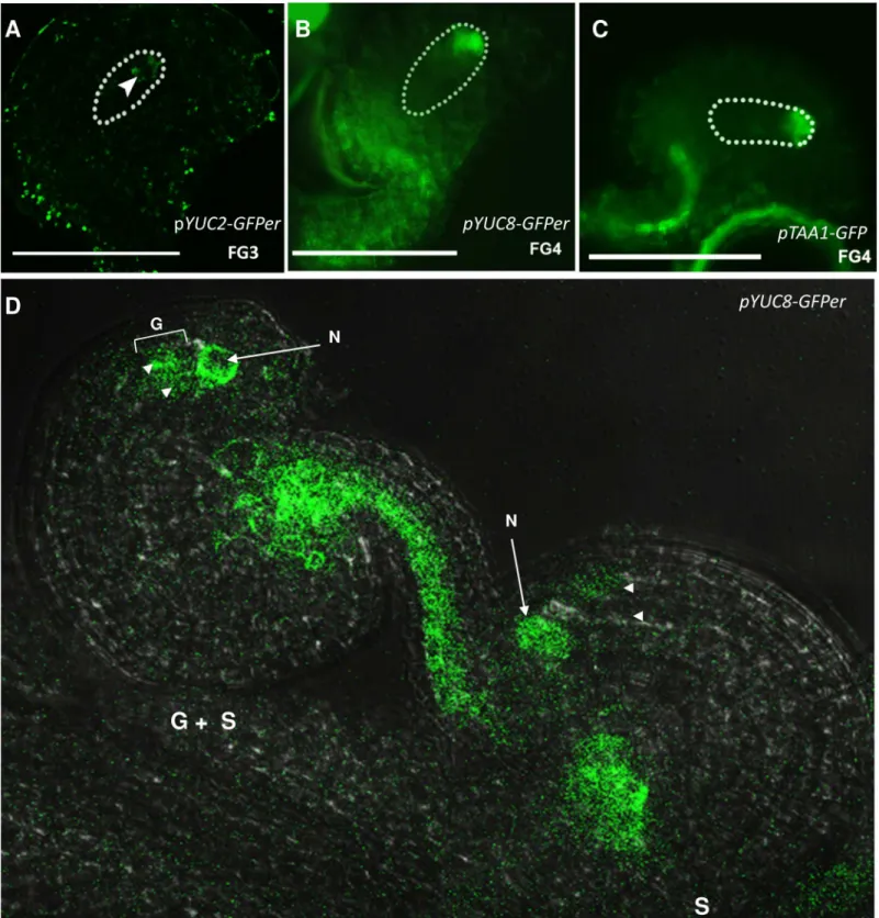
Fig 1. Expression pattern of auxin biosynthetic genes in the developing embryo sac.A, pYUC2-GFPerexpression at FG3 stage. B,pYUC8-GFPer
expression in the gametophyte at FG4 stage. C,pTAA1expression at FG4 stage of gametophyte development. D, Segregation of GFP signal in a line hemizygous forpYUC8::GFPer. From the two ovules shown, only one presents GFP detectable inside the embryo sac (marked as G + S). S, indicates sporophytic signal; G, indicates gametophytic signal. N, nucellus. Arrowheads point at embryo sac nuclei.
upstream of the YUC proteins [15,16,18]. Analysis of promoter activity using pTAA1::GFPand pTAA1::GUSlines indicated strong expression ofTAA1at the micropylar pole of the embryo sac from FG3 onwards (Fig 1andS2G–S2I Fig). The expression starts out as a faint signal at FG3 building up to a strong signal as the embryo sac reaches FG5 stage, persisting beyond this stage to FG6. At FG6 stage, we also detectedTAA1expression within the endothelial cells of the ovule adjacent to the micropylar region of the embryo sac (S2 Fig).pTAA1is also active in the inner integuments and funiculus from FG0 to FG3. Using a pTAR2::GUSfusion, we found thatTAR2is also expressed at the micropylar end of the embryo sac, starting slightly later than
TAA1, at about the FG4 stage (Fig 1).TAR2is expressed in the tip of the inner integument as well from the FG5 through FG7 stages. We were not able to detect anyTAR1expression in the developing ovules, either in the sporophytic tissues or the embryo sac, using a pTAR1::GUS fu-sion [10,12–14].
Loss of
YUC1
and
YUC2
functions affects specification of micropylar
cells
To uncover the contributions of individualYUCgenes to female gametophyte development, we examined insertional mutants inYUCgenes expressed in the embryo sac. The earliest gametophytically expressedYUCgenes areYUC1andYUC2. Single mutants in either gene had no detectable defects in embryo sac development. As the overlapping expression patterns of
YUC1andYUC2might result in functional redundancy, we examinedyuc1 yuc2double
mu-tants. Although sporophytic development appeared normal in theyuc1 yuc2mutant plants, marked defects were visible in embryo sac development. In a mature wild-type embryo sac, clei of synergid cells usually take up a distal position close to the micropylar end, while the nu-cleus of the egg cell occupies a somewhat more proximal position (S1 Fig, FG6 and FG7). In
yuc1 yuc2mutant plants, DIC analysis of emasculated pistils showed 14% of embryo sacs (N = 220) with mis-positioning of the nuclei within the cells at the micropylar end, with two cells containing nuclei at the normal position of the egg cell and only one at the synergid posi-tion (Fig 2J). In contrast, wild-type ovules exhibited only 1% of the embryo sacs (N = 550) with this defect. Theyuc1 yuc2double mutant displayed 94% normal seeds, 1% aborted seeds and 5% undeveloped ovules (N = 447). These numbers are very similar to what is observed in wild-type plants indicating that theyuc1 yuc2mutations did not affect the seed set significantly.
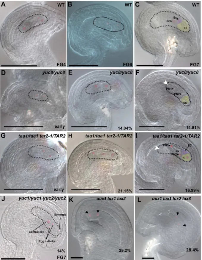
Fig 2. Embryo sac developmental defects in mutants impaired in auxin biosynthesis and import. A-C,WT;D-F,yuc8/yuc8;G-I,taa1/taa1 tar2/TAR2; J,yuc1/yuc1 yuc2/yuc2;aux1 lax1 lax2;K,aux1 lax1 lax2 lax3.A, WT 4-nucleate embryo sac at FG4 stage.B,WT 8-nucleate embryo sac prior to
Loss of
YUC8
function affects mitotic divisions, cell expansion and
nuclear migration during embryo sac development
Our studies usingpYUC8::GUSandpYUC8::GFPershowed thatYUC8is the primaryYUC
gene expressed from FG3 to FG6 stages of embryo sac development. Therefore, we examined theyuc8mutant for possible gametophytic functions. Homozygousyuc8plants were recovered stage with a 2-nucleate arrested embryo sac.F,Matureyuc8mutant embryo sac showing defective polar nucleus migration, no antipodals are visible at this stage.G,A mutant ovule, with only 2 nuclei in the coenocytic embryo sac.H,Mutant ovule with 3-nucleate gametophyte. Unlike clearly polarized nuclei in the WT, these nuclei are scattered in the cytoplasm.I,Mature mutant embryo sac showing defective polar nucleus migration, antipodals are completely degenerated by this stage.J,Ayuc1 yuc2double mutant showing miss-polarized micropylar nuclei, giving rise 2 egg cell-like structures instead of 1.K, Ovule from a triple mutantaux1 lax1 lax2showing an embryo sac arrested at FG2 stage. The arrows point at the nuclei inside the embryo sac.L, Ovule from a quadruple mutantaux1 lax1 lax2 lax3showing a collapsing embryo sac containing only two nuclei (arrows). Except for the phenotypes shown in panels D and G, all mutant phenotypes shown are terminal and observed at late stages of ovule development as indicated inTable 1and inS1 Table. Cv, central vacuole; Ec, egg cell; PN1, polar nucleus 1; PN2, polar nucleus 2; Sc, synergid cell. Scale bar, 50μM for A-J and 20μM for K-L.
doi:10.1371/journal.pone.0126164.g002
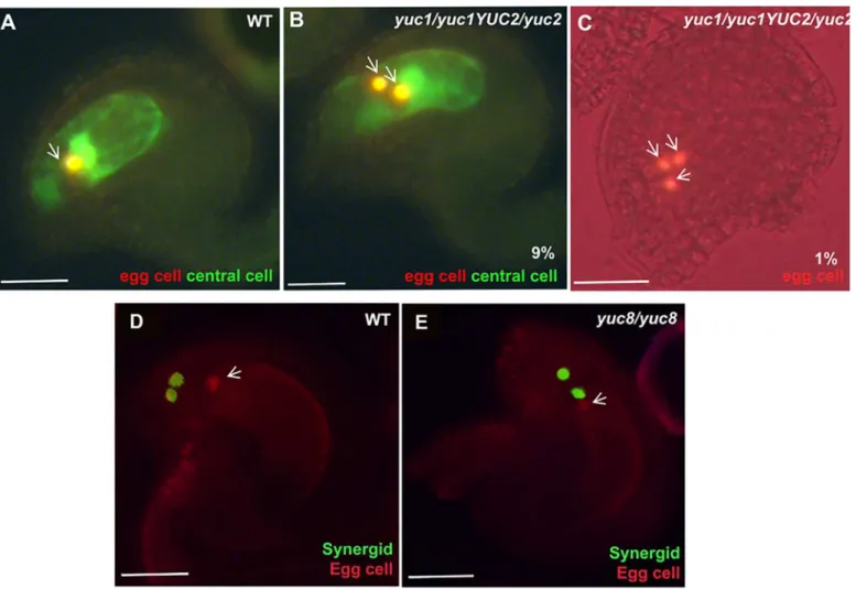
Fig 3. Changes in auxin homoeostasis lead to altered cell specification. A, D,WT.B and C,yuc1/yuc1 yuc2/YUC2.E,yuc8/yuc8.A-C,green signal indicates the expression of central cell specific marker, and red indicates egg cell specific marker. The yellow color observed results from the overlapping of the red colour (egg or egg-like nuclei) on the green background of the central cell in the confocal image.D-E,green signal indicates synergid cell-specific marker, and red indicates egg cell-specific marker.A,WT ovule showing a single egg cell (Red) and a central cell (green) at FG7 stage.B,A mutant ovule at FG7 stage showing 2 egg cells and 1 central cell.C,an FG7 mutant ovule showing 3 egg cells.D,A WT ovule showing normal polarization of synergid nuclei towards the micropylar end.E,merged image of ayuc8mutant ovule, showing miss-positioned synergids. The white arrows indicate the egg cell marker expression. Scale bar, 20μM.
from an F2 population indicating that the mutation can be transmitted through the gameto-phytes. Sporophytic development appeared normal and ovules displayed proper initiation and
growth of integuments. However, cleared ovules showed thatyuc8/YUC8andyuc8/yuc8
homo-zygous mutants are defective in the female gametophyte development (Fig 2D–2F). Mutant ga-metophytes looked normal from FG1 to FG3 stages of development, consistent with the absence of wild-typeYUC8expression at these stages. Female gametophyte defects were visible from stage FG4 onwards. Due to significantly delayed gametophyte development in the mu-tant, we have used ovule stages defined according to Schneitz et al [19] to make comparisons between wild-type and mutant embryo sacs when necessary. The observed phenotypes can be divided into 3 classes, i) defects in mitotic division, ii) defective embryo sac expansion and vac-uole development, and iii) defects in nuclear migration. All the mutant gametophytes had a combination of 2 or more defects belonging to the above described phenotypic classes. Game-tophytes showing the first class of defects exhibited a delayed gametophyte development as compared to normal ovules. A wild-type ovule at stage3-IVusually contains a 4-nucleate FG4 gametophyte (Fig 2A), while allyuc8homozygous ovules at stage3-IVcontained only 2 or 3 nu-cleate embryo sacs (Fig 2D, N>200). By the3-Vovule stage, theyuc8mutant displayed a
de-layed development in 14% of the gametophytes (N = 228), with 2 to 4 large nuclei, compared to wild type with distinct 8-nucleate FG5 embryo sacs (Fig 2E,S1 Table). The size of the nuclei was abnormally larger than those of wild type embryo sacs. In wild-type, the embryo sac usual-ly attains near-maximum size by FG5, accompanied by rapid expansion of the central vacuole. In theyuc8mutant gametophytes showing defects corresponding to class 2, we observed an unexpanded embryo sac with a small vacuole in the center (Fig 2D and 2E), indicating that
rapid growth of embryo sac is compromised. Another*15% of gametophytes showed defects
corresponding to class 3, in which the two polar nuclei were found at either end of the central vacuole (Fig 2F and 2I). This phenotype is clearly distinct from that of the previously reported
“unfused polar nuclei”phenotype, where the 2 polar nuclei remain in close proximity at the micropylar end just outside the central vacuole (e.g. Pagnussat et al. 2005 [20]). Embryo sacs with aberrant polar nuclear migration often had a very well developed vacuole as well as the correct number of nuclei, suggesting that the nuclear migration defect is not a consequence of the defects in vacuole formation or mitotic divisions.
To ascertain whether these functions ofYUC8are gametophytic or sporophytic, we also un-dertook observations onyuc8/YUC8heterozygous plants. As expected from a gametophytic function forYUC8, we observed defects similar to that of theyuc8homozygous mutant in the
yuc8/YUC8plants as well, although at a lesser proportion. Compared toyuc8homozygous mu-tant, theyuc8/YUC8plants showed 8.92% (vs. 14% inyuc8/yuc8, p-value = 0.08734) ovules with 2 to 4-nucleate embryo sacs at FG5 stage, roughly half of what is observed in the homozy-gous mutant (Fig 4,S1 Table). Also, 14.50% of gametophytes showed phenotypes correspond-ing to class 3.
Because auxin produced byYUC1andYUC2might be alleviating the effects of theyuc8 mu-tation, we constructed ayuc1 yuc2 yuc8triple mutant by double recombination, asYUC8lies
between theYUC1andYUC2genes on Chromosome 4 (seeMaterials and Methods). Theyuc1
yuc2 yuc8triple mutant phenotypes were similar to those of theyuc8mutant alone, and we did not obtain any additive phenotype, suggesting thatyuc8is epistatic toyuc1 yuc2(Fig 4,S1 Table).
To determine if the embryo sac phenotypes observed are terminal, we emasculatedyuc8
yuc8mutant is significantly less than the percentage of gametophytes showing visibly defective development in theyuc8/yuc8mutant (29%;S1 Table,S3 Fig). Therefore, it is likely that a sig-nificant fraction of the mutant gametophytes with developmental defects at the earlier stages were able to recover and form mature functional embryo sacs.
Next, we investigated possible alterations in the gametophytic cell specification in theyuc8
mutant using the FGR6.0 marker line (seematerials and methods), which specifically labels egg cell in red, central cell in yellow, and synergid cell in green. In the wild-type gametophytes we never observed any mis-expression of cell type specific markers. Although no misexpression of the markers was detected in theyuc8homozygous mutants, the position of one of the synergid cell´s nucleus was occasionally observed at a chalazal position, resembling the morphology of an egg cell (Fig 3E).
The
TAA
genes for auxin biosynthesis are expressed during female
gametophyte development and required for mitosis and nuclear
migration
Analysis of promoter activity usingpTAA1::GFPandpTAA1::GUSlines indicated strong ex-pression ofTAA1at the micropylar pole of the embryo sac from FG3 onwards (Fig 1Cand S2G—S2I Fig).TAR2was also found to be expressed at the micropylar end of the embryo sac, starting a little later thanTAA1, at about the FG4 stage (S2J—S2L Fig).TAR2is expressed in the tip of the inner integument as well from the FG5 through FG7 stages. To explore the game-tophytic function of TAA/TAR auxin biosynthetic genes, we examinedtaa1,tar1, andtar2-1
mutants [14]. These single mutants show sporophytic plants of reduced size, but the gameto-phytes were indistinguishable from those of wild-type plants (not shown). AsTAA1andTAR2
show overlapping expression in the embryo sac from FG4 through FG7 we examinedtaa1
Fig 4. Bar diagram indicating percentage of aberrant embryo sacs in various mutant backgrounds.SeeS1 Tablefor details. 2n-4n indicates the percentage of gametophytes arrested with 2, 3 or 4 nuclei in an embryo sac that corresponds to a FG6 ovule.“Polar nuclei apart”refers to the percentage of gametophytes with polar nuclei arrested at either end of the central vacuole in a FG7 embryo sac. Extra egg cells and abnormal expression of syn marker were studied in FG7 embryo sacs.
tar2-1double mutants. We used thewei8-1mutation ofTAA1as thetaa1mutant, and the
tar2-1mutation ofTAR2, both of which are presumptive null mutants [14]. Thetaa1 tar2-1
double homozygous plants did not produce any embryo sacs, being tiny and completely sterile as previously described [14]. So we examinedtaa1/taa1tar2-1/TAR2plants, which revealed a ~12% (N = 344) reduction in seed set in mature siliques (S3 Fig). DIC microscopy of mature
ovules showed that*38% of them contained abnormal gametophytes. 21% of the ovules in
thetaa1/taa1tar2-1/TAR2mutant show 2 to 4 nucleate embryo sacs (Fig 2H). When pistils were examined at earlier stages, we found that while 74% of the embryo sacs have reached an FG4 stage, the remaining ~26% showed only two nuclei inside the embryo sacs (Fig 2G, N = 208). Also, about 17% of the ovules displayed embryo sacs with defects in polar nuclei mi-gration (Fig 2I). Similar phenotypes were observed intaa1/TAA1tar2-1/TAR2double heretero-zygous plants as well (S1 Table), indicating that the defects are unlikely to arise from the loss of a sporophytic function. In addition, there is a reduction in the number of defective gameto-phytes by almost half in thetaa1/TAA1tar2-1/TAR2double heterozygotes (S1 Table, p-value = 7.175e-06), as compared to thetaa1/taa1tar2-1/TAR2plants. Taken together, these ob-servations suggest that expression ofTAA1andTAR2inside the gametophyte is an important requirement for its normal development.
The phenotypes observed in the female gametophytes oftaa1/taa1 tar2-1/TAR2plants are similar to those observed in theyuc8mutant, but at a higher frequency. A triple mutant was generated by crossingyuc8,taa1andtar2-1mutants. BecauseYUC8andTAR2are linked genes on chromosome 4,yuc8 tar2-1recombinants were first recovered in the F2. Subsequent-ly,yuc8/yuc8 taa1/taa1 tar2-1/TAR2plants were obtained (seeMaterials and Methods), and examined for embryo sac defects. As expected for proteins that work in the same biosynthetic pathway [18], the triple mutant showed phenotypes comparable to those oftaa1/taa1 TAR2/ tar2-1double mutants, both in severity and frequency (S1 Table) and no significant additive ef-fects due toyuc8were observed. Taken together, these results are consistent with a role for auxin synthesis through theTAA/TAR/YUCpathway in cell division, cell expansion and syner-gid specification during female gametogenesis.
Auxin import carriers are required for embryo sac development
Our previous study together with another independent study reported thatPIN1is expressed in the nucellus of developing ovule primordia, and restricted later to the inner integument of the ovule [5,8]. No expression inside the female gametophyte was detected neither forPIN1
nor for the other members of thePINfamily [5,8]. PIN1 localization however, indicates that auxin is accumulated in specific nucellar cells, suggesting that auxin could be transported into the developing embryo sac where might regulate gametophytic progression. Supporting this idea, it was reported that downregulation ofPIN1results in megagametogenesis arrest [8]. To investigate whether auxin import might also be implicated in female gametophyte
develop-ment, we studied the expression ofAUX1andLAX1, members of theAUXIN1/LIKE-AUX1
(AUX/LAX) family of auxin transporters which are the major influx carriers inArabidopsis
[21]. As can be observed inFig 5, using apAUX1::AUX1::YFPconstruct we were able to detect AUX1 from stage FG4 onwards at the micropylar pole of the embryo sac (A-C). After cellulari-zation, at stage FG6, AUX1 is located to egg cell and synergid cell membranes. Moreover,
AUX1::YFPsignal is segregating in plants hemizygous forAUX1::YFP, confirming its gameto-phytic expression (Fig 5C). While this study was in progress, AUX1 was independently shown to be expressed inside the female gametophyte and the protein to accumulate in the micropylar pole of the embryo sac [7]. We also examined the expression of the related influx carrierLAX1
expression was detected early in ovule development in sporophytic tissues of the nucellus as well as in the embryo sac, indicating activity of thepLAX1 promoter both outside and inside the embryo sac (Fig 5D). The LAX1::VENUS signal was visible in the sporophytic tissues of the nucellus, surrounding the embryo sac micropylar pole, but in contrast to pLAX1::GUS expres-sion, the LAX1::VENUS signal was not detectable inside the embryo sac (Fig 5E). However, we noted that the signal from the LAX1::VENUS fusion was weak compared to the AUX1::YFP fu-sion even in sporophytic tissues, suggesting that low levels of pLAX1expression might not be detectable using this reporter fusion. We were not able detect neitherLAX2norLAX3 expres-sion using the available reporter constructs [21].
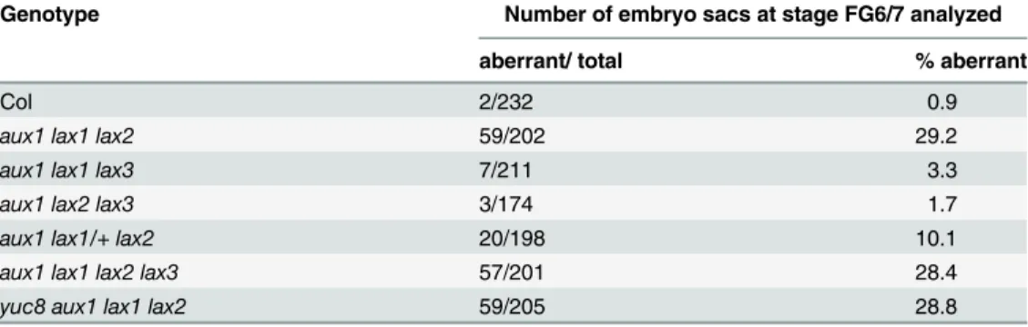
To assess the possible functions of these auxin influx carriers and of the other members of theAUXIN1/LIKE-AUX1 (AUX/LAX)familyLAX2andLAX3in embryo sac development, we examined single, double, triple and quadruple mutants for these genes. The single mutants for
aux1,lax1,lax2,lax3, the double mutant combinationsaux1 lax1,aux1 lax2andaux1 lax3and the triple mutant combinationsaux1 lax2 lax3andaux1 lax1 lax3showed no phenotypes and were fertile. However, the triple mutantaux1 lax1 lax2shows*29% of ovules containing
em-bryo sacs arrested at two nuclear stage. The sporophytic tissues of the ovule look normal (Table 1,Fig 2K). Quadruple mutantaux1 lax1 lax2 aux1 lax1 lax3shows a phenotype similar to the one observed foraux1 lax1 lax2triple mutant, with*28% of aberrant embryo sacs that
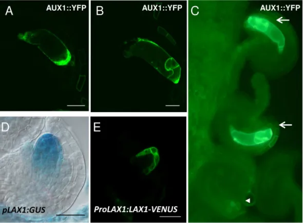
Fig 5. Expression pattern of the auxin influx carriers AUX1 and LAX1 in the developing embryo sac. A-C, Detection of AUX1::YFP.D-E, Expression of
LAX1by using thepLAX1:GUSconstruct (D) orProLAX1:LAX1-VENUS(E).A,YFP fluorescence is detected at stage FG4 at the micropylar side of the embryo sac.B, After cellularization, at stage FG6, AUX1 is located to egg cell and synergid cell membranes.C, Segregation of YFP signal in a line hemizygous forpAUX1:AUX1:YFP. From the four ovules shown, only two present YFP detectable inside the embryo sac (arrows). The arrowhead indicates an ovule in the same pistile without detectable YFP inside the embryo sac.D,GUS detection driven by theLAX1promoter shows expression at FG1 stage in the functional megaspore and in the nucellus.E, Expression ofLAX1-VENUSin the sporophytic tissues of the nucellus, surrounding the embryo sac micropylar pole at FG2. Scale bar, 20μM.
collapse at FG2 stage (Table 1,Fig 2L). Moreover, when theaux1 lax1 lax2triple mutant com-bination was heterozygous for theLAX1wild-type allele, the arrested embryo sac phenotype was still present but at a reduced frequency (10%;Table 1), indicative that the mutations act on the gametophyte and not on the sporophyte. These results suggest that auxin influx carriers
AUX1,LAX1andLAX2are redundantly required for normal female gametophyte
develop-ment.LAX3on the other hand is not required for embryo sac development.
YUCCA1
over-expression with upregulated auxin response restricted to
the embryo sac is sufficient to alter chalazal cell fates
In a previous paper, we reported that overexpression ofYUC1in the embryo sac was able to alter chalazal cell fates, resulting in cells at the antipodal location exhibiting egg cell or synergid cell attributes [5]. However, as auxin response was also observed in the sporophytic tissues sur-rounding the embryo sac in the lines analyzed, an indirect effect of auxin affecting embryo sac development from the sporophytic tissues could not be ruled out. In a recent study it was pos-tulated that the effects of YUC over-expression might be due to auxin in the sporophytic cells affecting gametophytic cell-specification via a non-cell-autonomous signal [7]. Moreover, the authors reported that the auxin response signals in wild-type ovules were entirely sporophytic, and not gametophytic. However, the gametophytic expression and phenotypes exhibited by the auxin biosynthetic genes and auxin influx carriers characterized in this study implied that auxin signaling is functioning within the female gametophyte. Therefore, the evidence for ga-metophytic auxin signaling was re-examined in wild-type plants carrying aDR5- GFPerauxin response reporter [22] by confocal microscopy, instead of the cytoplasmic GFP reporter used previously. The ER-localized GFPer reporter reduces the possibility of intercellular movement of GFP, and permits more accurate imaging of the localization of the signal. To aid proper stag-ing of ovules durstag-ing fluorescent confocal microscopy, we crossed wild-type (WT) plants carry-ing theDR5::GFPerconstruct to a marker line that labels all the embryo sac nuclei, as contains theAKV-NLS:Mcherry-AKVTconstruct [23]. Additionally, the amphiphilic styryl dye FM4-64, that produces a bright red fluorescence in membranes, was used to delineate the embryo sac at early stages. As can be observed inS4 Figand as previously reported [5,8], at FG1 stage, right after meiosis, theDR5::GFPersignal was detected at the distal tip of the nucellus outside the ga-metophyte (S4A Fig). This signal is retained exclusively in the nucellus up to the end of FG1. After that, a DR5::GFPer signal starts to appear within the developing gametophyte at FG2 stage and gets more noticeable at FG3 stage, at the micropylar pole (S4B and S4C Fig,S3 Movie). At FG4 stage, the DR5::GFPer signal could be clearly observed inside the female game-tophyte at a more central position, which is maintained before fading at maturity (S4D–S4F Fig,S4–S6Movies). The distribution of auxin response at the micropylar pole of the embryo Table 1. Quantification of aberrant embryo sacs detected in auxin influx mutants.
Genotype Number of embryo sacs at stage FG6/7 analyzed
aberrant/ total % aberrant
Col 2/232 0.9
aux1 lax1 lax2 59/202 29.2
aux1 lax1 lax3 7/211 3.3
aux1 lax2 lax3 3/174 1.7
aux1 lax1/+ lax2 20/198 10.1
aux1 lax1 lax2 lax3 57/201 28.4
yuc8 aux1 lax1 lax2 59/205 28.8
sac observed at the FG2-FG3 stages using confocal microscopy corresponds to the one previ-ously reported using a DR5::GFP reporter and epifluorescence microscopy until the FG3 stage [5]. From FG4 stage to FG5 however, the signal appears to be discretely localized at a more cen-tral position than the micropylar localization we reported in the earlier study. It is important to note that the fluorescence signal does not correspond directly to auxin molecules but indirectly measures the auxin response. The observed fluorescence from the GFPer protein is likely to be a reflection of the distribution of ER in the uncellularized embryo sac, and its utilization here is primarily to confirm the presence of auxin signaling inside the female gametophyte. At the later stages, FG6 and FG7, the auxin signal is weak or absent inside the wild-type embryo sac, as previously reported.
To determine whether manipulation of auxin signaling restricted to within the female game-tophyte can alter cell fates, a new set of transformant lines were constructed carrying the Op-LhG4transactivation system driving the expression ofYUC1with thepES1promoter, which is active from FG1 to FG7 in the whole embryo sac [24]. Transformants were crossed to lines car-rying a DR5-GFPer auxin reporter [22], which was preferred over the cytoplasmic GFP report-er used previously to exclude the possibility of intreport-ercellular movement. Two independent lines were selected that exhibited auxin response inside the embryo sac without detectable sporo-phytic signal (Fig 6A and 6B,S5A and S5B Fig). Although the signal was widely distributed along the embryo sac at earlier stages of development (S5 Fig), the signal detected was observed very strong in synergids and egg cell, with less intensity in central cell and in the antipodal cells when embryo sacs reached stage FG6 (Fig 6A and 6B). The fact thatYUCoverexpression trig-gers a strong auxin response in the embryo sac indicates that not only the rest of the gene prod-ucts involved in the auxin biosynthesis pathway are present in the female gametophyte, but also that the signaling pathway could be activated in response to auxin. To examine if these em-bryo sacs that show strong auxin response have cell identity abnormalities,YUC overexpres-sing lines were crossed to egg cell and synergid marker lines. In the F1, 25% of the embryo sacs are predicted to overexpressYUC1and 12.5% are predicted to overexpressYUC1and to carry the GUS reporter. As can be observed inFig 6DandTable 2, around 12% of the ovules in the F1 presented abnormal expression of a cytoplasmic synergid marker, which was observed at central and chalazal positions. For WT plants, aberrant patterns were observed in 2 embryo sacs out of 532 ovules analyzed. However, aberrant expression of a different synergid cell mark-erpSYN::NLS-GUSwas not detected, suggesting that conversion to synergid cell fate might be incomplete in these lines (Table 2). In the case of the egg cell markers, the percentage of aber-rant patterns was around 10% for an egg cell nuclear marker and 2.6% for a cytoplasmic egg cell marker (Fig 6F,Table 2andS5 Fig). In the case of WT plants, no abnormalities were de-tected in 455 ovules analyzed. All together, these results show that high levels of auxin inside the embryo sac are sufficient for mis-expression of micropylar cell type markers in chalazal cells, and support the model that gametophytic auxin can direct cell specification.
Discussion
Auxin biosynthetic genes active at specific developmental stages are
required for normal female gametophyte development
developmental transitions during its short span of development. Moreover, the asymmetrically localized gene expression patterns within the embryo sac suggest that nuclei in the developing embryo sac make different position-dependent decisions at these temporal transitions, imply-ing the existence of spatial cues. This would be possible if the gametophytic nuclei occupy func-tionally distinct cytoplasmic compartments within the coenocyte, responding to positional signals. Proximo-distal polarity is established early, with the two poles of the embryo sac show-ing differential expression from the 2-nuclear stage, implyshow-ing that spatial signals must act by this stage. The first spatial clue perceived by the developing female gametophyte might be Fig 6.YUC1overexpressing embryo sacs show extra egg cells and synergid cells at abnormal positions. A,Confocal image showing DR5::GFP activity localized inside the embryo sac ofYUC1
overexpressing female gametophytes.B, GFP signal in A is overlapped with a DIC image.C, A WT embryo sac showing the expression of a specific synergid marker.D, aYUC1overexpressing embryo sac showing specification of two extra synergid-like cells at a chalazal position (arrows).E, WT embryo sac showing the expression of an egg cell-specific marker.F, AYUC1overexpressing embryo sac showing expression of the marker in two extra cells at a central position (arrows). Scale bar 20μM.
sporophytic auxin, which is transported to the nucellus by PIN1 prior to meiosis [5]. In agree-ment with a critical role of PIN1 early in embryo sac developagree-ment, it was recently reported that maternal control ofPIN1is required for female gametophyte development, as PIN1 down-reg-ulation results in embryo sacs arrested at the mono or bi-nuclear stages [8].
Embryo sacs fromyuc1,yuc2,taa1andtar2-1single mutants were similar to wild-type in all aspects, indicating that these genes act redundantly in the gametophyte. This was confirmed
when double mutants foryuc1andyuc2showed gametophytic phenotypes, although the
pene-trance was low. In addition, compensation by other auxin biosynthetic genes or by imported auxin might occur when individual genes are mutated. For example, mutants in multipleYUC
genes do not show significant differences in auxin levels in sporophytic tissues, even though sporophytic phenotypes could be observed [12,25]. Thus, de-repression of one or more of the otherYUCgenes that are normally not expressed in female gametophyte development, or the auxin transporters, might be compensating for the absence ofYUC1andYUC2inyuc1 yuc2
mutant embryo sacs.YUC8expression is the most delayed of the gametophyticYUCgenes. Theyuc8mutant displayed distinct growth and developmental defects during embryo sac de-velopment, consistent with a requirement for local auxin biosynthesis during megagametogen-esis. The percentage of defective embryo sacs inyuc8/YUC8was reduced to nearly a half that observed in the homozygousyuc8mutant, which is consistent with gametophytic segregation ofYUC8. Thetaa1/taa1tar2-1/TAR2mutant also exhibited gametophytic defects similar to those observed in theyuc8mutant, although at higher frequencies. These results strongly sug-gest that local auxin biosynthesis through theYUCCA/TAApathway is essential for different key processes that take place during embryo sac development. The most frequent defects ob-served inyuc8andtaa1/taa1tar2-1/TAR2mutant embryo sacs were slower nuclear divisions, Table 2. Expression of cell specific markers in embryo sacs frompES1::LhG4/+; Op::YUC1/+; GUS/+plants.
Pistils studied GUS positive WT expression pattern GUS negative GUS positive Abnormal expression pattern (maximum possible = 12.5%†
)
Total (100%)
P values*
pES1>>YUC1x cytoplasmic Egg cell marker line
106 (36.9%) 163 (60.5%) 7 (2.60%) 269 (100%) 0.01399321
WT x cytoplasmic Egg cell marker line
117 (45.8%) 138 (54.2%) 0 (0%) 255 (100%) 0.352488
pES1>>YUC1x nuclear Egg cell marker line
79 (34.80%) 125
(55.06%)
23 (10.13%) 227 (100%) 0.00120237
WT x nuclear Egg cell marker line
88 (44%) 112 (56%) 0 (0%) 200 (100%) 0.229299
pES1>>YUC1x cytoplasmic Synergid cell marker line
52 (23.11%) 143
(63.55%)
30 (13.33%) 225 (100%) <0.00001
WT x cytoplasmic Synergid cell marker line
244 (45.9%) 286 (53.7%) 2 (0.4%) 532 (100%) 0.196272
pES1>>YUC1x nuclear Synergid cell marker line
88 (26.99%) 238 (73%) 0 (0%) 326 (100%) <0.00001
WT x nuclear
Synergid cell marker line
98 (46%) 115 (54%) 0 (0%) 213 (100%) 0.467737
*χ2-Test from the expectation that 50% of the gametophytes will be GUS positive, WT expression pattern (i.e. express the cell-specific marker in the correct cells), due to segregation of the GUS reporter.
†Of the total embryo sacs, 25% are predicted to overexpressYUC1, and 12.5% are predicted to be both overexpressingYUC1and carrying the GUS
reporter. Thus, full penetrance would result in 12.5% abnormal marker expression.
reduced cell expansion, and failure of the polar nuclei to migrate towards each other. During the early phase of development corresponding to stages FG3 and FG4, the coenocytic embryo sac undergoes a rapid expansion accompanied by the growth of the central vacuole. Auxin is known to induce rapid cell expansion in sporophytic tissues such as stem, coleoptiles or hypo-cotyls within minutes of treatment [26]. In addition, we also found that in both theyuc8and thetaa1/taa1tar2-1/TAR2mutant embryo sacs, the size of the vacuole was highly reduced compared to the wild-type counterpart. These observations suggest that the auxin present within the gametophyte at FG3 to FG4 transition (S4 Fig) could be a trigger for the rapid ex-pansion and growth of the embryo sac. The size of the embryo sac is not affected inyuc1 yuc2
mutants, presumably because these genes are active at earlier stages (FG1 to FG3) prior to the transition to rapid enlargement. The fact thatyuc8mutants show a phenotype less severe than thetaa1 tar2double mutant might be accounted for by redundancy with otherYUCgenes that might be expressed at low levels, and not detected by reporter fusions due to technical limita-tions. Alternately, or additionally, compensatory mechanisms might result in the upregulation of otherYUCgenes in theyuc8mutant embryo sacs. In addition to the defects in vacuole for-mation, theyuc8and thetaa1 tar2-1mutant gametophytes were also asynchronous in post-meiotic mitosis, and 3-nucleate embryo sacs were observed while the WT is at the 4–8 nucleate stages. This is likely due to the effect of auxin on cell cycle progression. For example, the auxin
inducible geneARGOSprevents the degradation of CYCLIN D3 when over-expressed, thereby
extending the proliferative period [27]. Also, the synergistic action of auxin and cytokinin has been shown to be required for the expression of the cell cycle genesCDKA,CYCD3and
CDKB1.1in Arabidopsis leaf calli and Tobacco BY2 cells [28].
Auxin influx genes are required for early growth of the female
gametophyte
Auxin influx carriers are also required for normal embryo sac development. The triple mutant
aux1 lax1 lax2show embryo sacs arrested at FG2 stage, while the development of the ovules is not affected (Table 1,Fig 2). Moreover, the frequency of defective embryo sacs in triple mutants heterozygous for the wild-typeLAX1allele is approximately half that of triple homozygotes, consistent with gametophytic defects. Double mutant combinations however are fully fertile. These results indicate that auxin influx carriersAUX1,LAX1andLAX2have overlapping func-tions early in embryo sac development and that their activity is essential for normal female ga-metophyte development.AUX1expression is detectable from stage FG4 and the protein shows a polarized distribution, at the micropylar side of the embryo sac (Fig 2). The expression of other members of theAUX/LAXfamily could not be detected in the developing embryo sac with the fluorescent reporter fusions, althoughLAX1expression could be detected using a GUS reporter. The genetic analysis and triple mutant phenotypes suggest that auxin import is func-tional at earlier stages, before expression can be detected using these reporter fusions. The ex-pression ofAUX1is strongly maintained until embryo sac maturity, but remains localized to the membranes of the micropylar cells. Thus, it remains possible that auxin import might be also have additional roles at later stages in embryo sac development during cellularization and cell specification, as was shown for sporophytic development where the AUX-LAX family of auxin influx facilitators is involved in cell type patterning in the apex of the embryonic root in Arabidopsis [29].
inside the embryo sac [7]. The differences between these studies might arise due to different sensitivities of the reporter constructs used, combined with an overall low level of auxin re-sponse signal compared to sporophytic tissues. Using the same degron-based reporter system as in Lutiev et al. [7], which relies on a reduction of GFP fluorescence in the presence of auxin, we were unable to detect even the nucellar auxin signal, which is very clearly observed when using DR5-based reporters ([5,8], and this study). We note that there are also differences be-tween the DR5::GFP signal localization described here with the ER-targeted GFP reporter and our previous report using a cytoplasmic GFP reporter [5], with a more centrally located signal observed at FG4 and early FG5 perhaps reflecting the organization of the ER within the embryo sac at these stages (seeResults). The different conclusions reached in previous studies on the presence of auxin within the syncitial embryo sac need to be reconciled with the genetic evi-dence provided in this study. The simplest and most direct explanation for the mutant pheno-types, segregation analyses and expression data presented here, is that auxin must be acting inside the embryo sac.
Effect of auxin synthesis genes on cell specification in the female
gametophyte
A fraction of the embryo sacs inyuc1 yuc2mutants (10%; Figs3and4) showed a shift in cell fate from synergids towards egg cells. However,YUC1andYUC2show detectable expression only at the early stages (FG1 through FG3), whereas observable cell-specification takes place only at the end of FG5. The loss of these early expressed genes resulting in defects that are man-ifested at a later stage was therefore unexpected. One possibility is that determinants for em-bryo sac polarity,e.g., sequestered factors, are established early, prior to the FG4 stage, and might be induced by the auxin signaling observed at FG2-FG3 stage at the micropylar pole of the female gametophyte and adjacent sporophytic cells. This finding implies that cell-specifica-tion might require a more complex mechanism involving other undetermined factors that may be auxin inducible, rather than the simple auxin gradient model that we proposed previously [5]. Another possible explanation for this result might be that there are posttranscriptional
mechanisms regulatingYUC1andYUC2mRNA or proteins.
Nevertheless, the previous conclusions that micropylar cell fates are promoted by increased auxin are supported by this study. When auxin distribution is perturbed inside the female ga-metophyte by overexpressingYUC1, embryo sacs show extra cells exhibiting micropylar identi-ties (i.e. synergid and egg cell) at the chalazal pole (Fig 6andS5 Fig). It has been suggested that the changes in cell-fate byYUC1over-expression might be the consequence of auxin that has moved to surrounding sporophytic cells [7]. In this study, transgenic overexpression lines where no auxin signal is evident in the sporophytic cells were utilized (Fig 6andS5 Fig), yet ec-topic expression of egg cell and synergid markers were observed in cells at the location of the antipodals, indicating that chalazal cells had acquired attributes of micropylar cells. These re-sults support the hypothesis that auxin is capable of directing specification of synergids and egg cell fates from inside the female gametophyte.
Growth dependence on auxin and developmental autonomy of the
embryo sac
plants have been relatively unexplored, but recent reports have shown that polarization and auxin-mediated patterning mechanisms are present in moss gametophytes. InPhyscomitrella,
AUX/IAAandAFBmutants defective in auxin signaling exhibit developmental defects at the chloronema-to-caulonema transition [11]. Additionally, it was recently reported that PIN-de-pendent intercellular auxin transport inPhyscomitrellamediates growth of filaments and dif-ferentiation in leaf-like structures [30,31]. A highly reduced female gametophyte is one of the defining characteristics of flowering plant evolution [32], and represents an evolutionary ex-treme in terms of the dependence of the gametophyte on the sporophyte. Thus, the findings that the embryo sac relies upon local spatial and temporally regulated biosynthesis and trans-port of a hormone that is usually transtrans-ported long distances to its sites of action during sporophtyic development, could reflect the persistence of developmental autonomy in the fe-male gametophyte of flowering plants, a feature that was a part of the free-living ancestral gametophytes.
Materials and Methods
Plant materials and growth conditions
All plants were grown in soil (Sunshine Professional Peat-Lite mix 4, Sun Gro Horticulture, Vancouver, BC) in a growth room lit by fluorescent lamps (model TL80; Phillips, Sunnyvale, CA) at 22° ± 3° with a 16 h:8 h light:dark photoperiod. Theyucmutants used in this study are all null mutants that were previously characterized and are as described [12,13]. Mutants for
the genesYUC1(At4g32540) andYUC2(At4g13260) were SALK_106293 and SALK_030199
respectively. The mutant forYUC8(At4g28720) is a dSpm insertion from the SLAT collection (Sainsbury Laboratory, UK). T-DNA insertion lines CS16413 (tar2-1/TAR2 wei8-1/wei8-1), CS16407 (wei8-1) andpTAA1::GFP(CS16432),pTAR2::GUS(CS16434) are lines that were pre-viously characterized as represent loss-of-function alleles [14] and were obtained from ABRC, Ohio. All genotypes were confirmed by genomic PCR using the following primers: Gene
YUCCA1WT allele: 5’CCTGAAGCCAAGTAGGCACGTT’3 and 5’CGTTCATGTGTTGC
CAAGGGAGATAC’3; T-DNA allele 5’CCTGAAGCCAAGTAGGCACGTT’3 and 5’GGCAA
TCAGCTGTTGCCCGTCTCACTGGTG’3; Gene YUCCA2 WT allele: 5’CGTCCAATACCT
TGAGTCTTACGC’3 and 5’CTGCATACAATCCGCTTTCGC’3; T-DNA allele: 5’GGCAAT
CAGCTGTTGCCCGTCTCACTGGTG’3 and 5’CTGCATACAATCCGCTTTCGC’3. Gene
YUCCA7 WT allele: 5’CATGGAGTGGGCTTATCTCTTTG’3 and 5’ACGAAAAACAGAG
CACCCTGA3’; T-DNA allele: 5’CATGGAGTGGGCTTATCTCTTTG’3 and 5’GGCAATCA
GCTGTTGCCCGTCTCACTGGTG’3. Gene YUCCA8 WT allele: 5’CTAGTGCTCAACCGT
CACAAACCCC’3 and 5’AACGTTGATTTACCCATTACTTCCCTCGG’3; T-DNA allele:
5’TACGAATAAGAGCGTCCATTTTAGAGGA’3 and 5’GAACTGACGCTTCGTCGGGT
AC’3. taa1 and tar2-1 mutants were genotyped as described [14]. Promoters used for the pYUC::GUS and pYUC::GFP fusions were already described [12] and were a gift from Youfa Cheng and Yunde Zhao, pSAV3::GUS, and DR5::GFPer are gifts from Joanne Chory [13], and Klaus Palme [33] respectively. The FGR 1.0 and FGR 6.0 markers were obtained from Rita Groß-Hardt (Groß-Hardt and Völz, unpublished). The nuclear synergid and egg cell markers (pSYN::NLS-GUS and pEC1::NLS-GUS) were obtained from Rita Groß-Hardt (unpublished).
Cleared whole-mount preparations
phosphate buffer, pH 7.0) for 24–48hrs at 37°C. The dissected pistils were observed on a Zeiss Axioplan imaging 2 microscope under DIC optics. Images were captured on an Axiocam HRC CCD camera (Zeiss) using the Axiovision program (version 4.2).
Fluorescence microscopy and image analysis
Pistils were dissected in 10mM phosphate buffer on a microscopic slide and immediately ob-served under fluorescence microscope. Fluorescence detection was done on a Zeiss Axioplan 2 Imaging microscope equipped with epifluorescence illumination and distinct filters for DIC and FITC and RFP using a 63X oil immersion objective. The images were captured with an Axioplan CCD camera using Axiovision software (Zeiss, AxiovisionRel 4.2). Confocal sections were obtained using Olympus FV 1000 Laser Scanning Confocal system. Image processing was done using Imaris image analysis software. All the images were processed using Adobe Photo-shop CS3.
FGR 1.0 and FGR 6.0 marker lines
FGR 1.0 and FGR 6.0 markers were kindly provided by Rita Groß-Hardt. FGR 1.0 comprises of egg cell specific promoter pEC1 [34] fused to NLS::3xdsRED:tNOS, and a central cell specific promoter pDD22 (Steffen et al., 2007) fused to YFP::tNOS. FGR 6.0 is amodified version of FGR1.0 with synergid-specific marker pDD2::NLS::3xGFP::tWUS.
Construction of mutant combinations
To generateyuc1 yuc2double mutants, theyuc1 yuc2 yuc7triple mutant [12] was first crossed toyuc8, and the F2 segregant sscreened by PCR foryuc1yuc2YUC7 YUC8progeny.YUC2,
YUC8andYUC1are all on chromosome 4, the genetic distances being approximately
32.5cMforYUC2andYUC8, and 7.5cMforYUC8andYUC1. Because of the close linkage
betweenYUC8andYUC1, to generate the triple homozygous mutantyuc1 yuc2 yuc8, we first obtainedyuc1yuc8recombinants. We screened F2 progeny of the triple mutantyuc1yuc2 yuc7
crossed toyuc8,for plants that wereyuc8homozygous, and which also carriedyuc1as well as
yuc2. We further self-pollinated the F2 recombinant (yuc2 yuc8yuc1/YUC2 yuc8 YUC1; yuc7/ YUC7)to generate the triple homozygous mutantyuc1 yuc2 yuc8with WTYUCCA7gene. To generatetaa1/taa1 tar2-1/TAR2 yuc8/yuc8triple mutant,tar2-1/TAR2 wei8-1/wei8-1was crossed toyuc8. We did not obtain any triple mutant combinations in the F2 due to the genes
TAR2andYUC8being linked on chromosome 4. So, thetaa1/taa1 tar2-1/TAR2 yuc8/yuc8was
obtained by screening the F3 progeny oftaa1/taa1 tar2-1/TAR2 YUC8/yuc8plants for
recom-binants betweenTAR2andYUC8.
Constructs and plant transformation
10op::YUC1was constructed by insertingYUC1cDNA behind anOParray
(10OP-TA-TA-BJ36) and subsequently subcloned into the binary vector pCAMBIA 1300 (CAMBIA,
Can-berra, Australia). The plasmid was introduced intoAgrobacterium tumefaciensstrain GV3101 by electroporation intopES1:LhG4carrying plants with the floral dip method.
Supporting Information
S1 Fig. Diagrammatic representation of the female gametophyte development in Arabidop-sis thaliana.Ant, antipodal; Ccn, central cell nucleus; Ec, egg cell; Pn, polar nucleus; Syn, synergid cell.
S2 Fig. Expression pattern of various auxin biosynthetic genes in the developing embryo sac., ApYUC1::GUS; B-C,pYUC2::GUSD-F,pYUC8::GUS; G-I,pTAA1::GUS; J-L,pTAR2::
GUS.A, Embryo sac at FG1 stage showing the expression pattern ofpYUC1. B, Expression of
pYUC2in an embryo sac at stage FG2. C, Expression ofpYUC2in the embryo sac at stage FG4. D, A 2-nucleate embryo sac showing weak expression ofpYUC8. E, An 8-nucleate embryo sac prior to cellularization, GUS expression can be seen at the micropylar tip of the embryo sac as well as inner integuments. F, A cellularized gametophyte, GUS expression inside the embryo sac has reduced significantly, but the integuments retain a strong GUS signal. G, A 2-nucleate embryo sac showing light expression of GUS, the signal is more concentrated inside the em-bryo sac. H, An 8-nucleate emem-bryo sac prior to cellularization, a strong signal can be found at the micropylar end of embryo sac. No signal in the integuments as opposed topYUC8-GUSat a similar stage. I, A cellularized embryo sac showing localized GUS signal at the micropylar re-gion, although less intense than FG5 stage. J, A 4-nucleate embryo sac showing very faint GUS expression inside. K, An 8-nucleate embryo sac, prior to cellularization, showing localized GUS activity at the micropylar region and at the tip of the inner integument. L, A mature embryo sac after cellularization showing polarized GUS signal at the micropylar end. Scale bar, 50μM
(TIF)
S3 Fig. Sterility in various auxin biosynthetic mutants. Sterility was determined by scoring aborted ovules in a mature silique.taa1/taa1, N = 345. taa1/taa1 tar2-1/TAR2 N = 344. yuc8/ yuc8, N = 284
(TIF)
S4 Fig. Expression of the synthetic ER-targeted auxin reporter DR5::GFPer during female gametophyte development.The ovules analyzed are from wild-type plants carrying the pAKV-NLS:Mcherry-AKVT construct in order to label all the embryo sac nuclei in addition to the DR5::GFPer reporter (A-F). Additionally, the amphiphilic styryl dye FM4-64 was used to delimit the embryo sac at early stages (A-C). A, At FG1 stage, the signal is strongly detected at the distal part of the nucellus, outside the gametophyte. B, at FG2 stage the signal is now detect-able inside the developing embryo sac, at the micropylar pole. C, at FG3 a strong signal is de-tected at the micropylar pole. See alsoS3 Movie. D, As the embryo sac continues to develop, at FG4 stage the DR5::GFPer signal is now localized at a central position. See alsoS4 Movie. E, at late FG5, a DR5 signal is associated with the endothelium, while the signal inside the embryo sac appears to be weaker and localized to a more chalazal position. See alsoS5 Movie. F, After cellularization but before polar nuclei fusion, the signal inside remains weak. See alsoS6 Movie. Ant, antipodal cells nuclei; Cc, central cell nucleus; Ec, egg cell nucleus; Fg, indicates the female gametophyte; Fm, functional megaspore; nu, nucellus; oi; Syn, synergid. Scale bar: 20μm.
(TIF)
S5 Fig. YUC1 overexpressing embryo sacs show abnormal expression of specific markers.
A, Confocal image showing DR5::GFP activity at FG3 stage. B, GFP signal in A is overlapped with a DIC image C, WT embryo sac showing the expression of a nuclear egg cell-specific marker. D, YUC1 overexpressing embryo sac showing expression of the nuclear egg cell marker in three chalazal nuclei, where antipodal cells are usually specified (arrows).
(TIF)
S6 Fig. A diagrammatic sketch of developing ovules summarizing the sequential activation of YUC and TAA/TAR genes in the ovule and embryo sac.
S1 Movie. Segregation of the GFP signal inside the embryo sac in a line hemizygous for
pYUC8::GFPer.
(AVI)
S2 Movie. Segregation of the GFP signal inside the embryo sac in a line hemizygous for
pYUC2::GFPer.
(WMV)
S3 Movie. Expression of the synthetic ER-targeted auxin reporter DR5::GFPer at FG3 stage.
(AVI)
S4 Movie. Expression of the synthetic ER-targeted auxin reporter DR5::GFPer at FG4 stage.
(AVI)
S5 Movie. Expression of the synthetic ER-targeted auxin reporter DR5::GFPer at late FG5 stage.
(AVI)
S6 Movie. Expression of the synthetic ER-targeted auxin reporter DR5::GFPer at FG6 stage.
(AVI)
S1 Table. Frequencies of embryo sac mutant phenotypes in auxin biosynthetic mutants.
(DOCX)
Acknowledgments
We thank R. Groß-Hardt and R. Völz for cell-type marker lines, J. Chory for the SAV3::GUS fusion, K. Palme for the DR5:GFPer reporter, Y. Cheng and Y. Zhao foryucmutants and YUC promoter fusions and extensive helpful advice, and J. Alonso and A. Stepanova for reporter fu-sions to TAA genes.
Author Contributions
Conceived and designed the experiments: AP GCP VS. Performed the experiments: AP MVM MA-S MS CN AB. Analyzed the data: AP MVM MA-S MS GCP VS. Contributed reagents/ma-terials/analysis tools: RS LY. Wrote the paper: AP MVM GCP VS.
References
1. Drews G, Koltunow A. The female gametophyte. Arabidopsis Book 2011; 9: e0155.doi:10.1199/tab. 0155PMID:22303279
2. Yang W-C, Shi D-Q, Chen Y-H. Female Gametophyte Development in Flowering Plants. Annual Re-view of Plant Biology. 2010; 61: 89–108. doi:10.1146/annurev-arplant-042809-112203PMID:
20192738
3. Kägi C, Groß-Hardt R. Analyzing female gametophyte development and function: There is more than one way to crack an egg. European Journal of Cell Biology. 2010; 89: 258–261. doi:10.1016/j.ejcb. 2009.11.005PMID:20018400
4. Berger F, Twell D. Germline Specification and Function in Plants. Annual Review of Plant Biology. 2011; 62: 461–484. doi:10.1146/annurev-arplant-042110-103824PMID:21332359
6. Sundaresan V, Alandete-Saez M. Pattern formation in miniature: the female gametophyte of flowering plants. Development. 2010; 137: 179–189. doi:10.1242/dev.030346PMID:20040485
7. Lituiev DS, Krohn NG, Müller B, Jackson D, Hellriegel B, Dresselhaus T, et al. Theoretical and experi-mental evidence indicates that there is no detectable auxin gradient in the angiosperm female gameto-phyte. Development. 2013; 140: 4544–4553. doi:10.1242/dev.098301PMID:24194471
8. Ceccato L, Masiero S, Sinha Roy D, Bencivenga S, Roig-Villanova I, Ditengou Frank A, et al. Maternal Control of PIN1 Is Required for Female Gametophyte Development in Arabidopsis. PLoS ONE. 2013; 8: e66148. doi:10.1371/journal.pone.0066148PMID:23799075
9. Zhao Y, Hull AK, Gupta NR, Goss KA, Alonso J, Ecker JR, et al. Trp-dependent auxin biosynthesis in Arabidopsis: involvement of cytochrome P450s CYP79B2 and CYP79B3. Genes Dev. 2002; 16: 3100–3112. PMID:12464638
10. Zhao Y, Christensen SK, Fankhauser C, Cashman JR, Cohen JD, Weigel D, et al. A role for flavin monooxygenase-like enzymes in auxin biosynthesis. Science. 2001; 291: 306–309. PMID:11209081
11. Prigge MJ, Lavy M, Ashton NW, Estelle M. Physcomitrella patens Auxin-Resistant Mutants Affect Con-served Elements of an Auxin-Signaling Pathway. Current Biology. 2010; 20: 1907–1912. doi:10.1016/ j.cub.2010.08.050PMID:20951049
12. Cheng Y, Dai X, Zhao Y. Auxin biosynthesis by the YUCCA flavin monooxygenases controls the forma-tion of floral organs and vascular tissues in Arabidopsis. Genes & Development. 2006; 20: 1790–1799. 13. Tao Y, Ferrer JL, Ljung K, Pojer F, Hong F, Long JA, et al. Rapid synthesis of auxin via a new
trypto-phan-dependent pathway is required for shade avoidance in plants. Cell. 2008; 133: 164–176. doi:10. 1016/j.cell.2008.01.049PMID:18394996
14. Stepanova AN, Robertson-Hoyt J, Yun J, Benavente LM, Xie D-Y, Doležal K, et al. TAA1-Mediated Auxin Biosynthesis Is Essential for Hormone Crosstalk and Plant Development. Cell. 2008; 133: 177–
191. doi:10.1016/j.cell.2008.01.047PMID:18394997
15. Mashiguchi K, Tanaka K, Sakai T, Sugawara S, Kawaide H, Natsume M, et al. The main auxin biosyn-thesis pathway in Arabidopsis. Proc Natl Acad Sci USA 2011; 108: 18512–18517. doi:10.1073/pnas. 1108434108PMID:22025724
16. Won C, Shen X, Mashiguchi K, Zheng Z, Dai X, Cheng Y, et al. Conversion of tryptophan to indole-3-acetic acid by TRYPTOPHAN AMINOTRANSFERASES OF ARABIDOPSIS and YUCCAs in Arabidop-sis. Proc Natl Acad Sci USA 2011; 108: 18518–18523. doi:10.1073/pnas.1108436108PMID:
22025721
17. Zhao YD (2010) Auxin Biosynthesis and Its Role in Plant Development. Annual Review of Plant Biolo-gy, Vol 61. Palo Alto: Annual Reviews. pp. 49–64. doi:10.1146/annurev-arplant-042809-112308
PMID:20192736
18. Hofmann NR. YUC and TAA1/TAR Proteins Function in the Same Pathway for Auxin Biosynthesis. The Plant Cell Online. 2011; 23: 3869. doi:10.1105/tpc.111.231112PMID:22108405
19. Schneitz K, Hülskamp M, Pruitt RE. Wild-type ovule development in Arabidopsis thaliana: a light micro-scope study of cleared whole-mount tissue. The Plant Journal. 1995; 7: 731–749.
20. Pagnussat GC, Yu HJ, Ngo QA, Rajani S, Mayalagu S, Johnson CS, et al. Genetic and molecular iden-tification of genes required for female gametophyte development and function in Arabidopsis. Develop-ment. 2005; 132: 603–614. PMID:15634699
21. Péret B, Swarup K, Ferguson A, Seth M, Yang Y, Dhondt S, et al. AUX/LAX Genes Encode a Family of Auxin Influx Transporters That Perform Distinct Functions during Arabidopsis Development. The Plant Cell Online. 2012; 24: 2874–2885. doi:10.1105/tpc.112.097766PMID:22773749
22. Ottenschläger I, Wolff P, Wolverton C, Bhalerao RP, Sandberg G, Ishikawa H, et al. Gravity-regulated differential auxin transport from columella to lateral root cap cells. Proceedings of the National Academy of Sciences. 2003; 100: 2987–2991. PMID:12594336
23. Escobar-Restrepo JM, Huck N, Kessler S, Gagliardini V, Gheyselinck J, Yang W-C, et al. The FERO-NIA receptor-like kinase mediates male-female interactions during pollen tube reception. Science. 2007; 317: 656. PMID:17673660
24. Yu HJ, Hogan P, Sundaresan V. Analysis of the female gametophyte transcriptome of Arabidopsis by comparative expression profiling. Plant Physiol. 2005; 139: 1853. PMID:16299181
25. Won C, Shen X, Mashiguchi K, Zheng Z, Dai X, Cheng Y, et al. Conversion of Tryptophan to Indole-3-acetic acid by TAAs and YUCs in Arabidopsis. Proceedings of the National Academy of Sciences. 2011; In Press.
26. Rayle DL, Cleland RE. The Acid Growth Theory of auxin-induced cell elongation is alive and well. Plant Physiology. 1992; 99: 1271–1274. PMID:11537886
28. Perrot-Rechenmann C. Cellular Responses to Auxin: Division versus Expansion. Cold Spring Harbor Perspectives in Biology. 2010; 2.
29. Ugartechea-Chirino Y, Swarup R, Swarup K, Péret B, Whitworth M, Bennett M, et al. The AUX1 LAX family of auxin influx carriers is required for the establishment of embryonic root cell organization in Ara-bidopsis thaliana. Annals of Botany. 2010; 105: 277–289. doi:10.1093/aob/mcp287PMID:19952011
30. Viaene T, Landberg K, Thelander M, Medvecka E, Pederson E, Feraru E, et al. Directional Auxin Trans-port Mechanisms in Early Diverging Land Plants. Current Biology. 2014; 24: 2786–2791. doi:10.1016/ j.cub.2014.09.056PMID:25448004
31. Bennett Tom A, Liu Maureen M, Aoyama T, Bierfreund Nicole M, Braun M, Coudert Y, et al. Plasma Membrane-Targeted PIN Proteins Drive Shoot Development in a Moss. Current Biology. 2014; 24: 2776–2785. doi:10.1016/j.cub.2014.09.054PMID:25448003
32. Friedman WE, Williams JH. Modularity of the angiosperm female gametophyte and its bearing on the early evolution of endosperm in flowering plants. Evolution. 2003; 57: 216–230. PMID:12683519
33. Ottenschläger I, Wolff P, Wolverton C, Bhalerao RP, Sandberg Gr, Ishikawa H, et al. Gravity-regulated differential auxin transport from columella to lateral root cap cells. Proceedings of the National Academy of Sciences. 2003; 100: 2987–2991. PMID:12594336