PREGNANCY IN CHRONIC ARTHRITIS:
ONLY A MATTER OF PLANNING
*Paloma Vela
Sección de Reumatología, Hospital General Universitario de Alicante; Departamento de Medicina, Universidad Miguel Hernández de Elche, Alicante, Spain
*Correspondence to [email protected]
Disclosure: The author has received support for assistance to scientiic meetings from AbbVie, Pizer, and Roche; honoraria for consulting and delivering of presentations from AbbVie, BMS, MSD, Pizer, Roche, and UCB; and funding for training projects from BMS.
Received: 19.01.15 Accepted: 09.03.15 Citation: EMJ Rheumatol. 2015;2[1]:66-74.
ABSTRACT
Chronic arthritis often afects women of childbearing age. The old concept that having chronic arthritis constitutes a major obstacle to women when planning a pregnancy is now obsolete. Thanks to our current capacity to control the activity of rheumatoid arthritis and other chronic inlammatory conditions, and due to the availability of highly efective drugs such as tumour necrosis factor inhibitor agents and other biological agents, many women with these diseases are now able to consider the challenge of childbearing and raising children. Careful pre-conceptional evaluation and risk assessment constitutes the irst step of proper care, which can be individualised according to the disease. More than ever, rheumatologists must know how to deal with this situation, and must be able to provide adequate counselling regarding the control of arthritis during conception and pregnancy.
Keywords: Counselling, pregnancy outcome, rheumatoid arthritis (RA), ankylosing spondylitis, psoriatic arthritis (PsA), teratogenicity, biological therapies, disease-modifying anti-rheumatic drugs (DMARDs), glucocorticoids (GCs), non-steroidal anti-inlammatory drugs (NSAIDs).
INTRODUCTION
The old concept that having chronic arthritis constitutes a major obstacle to women when planning a pregnancy is now obsolete. Thanks to our current capacity to control the activity of rheumatoid arthritis (RA) and other chronic inlammatory conditions, and due to the availability of highly efective drugs such as tumour necrosis factor inhibitor (anti-TNF) agents and other biological agents, many women with these diseases are now able to consider the challenge of childbearing and raising children. Careful pre-conceptional evaluation and risk assessment is important. More than ever, rheumatologists must know how to deal with this situation and must be able to provide adequate counselling regarding the control of arthritis during conception and pregnancy.
Prevention of unplanned pregnancy is an essential step to success: all medication must be reviewed
before pregnancy in order to prevent risks to the unborn child due to drug exposure. Maintaining remission or low disease activity before and during pregnancy is crucial for good outcomes: active disease at conception usually continues to be active throughout pregnancy and increases the risk of a post-partum lare.1 In RA patients, it
is important to carry out an assessment of certain antibodies that may require special monitoring, such as anti-Ro, anti-La, and antiphospholipid antibodies.2,3
The Efect of Pregnancy on Arthritis
Although a traditional belief considers that RA improves during pregnancy, recent literature has shown that this is not so. A study of approximately 90 pregnancies reported by de Man et al.4
criteria,5 and almost 50% overall had at least
moderate disease activity during the third trimester. The same author noticed a higher probability of improvement during pregnancy in seronegative women (negative for rheumatoid factor and anticitrullinated peptide antibodies).6
Patients with low disease activity usually remain stable throughout pregnancy, whereas those with high disease activity at conception have less chance of improvement during pregnancy.7
Not all forms of chronic arthritis behave similarly during pregnancy: only one-third of women with ankylosing spondylitis show improvement, while another one-third remain stable and the rest worsen. After delivery, 60% of the women will sufer an outbreak.8 Women with psoriatic arthritis
(PsA) improve or experience disease remission in 80% of pregnancies, although approximately 70% experience a post-partum lare during the irst 3 months after delivery.9
The Efect of Active Arthritis on Pregnancy
It is likely that most rheumatologists tend to interrupt drug therapies when a pregnancy is planned because of concerns about possible detrimental efects on the fetus. However, growing evidence about the inconvenience of an active disease during pregnancy, with serious materno-fetal consequences, is forcing us to reconsider this position. An association between high disease activity during pregnancy and poor pregnancy outcomes has been demonstrated in several studies,10-12 with greater incidence of pre-term
delivery and lower birth-weight infants resulting in an increased risk of perinatal mortality.13 Moreover,
caesarean sections were performed signiicantly more often in a group with intermediate or high disease activity compared with a group with low disease activity (22% versus 10%, p=0.04 by chi-squared test).10 Recently, a relationship between
elevated RA disease activity during pregnancy and rapid post-natal catch-up in weight of the ofspring has been found, with the latter associated with a worse cardiovascular and metabolic proile in early adulthood.14 Finding the balance
between the side-efects of disease-modifying anti-rheumatic drugs (DMARDs) and the control of disease is a challenge; each case must be managed on an individual basis to provide the least possible risk to the mother and to the fetus and neonate, but at the same time minimising adverse events due to disease.
The Efect of Drugs on Mother and Child
For the mother, a drug can be considered safe during pregnancy if it produces no additional side-efects than when used in non-pregnant patients, if it does not increase the rate of complications such as miscarriage or prematurity, and may be considered safe in the child if it does not produce short or long-term adverse efects. A teratogen is an agent that has the potential to interfere with the normal functional or structural development of an embryo or fetus. During organogenesis (15-60 days after conception), the fetus is presumed to be most vulnerable to teratogens. Gross anatomical defects are no longer possible once the deinitive form and relationships within an organ system are established, although derangements in the function of organ systems and resulting physiological defects and fetal growth restriction may be present later in pregnancy.15 The USA FDA has established ive
categories that classify the safety of drugs when used during pregnancy (Table 1).16 However,
new requirements published in December 2014 (efective from 30th June, 2015) substitute for
these categories by providing a summary of the risks of using a drug during pregnancy and lactation, a discussion of the data supporting that summary, and relevant information to help healthcare providers make prescribing decisions and counsel women about the use of drugs during pregnancy and lactation.17
In patients planning a pregnancy, adjustment of therapy is mandatory. Patients must understand the importance of low disease activity before conception and throughout pregnancy, and therefore the need to maintain the use of certain drugs. On the basis of consensus opinion: corticosteroids and analgesics such as paracetamol can be used throughout pregnancy; non-steroidal anti-inlammatory drugs (NSAIDs) must be avoided during the conception cycle and used sparingly in the irst trimester, but can be administered safely until Week 30 of gestation; anti-malarials, sulfasalazine, azathioprine, and cyclosporine are compatible with pregnancy; and methotrexate (MTX), lelunomide (LEF), and biological agents should be withdrawn before a planned pregnancy.18 The most recent information
regarding certain biological agents, such as anti-TNF agents, suggests that these medications can be continued in certain circumstances.19
and experience with therapy often derives from conditions other than rheumatic diseases.20
SAFETY OF DRUG CLASSES MOST
COMMONLY USED IN CHRONIC
ARTHRITIS PATIENTS
DURING PREGNANCY
Non-Steroidal Anti-Inlammatory Drugs
The FDA considers these agents Category B (Table 2). Aspirin can be used throughout pregnancy. Other NSAIDs should be discontinued at the beginning of a conception cycle in order to increase the likelihood of ovulation and, if fertilisation occurs, subsequent implantation.21
These drugs can then be continued up to Week 30 of gestation since there are no data for teratogenic efects, but they should then be stopped because of the risk of premature closure of the ductus arteriosus.22 Constriction of the
ductus can be seen in approximately 10-50% of fetuses after Week 31. This efect can be avoided if the NSAID is withdrawn 8 weeks before delivery.23 There are limited data on the use of
COX-2 inhibitors during pregnancy. Constriction of the ductus appears to be less pronounced but a recent study suggests that they may increase the risk of congenital anomalies.24 Therefore, COX-2
inhibitors are considered Category C and should be avoided during pregnancy (Table 2).
Table 1: FDA risk-classiication system for drug safety during pregnancy.16
Category A: safety established
Controlled studies in women fail to demonstrate a risk to the fetus during the irst trimester, there is no evidence of risk during later trimesters, and the possibility of fetal harm appears remote.
Category B: safety likely
Either animal-reproduction studies have not demonstrated a fetal risk but there are no controlled studies in pregnant women, or animal-reproduction studies have shown an adverse efect (other than a decrease in fertility) that was not conirmed by controlled studies in women during the irst trimester and there is no evidence of a risk during later trimesters.
Category C: teratogenicity possible
Either studies in animals have revealed adverse efects on the fetus (teratogenic, embryocidal, or other) and there are no controlled studies in women, or studies in women and animals are not available. These drugs should be given only if the potential beneit justiies the potential risk to the fetus.
Category D: teratogenicity probable
There is positive evidence of human fetal risk, but the beneits from use in pregnant women may be acceptable despite the risk (e.g. if the drug is needed in a life-threatening situation or for a serious disease for which safer drugs cannot be used or are inefective).
Category X: teratogenicity likely – contraindicated in pregnancy
Studies in animals and humans have demonstrated fetal abnormalities and/or there is evidence of fetal risk based on human experience and the risk of the use of the drug in pregnant women clearly outweighs any possible beneit. These drugs are contraindicated in women who are, or may become, pregnant.
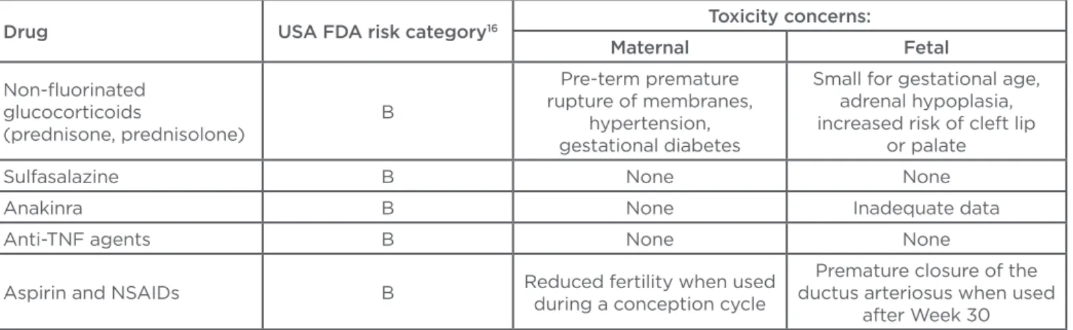
Table 2: Toxicities of drugs most commonly used in chronic arthritis patients during pregnancy.
Drug USA FDA risk category16 Toxicity concerns:
Maternal Fetal
Non-luorinated glucocorticoids
(prednisone, prednisolone)
B
Pre-term premature rupture of membranes,
hypertension, gestational diabetes
Small for gestational age, adrenal hypoplasia, increased risk of cleft lip
or palate
Sulfasalazine B None None
Anakinra B None Inadequate data
Anti-TNF agents B None None
Aspirin and NSAIDs B Reduced fertility when used
during a conception cycle
Premature closure of the ductus arteriosus when used
Glucocorticoids
Glucocorticoids (GCs) are frequently used in the management of pregnant patients with chronic arthritis and are considered Category B (Table 2). When active arthritis afects only one or a limited number of joints, intra-articular steroid injections can be very useful. Recently, prednisone use during pregnancy in RA patients has been associated with unfavourable outcomes as described by a study of 31 RA pregnancies followed prospectively in a single clinic. The patients with neither prednisone nor anti-TNF exposure had a mean irst trimester disease activity score — C-reactive protein (DAS-CRP) of 1.63, while patients exposed to prednisone or anti-TNF had a mean DAS-CRP of 3.92 or 3.19, respectively. An adverse outcome (pre-term birth and/or pre-eclampsia) occurred in 54.2% of patients exposed to prednisone compared with 14.3% of those with anti-TNF exposure, suggesting that the latter may be preferable to prednisone in pregnant women with active RA.25 There are
reports suggesting that GC exposure during the irst trimester may increase the risk of cleft lip or palate, but no other major congenital anomalies; women should be counselled about this potential
risk. Other adverse efects associated with prednisone use during pregnancy are pre-term premature rupture of membranes and children who are relatively small for their gestational age.
Disease-Modifying Anti-Rheumatic Drugs
Both MTX and LEF are considered as Category X (Table 2). MTX is teratogenic and embryotoxic, and its use during pregnancy is associated with multiple abnormalities, most often involving the central nervous system, cranial ossiication, and the palate (aminopterin syndrome).26 The drug
is contraindicated during pregnancy and should be prescribed to fertile women only under the condition of safe contraception. It has been proposed that conception should be postponed until 3 months after withdrawal of the drug,27
although this may be too conservative. Since MTX is a folic acid antagonist, folate supplementation should be continued antenatally and throughout pregnancy. There are no deinitive data on whether the ofspring of men receiving MTX have an increased risk of teratogenicity, although it is usually recommended that men discontinue this medication for 1-3 months prior to attempting conception.28
Anti-TNF agents: tumour necrosis factor inhibitor agents; NSAID: non-steroidal anti-inlammatory drug.
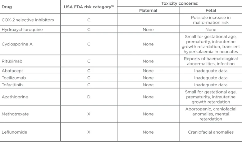
Drug USA FDA risk category16 Toxicity concerns:
Maternal Fetal
COX-2 selective inhibitors C Possible increase in
malformation risk
Hydroxychloroquine C None None
Cyclosporine A C None
Small for gestational age, prematurity, intrauterine growth retardation, transient
hyperkalaemia in neonates
Rituximab C None Reports of haematological
abnormalities, infection
Abatacept C None Inadequate data
Tocilizumab C None Inadequate data
Tofacitinib C None Inadequate data
Azathioprine D None
Small for gestational age, prematurity, intrauterine
growth retardation
Methotrexate X None
Abortogenic, craniofacial anomalies, mental
retardation
Lelunomide X None Craniofacial anomalies
LEF is a pyrimidine synthesis inhibitor that is both embryotoxic and teratogenic in animals. Women of childbearing potential for whom LEF is prescribed must be advised to avoid pregnancy. In a follow-up study of 45 women exposed to LEF prior to or during pregnancy, there was a 12.5% incidence of congenital anomalies in the 16 patients exposed during the irst trimester, which is higher than the generally reported background rate of 3%.29 Due to its long half-life and
protracted elimination from plasma, LEF-treated patients must follow a cholestyramine drug elimination procedure (‘wash out’) and then wait at least 90 days before attempting pregnancy: cholestyramine 8 g is given three-times daily for 11 days and plasma levels, checked twice 2 weeks apart, should be below 0.02 mg/l, with additional cholestyramine administration if the level is higher than this cut-of value.30 For those who
inadvertently become pregnant while taking LEF, this ‘wash out’ procedure early during pregnancy can allow the continuation of the pregnancy, with an absence of substantially increased risk of adverse pregnancy outcomes, as has been previously demonstrated.31
Sulfasalazine is considered to be Category B (Table 2). Although both the molecule and its metabolite, sulfapyridine, do cross the placenta, they do not have a signiicant clinical impact on the fetus. The main data describing pregnancies exposed to sulfasalazine were derived from the treatment of patients with inlammatory bowel disease (IBD), and no increases in birth defects, pathological jaundice, or babies who were small for their gestational age were detected.32-34
Because the drug is a strong inhibitor of the reduced folate carrier, folate supplementation before and throughout pregnancy must be ensured.35 Maternal doses should not exceed 2 g
daily in order to avoid the risk of neutropaenia in the newborn.36 Sulfasalazine impedes spermatogenesis,
causing azoospermia, and so men should discontinue this medication 3 months prior to attempting conception.37
Cyclosporine is also a treatment option, especially for RA and PsA patients, and is considered Category C (Table 2). More than 800 pregnancies exposed to cyclosporine for several weeks or throughout gestation have been reported, with the majority being in transplant recipients,28,38 and
there was no incremental increase in congenital malformations. Renal and liver functions were both normal in the neonates exposed.39 The major
problems associated with pregnancies exposed to cyclosporine treatment were prematurity in 40-46% and low birth weight (<2,500 g) in 44-65% of cases, but it has been diicult to ascribe a causative role to drug treatment or the underlying maternal disorder.
Azathioprine is not metabolised to its active form by the placenta and so hardly reaches the developing fetus. Pregnancy data from large transplant registries,40,41 as well as women with
systemic lupus erythematosus42,43 and IBD,44 found
no predominant or frequent birth defect. Although the drug is considered Category D (Table 2) for use during pregnancy, there is substantial evidence suggesting that it is not teratogenic. Intrauterine exposure to azathioprine may occasionally cause slight suppression of the bone marrow, as shown by decreased leukocyte counts and thrombocytopaenia at birth.45 Doses should be
maintained at 1.5-2 mg/kg/day in order to avoid neonatal depression of haematopoiesis. There are conlicting reports in the literature as to whether these medications increase the risk of children being small for their gestational age. Hydroxychloroquine is classiied as Category C (Table 2), but does not appear to cause fetal toxicity and can be used safely.46,47
Biological Agents
As in non-pregnant individuals, the use of biological agents increases the potential risk of infections. This is especially important for pathogens such as Listeria monocytogenes, for which the risk is already increased in pregnant women48 and can be further increased by
treatment.49 Current manufacturers’ guidelines
recommend the stopping of all currently licensed biological therapies prior to conception, primarily due to the lack of controlled studies in pregnant women. However, there is now a huge amount of information available regarding their use during conception, pregnancy, and breastfeeding.50
Much of this post-marketing experience has been obtained through registries, especially in the ield of IBD. In many cases, not treating IBD or discontinuing therapy prior to or during pregnancy may pose a greater risk to mother and fetus,20
while maintaining treatment appears to be of lower risk.
Tumour necrosis factor inhibitors
and inliximab (IFX), are considered Category B (Table 2). Although some case reports of congenital anomalies (VACTERL anomalies) following exposure to anti-TNF agents raised concerns about their safety proile when used during pregnancy, numerous case series of pregnancies exposed to anti-TNF agents, as well as several recent systematic literature reviews, have failed to ind maternal toxicity, embryotoxicity, or teratogenicity.51,52 In the pregnant woman,
maternal transfer of immunoglobulins across the syncytiotrophoblast of the chorionic villi provides fetal immunity and is mediated by the neonatal Fc receptor. Immunoglobulin G (IgG) concentrations in fetal blood increase steadily from the early stages of the second trimester until delivery.53
Small amounts of biological agents will pass into the fetal circulation during early pregnancy, when organogenesis is underway, suggesting that women can be reassured that the use of anti-TNF agents during the peri-conceptional period, until pregnancy is established, should be low risk. By continuing anti-TNF at least until established pregnancy is conirmed, women who are hoping to conceive should not need to risk disease lare during the period between stopping teratogenic medications, such as MTX, and successful conception.
However, most of the safety data available are derived from women discontinuing anti-TNF therapy during the irst trimester; few data exist regarding exposure throughout pregnancy. Due to the diferent composition of anti-TNF agents, some risks may depend on the individual agent rather than the drug class itself. Both IFX and ADA belong to the IgG1 subclass of antibodies and are capable of crossing the placental barrier, particularly during the second half of pregnancy. At birth, both infant and cord blood demonstrated higher concentrations of IFX and ADA. This raises concerns about infections, safety of vaccination, and, in general, about the development of the immune system of the child: serious infections in infants exposed to anti-TNF agents have been described,54 and a case of fatal disseminated
tuberculosis after Bacillus Calmette–Guérin (BCG) vaccination of a child exposed to IFX intra-utero has been described.55 In contrast, very low
concentrations of CZP were observed in infant and cord blood when mothers were treated with CZP.56
This is attributed to the absence of an Fc domain on the CZP molecule, which prevents it being bound by the neonatal Fc receptor.57 Therefore,
it is recommended that live vaccines (including rotavirus, intranasal inluenza vaccine, and BCG) be withheld for 6 months from birth in neonates who have had in utero exposure to anti-TNF agents, except CZP due to its minimal transfer. Other vaccines can be given on schedule.
Recent data from the multinational PIANO registry provide reassurance regarding the potential long-term efects on children exposed in utero to anti-TNF agents. A total of 501 women with IBD were followed through pregnancy and during the irst 4 years of the child’s life and exposure to biological therapy during the third trimester of pregnancy was not associated with increased infant infection rates,58 and children did not exhibit
developmental delay compared with infants not exposed to these agents, controlling for pre-term birth.59
Other biological agents
Anakinra is a recombinant interleukin (IL)-1 receptor antagonist. In animal studies, no harm to the fetus has been demonstrated. The drug is considered Category B but little has been reported about its safety in this setting (Table 2). Tocilizumab is a monoclonal antibody directed against IL-6 receptors and capable of blocking downstream signalling. The drug is considered Category C and no teratogenicity has been demonstrated in animal models, although at high dose there was increased risk of abortion (Table 2). Abatacept works by blocking interactions between antigen-presenting cells and T cells via binding to CD80/ CD86 on antigen-presenting cells, with subsequent inhibition of T cell activation. The drug is considered Category C and there are inadequate data to fully comment on its safety during pregnancy (Table 2), although animal studies saw no increased risk when exposed to the maximum recommended human dose. Current recommendations are to discontinue therapy at least 10 weeks before conception. Rituximab, a monoclonal antibody directed against CD20, depletes B cells. Animal studies are limited but show no teratogenic efect, although B cells have been demonstrated to be reduced in ofspring.60
REFERENCES
1. Förger F et al. Pregnancy mediated improvement of rheumatoid arthritis. Swiss Med Wkly. 2012;142:w13644.
2. Cavazzana I et al. Anti-Ro/SSA antibodies in rheumatoid arthritis: clinical and immunologic associations. Clin Exp Rheumatol. 2006;24:59-64.
3. Olech E, Merrill JT. The prevalence and clinical signiicance of antiphospholipid antibodies in rheumatoid arthritis. Curr Rheumatol Rep. 2006;8:100-8.
4. de Man YA et al. Disease activity of rheumatoid arthritis during pregnancy: results from a nationwide prospective study. Arthritis Rheum. 2008;59:1241-8. 5. van Gestel AM et al. Development and validation of the European League Against Rheumatism response criteria for rheumatoid arthritis. Comparison with the preliminary American College of Rheumatology and the World Health Organization/International League
Against Rheumatism Criteria. Arthritis Rheum. 1996;39:34-40.
6. de Man YA et al. Women with rheumatoid arthritis negative for anticyclic citrullinated peptide and rheumatoid factor are more likely to improve during pregnancy, whereas in autoantibody-positive women autoantibody levels are not inluenced by pregnancy. Ann Rheum Dis. 2010;69:420-3.
7. de Man YA et al. Disease activity of
Tofacitinib is a Janus kinase inhibitor recently approved by FDA for RA, although not approved in Europe. The drug is classiied as Category C (Table 2) and there are no available data describing its safety during pregnancy. Ustekinumab is considered Category B, with data in humans being limited to case reports. A recent abstract described 26 completed pregnancies that involved ustekinumab exposure in the course of treatment of psoriasis, and which documented a rate of spontaneous abortions comparable to that observed in the general population.61
CONCLUSION
Chronic arthritis often afects women of childbearing age. Counselling of the pre-pregnancy
patient constitutes the irst step of proper care. Complete information regarding risks must be ofered to the patient and her partner, and should be individualised according to the disease (Table 3). At the time of conception, the disease must be under control with medications compatible with pregnancy in order to minimise the risks. Ideally, drug treatment during pregnancy should control the mother’s disease activity, do no harm to the fetus, and ensure a healthy period of pregnancy for mother and child. An interdisciplinary approach between the rheumatologist, obstetrician, and eventually the paediatrician, is needed in order to ensure success. Currently, we can say that pregnancy in chronic arthritis patients is only a matter of planning.
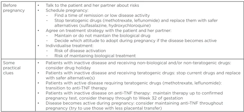
Table 3: Practical points to consider when treating women of childbearing age with chronic arthritis.
TNF: tumour necrosis factor.
Before pregnancy
• Talk to the patient and her partner about risks • Schedule pregnancy:
- Find a time of remission or low disease activity
- Stop teratogenic drugs (methotrexate, lelunomide) and replace them with safer alternatives (sulfasalazine, hydroxychloroquine)
• Agree on treatment strategy with the patient and her partner: - Maintain or do not maintain the biological drug
- Decide which attitude to adopt during pregnancy if the disease becomes active • Individualise treatment:
- Risk of disease activation
- Risk of maintaining biological treatment Some
practical clues
• Patients with inactive disease and receiving non-biological and/or non-teratogenic drugs: consider drug holiday
• Patients with inactive disease and receiving teratogenic drugs: stop current drugs and replace with safer alternative(s)
• Patients with active disease requiring teratogenic drugs (methotrexate, lelunomide): transition to anti-TNF therapy
• Patients with inactive disease on anti-TNF therapy: maintain therapy up to conirmed pregnancy test, consider therapy through to Week 32 of gestation
rheumatoid arthritis during pregnancy: results from a nationwide prospective study. Arthritis Rheum. 2008;59:1241-8. 8. Ostensen M, Ostensen H. Ankylosing spondylitis--the female aspect. J Rheumatol. 1998;25:120-4.
9. Ostensen M. The efect of pregnancy on ankylosing spondylitis, psoriatic arthritis, and juvenile rheumatoid arthritis. Am J Reprod Immunol. 1992;28:235-7.
10. de Man YA et al. Association of higher rheumatoid arthritis disease activity during pregnancy with lower birth weight results of a national prospective study. Arthritis Rheum. 2009;60:3196-206. 11. Chakravarty EF. Rheumatoid arthritis and pregnancy: beyond smaller and preterm babies. Arthritis Rheum. 2011;63:1469-71.
12. Langen ES et al. High rate of preterm birth in pregnancies complicated by rheumatoid arthritis. Am J Perinatol. 2014;31:9-14.
13. Wallenius M et al. Pregnancy and delivery in women with chronic inlammatory arthritides with a speciic focus on irst birth. Arthritis Rheum. 2011;63:1534-42.
14. de Steenwinkel FD et al. Rheumatoid arthritis during pregnancy and postnatal catch-up growth in the ofspring. Arthritis Rheumatol. 2014;66:1705-11.
15. Blackburn ST, “Pharmacology and Pharmacokinetics during the Perinatal Period,” Blackburn ST (ed.), Maternal, Fetal, & Neonatal Physiology: a Clinical Perspective (2013) 4th edition, Maryland Heights: Saunders, pp. 183-202.
16. U.S. Food and Drug Administration. CFR - Code of Federal Regulations Title 21. 2014. Available at: http://www. accessdata.fda.gov/scripts/cdrh/cfdocs/ cfCFR/CFRSearch.cfm?fr=201.57. Last accessed: 29 May 2015.
17. U.S. Food and Drug Administration. Content and format of labeling for human prescription drug and biological products; requirements for pregnancy and lactation labeling. April 2014. Available at: https://www.federalregister. gov/articles/2014/12/04/2014-28241/ content-and-format-of-labeling-for- human-prescription-drug-and-biological-products-requirements-for#page-72063. Last accessed: 29 May 2015.
18. Østensen M, Förger F. Management of RA medications in pregnant patients. Nat Rev Rheumatol. 2009;5:382-90.
19. Hyrich KL, Verstappen SM. Biologic therapies and pregnancy: the story so far. Rheumatology (Oxford). 2014;53:1377-85. 20. Hassid B, Mahadevan U. The use of biologic therapy in pregnancy: a gastroenterologist’s perspective. Curr Opin Rheumatol. 2014;26:347-53.
21. Pall M et al. Induction of delayed
follicular rupture in the human by the selective COX-2 inhibitor rofecoxib: a randomized double-blind study. Hum Reprod. 2001;16:1323-8.
22. Alano MA et al. Analysis of nonsteroidal antiinlammatory drugs in meconium and its relation to persistent pulmonary hypertension of the newborn. Pediatrics. 2001;107:519-23.
23. Ostensen M, Ostensen H. Safety of nonsteroidal antiinlammatory drugs in pregnant patients with rheumatic disease. J Rheumatol. 1996;23:1045-9.
24. Daniel S et al. Major malformations following exposure to nonsteroidal antiinlammatory drugs during the irst trimester of pregnancy. J Rheumatol. 2012;39:2163-9.
25. Chaudhary P, Clowse MEB. Prednisone use associated with worse outcomes in rheumatoid arthritis pregnancies. Arthritis Rheum. 2013;65 Suppl 10:1330.
26. Lloyd ME et al. The efects of methotrexate on pregnancy, fertility and lactation. QJM. 1999;92:551-63.
27. Donnenfeld AE et al. Methotrexate exposure prior to and during pregnancy. Teratology. 1994;49:79-81.
28. Bar Oz B et al. Pregnancy outcome after cyclosporine therapy during pregnancy: a meta-analysis. Transplantation. 2001;71:1051-5.
29. Cassina M et al. Pregnancy outcome in women exposed to lelunomide before or during pregnancy. Arthritis Rheum. 2012;64:2085-94.
30. Brent RL. Teratogen update: reproductive risks of lelunomide (Arava); a pyrimidine synthesis inhibitor: counseling women taking lelunomide before or during pregnancy and men taking lelunomide who are contemplating fathering a child. Teratology. 2001;63: 106-12.
31. Chambers CD et al. Birth outcomes in women who have taken lelunomide during pregnancy. Arthritis Rheum. 2010;62:1494-503.
32. Nørgård B et al. Population-based case control study of the safety of sulfasalazine use during pregnancy. Aliment Pharmacol Ther. 2001;15:483-6.
33. Mogadam M et al. Pregnancy in inlammatory bowel disease: efect of sulphasalazine and corticosteroids on fetal outcome. Gastroenterology. 1981;80:72-6.
34. Rahimi R et al. Pregnancy outcome in women with inlammatory bowel disease following exposure to 5-aminosalicylic acid drugs: a meta-analysis. Reprod Toxicol. 2008;25:271-5.
35. Hernández-Díaz S et al. Folic acid antagonists during pregnancy and the risk of birth defects. N Engl J Med. 2000;343:1608-14.
36. Levi S et al. Reversible congenital neutropenia associated with maternal sulphasalazine therapy (letter). Eur J Pediat. 1988;148:174-5.
37. Chatzinof M et al. Sulfasalazine-induced abnormal sperm penetration assay reversed on changing to 5-aminosalicylic acid enemas. Dig Dis Sci. 1988;33:108-10.
38. Lamarque V et al. Analysis of 629 pregnancy outcomes in transplant recipients treated with Sandimmun. Transplant Proc. 1997;29:2480.
39. Shaheen FAM et al. Long-term nephrotoxicity after exposure to cyclosporine in utero. Transplantation. 1993;56:224-25.
40. Registration Committee of the European Dialysis and Transplant Association. Successful pregnancies in women treated by dialysis and kidney transplantation. Report from the Registration Committee of the European Dialysis and Transplant Association. Br J Obstet Gynaecol. 1980;87:839-85.
41. Radomski JS et al. Outcomes of 500 pregnancies in 335 female kidney, liver, and heart transplant recipients. Transplant Proc. 1995;27:1089-90.
42. Meehan RT, Dorsey JK. Pregnancy among patients with systemic lupus erythematosus receiving immunosuppressive therapy. J Rheumatol. 1987;14:252-8.
43. Ramsey-Goldman R et al. Pregnancy outcome in women with systemic lupus erythematosus treated with immunosuppressive drugs. J Rheumatol. 1993;20:1152-7.
44. Casanova MJ et al. Safety of thiopurines and anti-TNF-α drugs during pregnancy in patients with inlammatory bowel disease. Am J Gastroenterol. 2013;108:433-40.
45. Davison JM et al. Maternal azathioprine therapy and depressed haemopoiesis in the babies of renal allograft patients. Br J Obst Gynecol. 1985;92:233-9.
46. Sperber K et al. Systematic review of hydroxychloroquine use in pregnant patients with autoimmune diseases. Pediatr Rheumatol Online J. 2009;7:9. 47. Bermas BL. Non-steroidal anti inlammatory drugs, glucocorticoids and disease modifying anti-rheumatic drugs for the management of rheumatoid arthritis before and during pregnancy. Curr Opin Rheumatol. 2014;26:334-40. 48. Southwick FS, Purich DL. Intracellular pathogenesis of listeriosis. N Engl J Med. 1996;21;334:770-6.
2008;26:854-9.
50. Vinet E et al. Biologic therapy and pregnancy outcomes in women with rheumatic diseases. Arthritis Rheum. 2009;61:587-92.
51. Nielsen O et al. Safety of TNF-α inhibitors during IBD pregnancy: a systematic review. BMC Med. 2013;11:174. 52. Marchioni RM, Lichtenstein GR. Tumor necrosis factor-α inhibitor therapy and fetal risk: a systematic literature review. World J Gastroenterol. 2013;19:2591-602. 53. Palmeira P et al. IgG placental transfer in healthy and pathological pregnancies. Clin Dev Immunol. 2012;2012:985646. 54. Johnsson A et al. Chicken pox infection in a three months old infant
exposed in utero to Adalimumab. J Crohns Colitis. 2013;7:e116-7.
55. Cheent K et al. Case Report: Fatal case of disseminated BCG infection in an infant born to a mother taking inliximab for Crohn’s disease. J Crohns Colitis. 2010;4:603-5.
56. Mahadevan U et al. Placental transfer of anti-tumor necrosis factor agents in pregnant patients with inlammatory bowel disease. Clin Gastroenterol Hepatol. 2013;11:286-92.
57. Clowse MEB et al. Retrospective analysis of certolizumab pegol use during pregnancy: update of impact on birth outcomes. Arthritis Rheum. 2013;65 Suppl 10:433.
58. Mahadevan U et al. Exposure to
anti-TNFα therapy in the third trimester of pregnancy is not associated with increased adverse outcomes: results from the PIANO Registry. Gastroenterol. 2014:146 suppl 1:S-170.
59. Mahadevan U et al. Achievement of developmental milestones among ofspring of women with inlammatory bowel disease: the PIANO Registry. Gastroenterol. 2014:146 suppl 1:S-1.
60. Chakravarty EF et al. Pregnancy outcomes after maternal exposure to rituximab. Blood. 2011;117:1499-506. 61. Cather JC et al. Pregnancy outcomes in women exposed to ustekinumab in the psoriasis clinical development program. Australasian Journal of Dermatology. 2014;55 suppl 1:1-58.