Bull Pan Am Health Organ 14(3), 1980.
THE USE OF SOLID-PHASE RADIOIMMUNOASSAY TECHNIQUES FOR SERODIAGNOSIS OF HUMAN PLAGUE INFECTION
B. W. Hudson,1 K. Wolff,’ and T. Butler2
The results of solid-phase radioimmunoassay tests conducted with
sera from confirmed and suspected human plague cases suggest
such testing could have significant advantages for the diagnosk of thzj disease.
Introduction
Solid-phase radioimmunoassay (SPRIA)
techniques have been valuable for quantify- ing antibody to bacteria (I) and viruses (2,3). In the indirect solid-phase radioimmunoassay technique, a purified antigen is usually at- tached directly to the solid phase. The at- tached antigen is then exposed to the human test serum; and after thorough washing, an- tibodies combined with the antigen are de- tected by means of radioisotopically labeled antibodies specific for human IgM or IgG.
Application of these techniques for deter- mining the presence of human antibody to
Ye&z&z pestis offers several apparent advan- tages. These are: the use of relatively moderate amounts of purified antigens; ex- quisite sensitivity- on the order of 0.5 pg of antibody per ml (I); and the ability to measure both IgM and IgG responses.
The soluble protein fraction 1A of Y. pestis (4), recommended for serodiagnosis of human plague infection (5), constitutes a highly purified specific antigen; but it is only available in limited quantities from noncom- mercial sources. The most sensitive serologic test now used for diagnosis of human plague, the passive hemagglutination (PHA) test (5),
lImmunochemistry Branch, Vector-Borne Diseases Division, Bureau of Laboratories, Center for Disease Control, United States Public Health Service, Depart- ment of Health, Education, and Welfare, P. 0. Box 2087, Fort Collins, Colorado 80522, U.S.A.
‘Division of Geographic Medicine, Department of Medicine, Case Western Reserve University, Cleveland, Ohio 44106, U.S.A.
sometimes fails to detect serologic responses in human sera collected shortly after the onset of disease (6,7). Of particular concern is the PHA test’s failure to detect titer increases in acute-to-convalescent sera from some vac- cinated primates (8) or from occasional recur- rent cases of human plague (7). Finally, it is difficult to adapt the PHA test to differentia- tion of human IgM and IgG responses, and a possibility exists that suitable measurement of these responses might have value in the serodiagnosis of human plague, particularly during early convalescence. For all of these reasons, we decided to use SPRIA techniques to examine a number of human sera available to us.
Materials and Methods
Sera
Most of the serum samples came from con- firmed cases of human plague in Vietnay). These included acute and convalescent sera from 14 patients and convalescent sera from four other patients. An additional eight sera were obtained from North American patients with confirmed plague. These latter included an acute serum and two convalescent sera from one patient, and five convalescent sera from five other patients.
As negative controls we used the following sera, all collected in North America: three convalescent sera from patients with sus- pected plague infections later shown to be tularemia; seven convalescent sera from pa-
tients with suspected but unconfirmed plague; and three sera from family contacts of patients with suspected but unconfirmed plague.
Antigen
Purified protein fraction IA of Y. pestis
strain A 1122 was prepared using the methods of Baker (4). This preparation was homo- genous when analyzed by sodium laurel sul- fate acrylamide disc gel electrophoresis (9) and showed no cross-reactivity when used in
the PHA test with hyperimmune Yersiniu
pseudotuberculosis or Yersinia enterocolitica
sera (5).
Solid-phase Radioimmunoassay
The technique was adapted from that of Trent et al. (IO). Y. pestis fraction IA (0.8 pg)-in 50 pl of phosphate-buffered saline (PBS) at pH 7.4, containing 0.1 per cent so- dium azide, 2mM MgCla, and 1mM CaCls- was placed in each well of 96-well microtiter plates treated for tissue culture (Linbro Scien- tific Co., Inc., Hamden, Conn.). 3 Plates con- taining antigen were air-dried overnight at room temperature. The next day 200 ~1 of 10 per cent normal rabbit serum in PBS (PBS- NRS) were added to each well and incubated 45 minutes at 37°C. The plates were then washed three times with PBS.
Sera to be tested were diluted I : 100 in PBS- NRS, and 50 pl of each specimen was added to each of four wells. For IgM determinations, the plates were then incubated for 5 hours at 37°C and 12 hours at 4%, while for IgG subclass determinations they were incubated 1 hour at 37°C. All the plates were then washed three times with PBS.
Goat antisera specific for the heavy chains of human IgM and IgG were obtained from Antibodies, Inc., in Davis, California. These
‘Use of trade names is for identification only and does not constitute endorsement by the Public Health Service
or the U.S. Department of Health, Education, and Welfare.
antisera had been purified on affinity col- umns by the manufacturer. After purifica- tion, the sera showed no cross-reactivity when
tested by radial immunodiffusion, im-
munoelectrophoresis. and Ochterlon tests.( The IgG-rich fractions of the antisera were prepared by ammonium sulfate precipitation (II), and protein concentrations were deter- mined by a modified Folin method (12). These IgG fractions were then radioiodinated
with la51 (Amersham Searle) using the
choramine T method (3). Antiglobulins were labeled with 1,000 microcuries of lz51 per 100 pg of protein in 1 ml volumes and were separated from free iodine on Sephadex G-25 columns. The resulting 5 to 6 ml amounts of iodinated globulin solution were diluted with an equal volume of normal rabbit serum
(NRS). These stock solutions were then
diluted in NRS for the SPRIA test (1:2 for IgM, 1:4 for IgG). Fifty d of the lz51-anti- globulin working dilution was added to each well of the microtiter plate involved. For IgM determinations the plates were incubated 5 hours at 37”C, and for IgG they were in- cubated 2 hours at 37°C. The plates then
were washed three times with PBS and
covered with plastic film; the wells were cut from the plate, placed in 20 ml scintillation vials, and counted in a Beckman Gamma-310 Counter. Means and standard deviations of
the four determinations were used as a
measure of lz51 antiglobulin binding for each serum.
Passive Hemagglutination (PHA)
In the PHA test, the purified fraction 1A
and the standard methods recommended by
the World Health Organization Expert Com- mittee on Plague (5) were used.
Results
The SPRIA results obtained for the various categories of sera tested are summarized in
246 PAHO BULLETIN . vol. 14, no. 3, 1980
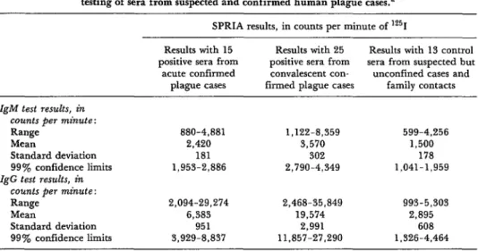
Table 1. In tests with convalescent sera from patients with confirmed plague, the average binding of lZ51-labeled antihuman IgM and IgG reagents was two to six times higher than that obtained in tests performed with negative control sera from patients with suspected but unconfirmed plague. The range of values ob- tained overlaps, however.
In Table 1, assuming normal distribution of lz51 binding values, we have estimated the variance of each group of determinations and the 99 per cent confidence limits. The upper confidence limits for SPRIA binding of lz51 obtained using the negative control sera (IgM, 1,959 counts per minute [CPM] ; IgG, 4,464 CPM) were found to lie below the lower confidence limits obtained using the convales- cent sera from patients with confirmed plague (IgM, 2,790 CPM: IgG, 11,857 CPM). The
CPM, IgG >4,464 CPM, and a PHA titer equaling or exceeding 1: 16 as positive results. The outcome of this classification is shown in Table 2. PHA results were positive for eight of the 15 acute sera tested, while SPRIA results were positive for 10 of the 15. In tests with the convalescent sera, 23 of the 25 sera tested were positive by PHA, while all 25 were positive by SPRIA.
. .
upper confidence limits obtained in tests us- ing the negative control sera therefore seem to provide reasonable criteria for categorizing sera as positive when tested by SPRIA.
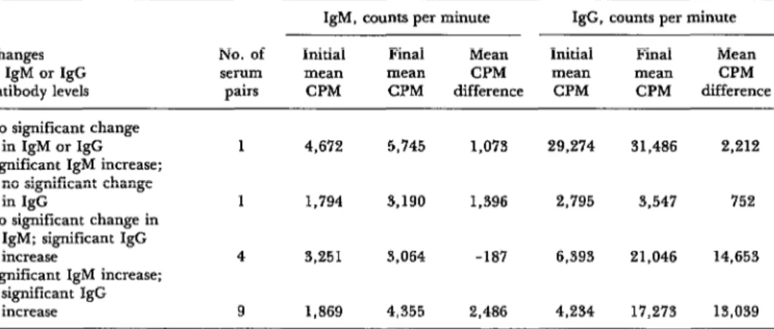
Since the sera were tested four times to ob- tain a measure of mean radioiodine uptake, we used Student’s t test to compare values for acute and convalescent sera. Mean values dif- fering at the 1 per cent significance level were classed as showing a rise in IgM or IgG levels. We analyzed data for the 15 cases in which both acute and convalescent sera were avail- able (Table 3). Only one serum pair failed to show a significant increase in IgM or IgG levels. Both sera of this pair showed high levels of IgM and IgG, and a moderate but not statistically significant increase in both levels.
We then classified all sera from patients with confirmed plague in terms of their positive or negative results when tested by SPRIA and PHA-considering IgM > 1,959
Four of the 15 serum pairs did not show fourfold increases in PHA titers. These sera are of particular interest in the comparative evaluation of SPRIA and PHA tests. Serum
Table 1. The results of IgM and IgG specific solid-phase radioimmunoassay (SPRIA) testing of sera from suspected and confirmed human plague cases.=
SPRIA results, in counts per minute of lz51 Results with 15
positive sera from acute confirmed
plague cases
Results with 25 Results with 13 control positive sera from sera from suspected but convalescent con- unconfined cases and firmed plague cases family contacts IgM test results, in
counts per minute: Range
Mean
Standard deviation 99% confidence limits IgG test results, in
counts per minute: Range
Mean
Standard deviation 99% confidence limits
880-4,881 1,122-8,359 599-4,256
2,420 3,570 1,500
181 302 178
1,953-2,886 2,790-4,349 1,041-1.959
2,094-29,274 2,468-35,849 993-5,303
6,383 19,574 2,895
951 2,991 608
$929-8.837 11,857-27,290 1,326-4,464
Table 2. A qualitative comparison of the test results obtained using solid-phase radioimmunoassay (SPRIA) and passive hemagglutination (PHA) on acute and
convalescent sera from 25 confirmed cases of human plague. Passive hemagglutination (PHA) results
Acute sera (15 samples) Convalescent sera (25 samples)
SPRIA results PHA- PHA + PHA- PHA +
Negative (IgM - , IgG - ) 4 1 0 0
Positive (IgM + , IgG - ) 3 1 0 I
Positive (IgM - , IgG + ) 0 2 2 4
Positive (IgM + , IgG + ) 0 4 0 18
Total SPRIA positive 3 7 2 23
Table 3. Results of SPRIA testing of 15 paired acute and convalescent sera. In this table the paired sera have been classified as showing either significant increases or no
significant increases in Y. pestis fraction IA IgM or IgG antibody by Student’s t test at the 1 per cent significance level.
Changes No. of
in IgM or IgG serum
antibody levels pairs
IgM, counts per minute IgG, counts per minute
Initial Final Mean Initial Final Mean
mean mean CPM mean mean CPM
CPM CPM difference CPM CPM difference
No significant change in IgM or IgG Significant IgM increase:
no significant change in IgG
No significant change in IgM; significant IgG increase
Significant IgM increase; significant IgG increase
1 4,672 5,745 1,073 29,274 31,486 2,212
1 1,794 3,190 1,396 2,795 3,547 752
4 3,251 3,064 -187 6,393 21,046 14,653
9 1,869 4,355 2,486 4,234 17,273 13,039
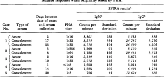
from one other case is also of interest, al- though acute serum was no longer available.
This serum came from a case in North
America that was confirmed bacteriologically and was followed serologically for 3 months after onset. None of the sera collected demon- strated a significant PHA titer. One serum collected 30 days after onset yielded a PHA titer of 1:8.
Data on these five cases are shown in Table 4. All five yielded significant SPRIA results. Case 2 is particularly interesting, because the paired sera were from one of two cases of re-
current plague infection showing a decrease
in PHA titer. Those two cases have been dis- cussed elsewhere (7). These data suggest that, in comparison to PHA results, certain human convalescent sera may yield unusually high SPRIA results.
248 PAHO BULLETIN . vol. 14, no. 3, 1980
Table 4. Passive hemagglutination (PHA) and SPRIA data from five confirmed cases of human plague. These cases were selected on the basis of low or
reduced responses when originally tested by PHA. SPIRA resultsa
Days between IgMb IgGb
date of onset
Case Type of and serum PHA Counts per Standard Counts per Standard
NO. serum collection titer minute deviation minute deviation
I
Acute 2 1:16 3,301 282 5,538 388
1 Convalescent 17 1:32 8,275 564 24,787 5,720
Convalescent 33 I:32 6,778 194 26,599 4,206
2 I Acute 3 1:256 1,209 91 8,109 555
Convalescent 8 1:64 1,122 126 29,453 3,573
3 i Acute 2 1:16 2,359 149 3,872 549
Convalescent 10 I:32 2,972 2.12 5,114 413
4 i Acute 1 <1:8 1,652 163 3,014 915
Convalescent 14 1:16 1,225 209 6,493 1,784
5 Convalescent 30 1:8 756 44 12,624 685
‘SPRIA results beyond the 99% confidence limit for unconfirmed cases are italicized (see text).
bCPM and the standard deviation for each serum were estimated on the basis of standard deviations from quadruplicate determinations.
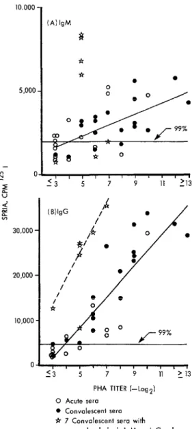
figure. All seven were convalescent sera - four being from three of the five cases presented in Table 4. Of these seven sera, SPRIA-IgG data from six seemed to have a distribution dif- ferent than data from the remaining sera. The regression line for these six appears as the dashed line in Figure 1B. If the data from these seven sera are deleted, analysis of data from the 33 remaining acute and convales- cent sera shows a correlation between the ob- served PHA titers and the IgM and IgG levels (the correlation coefficients being 0.658 for IgM and 0.894 for IgG). SPRIA IgG and PHA data are more closely correlated than SPRIA IgM and PHA.
Analysis of IgG and PHA data for the seven anomalous sera gives a correlation coefficient of 0.745. If the one serum showing high IgM levels but “normal” IgG levels is excluded from this sample, data from the remaining six sera yield a correlation coefficient of 0.984.
Discussion
Difficulties in using the PHA test for sero- diagnosis of plague infection have frequently
been pointed out. Thus, Marshall and
coworkers (6) emphasized that 35 of 69 Viet- namese patients, from each of whom two sera were obtained, showed no significant rise in titer. In addition, 50 of 71 single-serum specimens from patients with bacteriologic- ally proven plague were seronegative. The serum specimens used in these studies were collected during early convalescence, and the
authors indicate that during early con-
valescence the serum antibody response may be delayed. This delay may be complicated by actual decreases in titer in populations with preexisting titers. Two cases of recurrent plague infection studied by Butler (7) in Viet- nam showed actual drops in PHA titers. Similar results have been reported in vac- cinated primates when challenged with living plague bacteria (8). Obviously, this type of
result, although infrequent, could cause
diagnostic difficulties, particularly in vac- cinated individuals.
Some of these difficulties could perhaps be avoided by use of SPRIA techniques. Serum samples from one patient with recurrent plague showed highly significant increases in IgG levels when tested by SPRIA, and four
additional cases-samples from which were
Figure 1. A scatter diagram comparing data ob- tained by PHA and SPRIA analysis of acute and con- valescent sera from confirmed plague cases. (A) SPRIA-determined IgM levels compared to PHA titers; (B) SPRIA-determined IgG levels compared to PHA titers. The two horizontal lines represent the upper 99 percent confidence limits of the SPRIA data obtained from sera of unconfirmed suspected plague cases and family contacts (see text and Table 1). The two solid diagonal lines show the least squares linear regression fits for 33 sera. The dashed line in Figure 1B is the regression line for six convalescent sera with unusually high SPRIA binding values (see text and Table 4).
10,000
1
(Al IgM 8 *5,000
5
% Ok-n-ET-
- L13 5E 30,000 -
20,000 -
10,000 -
creases in PHA titer-were confirmable by SPRIA tests (Table 4).
A second consideration relates to data ob- tained by analyzing 13 sera from unconfirmed suspect plague cases. Two convalescent sera in this group of 13 gave SPRIA results above the estimated upper 99 per cent confidence limit for the group. One serum was obtained from a patient suspected on good epidemio- logic evidence of having contracted plague from rabbits in New Mexico. Serologic tests (PHA) and bacteriologic tests were negative for plague, but a moderate titer of 1:128 to tularemia was found. This serum yielded IgM SPRIA levels of 4,256 CPM and negative IgG results. Bacteriologic and serologic results for the second suspect case, a patient in Califor- nia, were negative. This serum gave negative IgM SPRIA results but moderately positive SPRIA IgG levels of 5,303 CPM. Naturally, this initial investigation does not provide grounds for accurately defining the criteria
for use of SPRIA in diagnosing human
plague; but it does suggest that the first- mentioned unconfirmed case may have been misdiagnosed.
The possibility that serologic diagnosis of human plague could be improved by suitable application of SPRIA techniques is of obvious importance to those engaged in control of this disease. In addition, conserving scarce an- tigen and serum samples is important for lab- oratories engaged in plague diagnosis. A total of 6.4 pgm of fraction 1 antigen and 4 hl of serum were needed to test the IgM and IgG content of each serum specimen. Although equipment costs may limit the use of SPRIA tests to better-equipped reference labora- tories, the potential for conserving antigen and serum specimens may prove an advan- tage to such laboratories.
(\ ’ ; * ; ’ ; ’ ;, ’ >_ ;3 PHA TITER I-tog2)
0 Acute sero 0 Convalescent sero t 7 Convalescent sew with
250 PAHO BULLETIN l vol. 14, no. 3, 1980
SUMMARY
Solid-phase radioimmunoassay (SPRIA) tech- Plague. These results suggest that use of SPRIA niques were used to analyze the levels of IgM and might help conserve the commercially unob- IgG antibody to purified Yersinia pestis fraction IA tainable fraction 1A antigen. In addition, it ap- in various human sera. The results were compared pears that certain human plague cases that fail to with data obtained by using the standard passive show significant PHA titers or fail to exhibit in- hemagglutination (PHA) test recommended by the creases in PHA titers can be diagnosed by suitable World Health Organization Expert Committee on use of SPRIA.
REFERENCES
(I) Zollinger, W. D., J. M. Dahymple, and M. S. Artenstein. Analysis of parameters affecting the solid phase radioimmunoassay quantitation of anti- body to meningococcal antigens. J Immunol 117:1788-1798, 1976.
(2) Rosenthal, J. D., K. Hayashi, and A. L. Notkins. Comparison of direct and indirect solid- phase micro-radioimmunoassays for the detection of viral antigens and antiviral antibody. Appl Microbial 25:567-573, 1973.
(3) Purcell, R. H., D. C. Wong, H. J. Alter, and P. V. Holland. Microtiter solid-phase radioim- munoassay for Hepatitis B antigen. Appl Microbial 26~478-484, 1973.
(4) Baker, E. E., H. Sommer, L. E. Foster, E. Meyer, and K. F. Meyer. Studies on immunization against plague: I. The isolation and characteriza- tion of the soluble antigen of Pasteurella pestis. J Immunol68:131-145, 1952.
(5) World Health Organization. WHO Expert Committee on Plague: Fourth Report. WHO Technical Report Series, No. 477. Geneva, 1970, pp. 23-25.
(6) Marshall, J. D., Jr., F. L. Gibson, T. C. Dung, and D. V. Quy, Early serological non- response in human plague infections. Bull WHO 3’7:495-497, 1967.
(7) Butler, T., and B. W. Hudson. The sero- logical response to Yersinia pestis infection. Bull WHO 55:39-42, 1977.
(8) McNeill, D., and K. F. Meyer. Pasteurella pestis antibody patterns in sera of plague- convalescent and vaccinated persons determined by immunoelectrophoresis and passive hemag- glutination. J Immunol94:778-784, 1965.
(9) Bennett, L. G., and T. G. Tournabene. Characterization of the antigenic subunits of the envelope protein of Yersinia pestis. J Bacterial 117:48-55, 1974.
(10) Trent, D. W., C. L. Harvey, A. Qureshi, and D. LeStourgeon. Solid-phase radioim- munoassay for antibodies to flavivirus structural and nonstructural proteins. Infect Immun 13:1325-1333, 1976.
(II) Herbert, G. A. Ammonium sulfate frac- tionation of sera: mouse, hamster, guinea pig, monkey, chimpanzee, swine, chicken and cattle. Appl MicTobiol27:389-393, 1974.