Influence of obstructive sleep apnea syndrome in the
fluctuation of the submaximal isometric torque of knee
extensors in patients with early-grade osteoarthritis
Andressa Silva1, Marco T. Mello2, Paula R. Serrão1, Roberta P. Luz1, Lia R. Bittencourt3, Stela M. Mattiello1
ABSTRACT | Objective: The aim of this study was to investigate whether obstructive sleep apnea (OSA) alters the luctuation of submaximal isometric torque of the knee extensors in patients with early-grade osteoarthritis (OA).
Method: The study included 60 male volunteers, aged 40 to 70 years, divided into four groups: Group 1 (G1) - Control (n=15): without OA and without OSA; Group 2 (G2) (n=15): with OA and without OSA; Group 3 (G3) (n=15): without OA and with OSA; and Group 4 (G4) (n=15) with OA and with OSA. Five patients underwent maximal isometric contractions of 10 seconds duration each, with the knee at 60° of lexion to determine peak torque at 60°. To evaluate the luctuation of torque, 5 submaximal isometric contractions (50% of maximum peak torque) of 10 seconds each, which were calculated from the standard deviation of torque and coeficient of variation, were performed. Results: Signiicant
differences were observed between groups for maximum peak torque, while G4 showed a lower value compared with G1 (p=0.005). Additionally, for the average torque exerted, G4 showed a lower value compared to the G1 (p=0.036). However, no differences were found between the groups for the standard deviation (p=0.844) and the coeficient of variation (p=0.143). Conclusion: The authors concluded that OSA did not change the parameters of the luctuation of
isometric submaximal torque of knee extensors in patients with early-grade OA.
Keywords: osteoarthritis; sleep apnea syndromes; muscle strength; knee; rehabilitation.
Clinical Trials Identifier: clinical trials.gov (NCT01422967).
HOW TO CITE THIS ARTICLE
Silva A, Mello MT, Serrão PR, Luz RP, Bittencourt LR, Mattiello SM. Inluence of obstructive sleep apnea syndrome in the luctuation of the submaximal isometric torque of knee extensors in patients with early-grade osteoarthritis. Braz J Phys Ther. 2015 July-Aug; 19(4):271-278. http://dx.doi.org/10.1590/bjpt-rbf.2014.0106
1 Departamento de Fisioterapia, Universidade Federal de São Carlos (UFSCar), São Carlos, SP, Brasil
2 Departamento de Esportes, Universidade Federal de Minas Gerais (UFMG), Belo Horizonte, MG, Brasil
3 Departamento de Psicobiologia, Universidade Federal de São Paulo (UNIFESP), São Paulo, SP, Brasil
Received: Aug. 05, 2014 Revised: Dec. 26, 2014 Accepted: Mar. 10, 2015
Introduction
Sleep complaints in osteoarthritis (OA) patients have been recently reported in the literature1
, and some
studies have described an increase in sleep disorders in this population, with Obstructive Sleep Apnea
(OSA) being the most common and most frequent1-3. It is estimated that in 2030, 20 to 30% of the world
population will present some type of OA4, most frequently found in the population over 60 years5, particularly in the knee joint, which accounts for approximately 7% of cases6. OA is characterized by loss of articular cartilage and thickening of the joint
capsule and is associated with changes in muscle function7, especially decreased quadriceps muscle
strength8. Clinically, OA patients generally present with complaints of pain, fatigue, crepitus, limitations
in performing activities of daily living9, and sleep complaints1.
The decrease in quadriceps muscle strength
has been associated with functional changes and neuromuscular functional impairments, also due to OA10,11. Neuromuscular function plays an important role in knee joint stability, which involves muscle strength, coordination and the knee joint position sense12. Neuromuscular function arises from the integration
of peripheral afferent signals of receptors located in
the muscles, tendons, joint capsule, ligaments and
The ability to produce and maintain a steady
submaximal force production (i.e. force steadiness)
has been previously studied13. Submaximal force assessment is a means to quantify deficits in
neuromuscular control14.
Submaximal force relects the deicits that might
affect an individual’s ability to reach the desired
force and to successfully produce movement. This characteristic might be compromised in knee OA
patients, and reports in the literature have suggested
that this deicit might play an important role in knee
OA pathogenesis15. However, the relationship between muscle strength and the luctuation of the submaximal force is not fully understood. It has been suggested that the decrease in submaximal force luctuation signiicantly contributes to the development and progression of knee OA12, but there is no current
evidence to support this relationship16.
The aforementioned neuromuscular changes
might affect the muscle strength of knee OA patients.
Knowing that the presence of OSA in these patients
leads to sleep deicit, these factors could inluence the
symptoms reported by the patients, such as pain and fatigue17. Moreover, the literature has shown that sleep deicit could induce muscle atrophy because of the
decreases in anabolic hormones, such as testosterone,
growth hormone and insulin-like growth factor 1 (IGF-1), and of the increases in catabolic hormones,
such as myostatin and glycocorticoid18,19.
Considering that knee OA patients present changes in
their sleep patterns, such as OSA, and both conditions compromise muscle function, this study’s hypothesis was that the presence of OSA associated with OA would compromise the motor and functional capacities of
patients and that OA alone would not. Therefore, this study aimed to assess if OSA affected the luctuation
of the submaximal isometric torque of knee extensors of patients with early-grade OA.
Method
VolunteersThe male volunteers were recruited via advertisements
in print and electronic local media. Following the
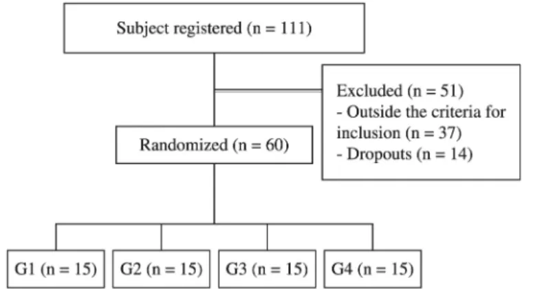
advertisement, a total of 111 individuals were initially enrolled, of whom 37 did not meet the inclusion criteria, and 14 dropped out (problems with the schedule
of work (6), health problems (4), travel (2) and
personal problems (2)) during the study (Figure 1). The study included men between 40 and 70 years of
age diagnosed with knee OA according to the clinical
criteria recommended by the American College of Rheumatology20 and with severity grade II according
to the Kellgren and Lawrence21 classiication through X-ray examination. In addition, to be included in
the study, the individuals could not have engaged in any regular physical activity in the last 6 months; could not have had any previous trauma, surgery or fracture of the lower limbs10; had not taken any pain
medication and presented with normal resting and
exercise electrocardiogram readings. All individuals underwent a polysomnography test and a sleep clinical.
Therefore, the inal sample consisted of 60 volunteers
separated into four groups based on the results of the data collected from the radiographs and sleep tests
(see below): Group 1 (G1) (Control): without OA and without OSA (N=15); Group 2 (G2): without OA and with OSA (N=15); Group 3 (G3): with OA and without OSA (N=15); and Group 4 (G4): with OA and with OSA (N=15). The participants’ characteristics are
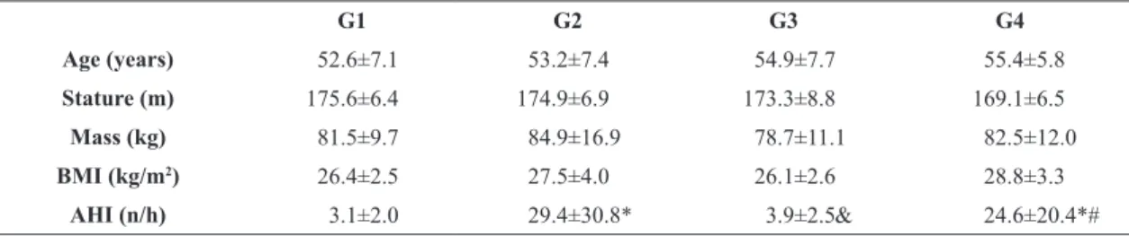
described in Table 1. The sample was homogeneous
because the variables age [F (3.56)=0.559; p=0.644],
height [F (3.56)=2.401; p=0.07], mass [F (3.56)=0.606; p=0.614] and body mass index (BMI) [F (3.56)=1.987; p=0.126] were not different between groups.
However, regarding the Apnea-Hypopnea Index (AHI) [F (3.56)=8.191; p=0.001; size effect=0.305; power=0.988], a higher AHI was detected in G4 and statistically differed from those in G1 (p=0.013) and G3 (p=0.017). A higher AHI was also observed in G2 and signiicantly differed from those in G1 (p=0.002) and G3 (p=0.002).
All of the participants signed an informed consent
form, and the Universidade Federal de São Carlos (UFSCar) Ethics Committee, São Carlos, state of São Paulo - SP, Brazil (CEP #109/2011) approved the study.
Procedures
The study was conducted at UFSCar, São Carlos city, and at the Centre for Studies in Psychobiology and Exercise (Centro de Estudos em Psicobiologia and Exercício – CEPE), São Paulo state, Brazil. First,
the principal investigator interviewed the volunteers,
and after, the resting and exercise electrocardiograms were scheduled with the cardiologist of the CEPE. If results of the electrocardiograms were normal, knee X-ray examinations were scheduled, and the results were analyzed by a rheumatologist of the CEPE.
Then, each volunteer was referred to a sleep doctor who scheduled the polysomnography testing, and after the results were received, a new appointment
was scheduled to diagnose OSA. Isokinetic testing
was performed on volunteers only after all of these
procedures were completed and the subjects were assigned to one of the 4 groups.
Measurements
Radiographic assessment
Anteroposterior and mediolateral X-rays were taken from both knees of all participants. The criteria for the
diagnosis of OA for Groups 3 and 4 was presence of
well-deined osteophytes, without narrowing of the intra-articular space, classiied as an early grade of the disease (grade II)21,22.
Polysomnography (PSG)
To record a full-night PSG, the Embla S7000 (Embla Systems, Inc., Reykjavik, Iceland) device was used at the
Sleep Laboratory (Sleep Institute [Instituto do Sono], São Paulo, Brazil). All recording sensors were attached to the patient in a non-invasive manner using adhesive tape or elastic bands. The following physiological variables
were continuously and simultaneously monitored:
4-channel electroencephalogram (EEG) (C3-A2, C4-A1, O1-A2, O2-A1), 2-channel electrooculogram (EOG) (EOG-left-A2, EOG-right-A1), 4-channel
surface electromyogram (muscle of the submentonian region, tibialis anterior muscle, region of the masseter muscle and 7th intercostal space) and 1-channel electrocardiogram (modiied V1 derivation). Airlow
detection was performed using 4 channels, namely one pair of thermal sensors (one channel) and nasal pressure (one channel), chest respiratory effort (one
channel) and abdominal respiratory effort (one channel). Inductance plethysmography, snoring (one channel), position (one channel), oxygen saturation (SaO2) and pulse oximetry were also recorded. Sleep staging, awakenings and respiratory events were analyzed
according to the criteria established in the American Academy of Sleep Medicine Manual23. OSA was diagnosed by a sleep specialist. To be diagnosed with
OSA for Groups 2 and 4, the volunteers presented with
an AHI score from 5 to 15 and at least one complaint of snoring, sleepiness or a report of apnea, or an AHI
score higher than 15 regardless of the symptoms24.
Based on the data from the radiographs and sleep tests, the volunteers were allocated to one of the
4 groups.
Table 1. Patient characteristics of the 4 groups.
G1 G2 G3 G4
Age (years) 52.6±7.1 53.2±7.4 54.9±7.7 55.4±5.8
Stature (m) 175.6±6.4 174.9±6.9 173.3±8.8 169.1±6.5
Mass (kg) 81.5±9.7 84.9±16.9 78.7±11.1 82.5±12.0
BMI (kg/m2) 26.4±2.5 27.5±4.0 26.1±2.6 28.8±3.3
AHI (n/h) 3.1±2.0 29.4±30.8* 3.9±2.5& 24.6±20.4*#
Data are presented as means±SDs. One-factor ANOVA followed by Tukey’s test. The assumed signiicance was alpha=0.05. *Different from
Assessment of maximum isometric peak torque and submaximal torque fluctuation
Maximum isometric peak torque and submaximal torque luctuation of both knee extensors were assessed using an isokinetic dynamometer (Biodex Multi Joint System 3, Biodex Medical Inc., Shirley, New York, USA). The isokinetic dynamometer was calibrated
according to the manual provided by the manufacturer.
Before the assessment, the volunteers performed a
warm-up on a stationary bicycle for 5 minutes, with 75 W load and 20 km/h constant speed, followed by stretching of the lower limb muscles (i.e. quadriceps,
hamstrings, gastrocnemius and soleus)8.
Isokinetic tests were conducted with the volunteer
seated on the device, stabilized with the belts that
crossed the trunk and pelvis. The dynamometer’s mechanical axis of rotation was aligned with the
lateral epicondyle of the femur, and resistance was
applied distally at the ankle, 5 cm above the medial
malleolus8. The volunteers were instructed to keep
their arms crossed in front of the trunk during the test to avoid compensation.
The assessment of the maximal isometric torque of the knee extensors was performed with 60º lexion (0º full extension). Initially, the volunteers performed 5 maximal isometric contractions to determine maximum peak torque, with each contraction lasting 10 seconds
and with 5 minutes of rest between contractions10.
Before each assessment, the volunteers performed
3 submaximal contractions for familiarization with the procedures. During maximal contractions, a standardized
verbal command was given to encourage the patients
to reach maximum force in all contractions8. For the torque luctuation test, the target torque was set at 50% of maximum isometric peak torque25. During the submaximal isometric torque luctuation test, the individuals received visual and verbal feedback. The participants were instructed to keep the produced torque line on the target torque line with the least possible oscillation for 10 seconds. Five attempts were made to maintain knee extensor torque, with 1
minute of rest between each attempt26.
Data processing
The isokinetic dynamometer data were collected with an acquisition frequency of 100 Hz and were
analyzed using a routine programmed in MatLab
software (version 7.0.1, MathWorks Inc., Natick, USA).
The variables used to express submaximal isometric torque luctuation were standard deviation (SD) and the coeficient of variation (CV) (CV=SD/mean submaximal
torque), calculated in an 8-second window16. Torque SD is an absolute measure of the submaximal torque luctuation amplitude, and the torque CV was used as a relative luctuation measure and was expressed as a percentage of the mean produced submaximal torque. The irst two seconds of contraction were excluded to avoid the initial adjustment phase, as suggested by Lavender and Nosaka27. The assessments were always
carried out in the period between the 2:00 pm and
6:00 pm and were executed by a researcher blinded.
Statistical analysis
For the statistical analysis, the limb affected by OA
or the most affected limb of the patients with bilateral OA was used28, and for the volunteers without OA, the limb to be statistically analyzed was selected by
drawing lots.
The Shapiro-Wilk test was used for assessing data normality, and variables with non-parametric distributions (CV and SD) were normalized by Z score. Levene’s test was used for assessing intra-group homogeneity. The analysis of the different parameters measured was performed using 1-factor analysis of variance (ANOVA; group factor) and Tukey’s test for multiple comparisons using PASW 18 software. The results are expressed as means±SDs, and the alpha signiicance level was set at 0.05.
Results
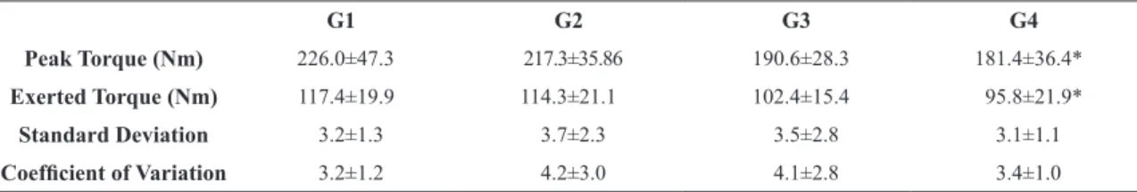
Table 2 shows the data regarding peak torque,
exerted torque (50%), the CV and the SD of the submaximal isometric peak torque curve of the knee extensors. The authors detected a signiicant difference in isometric torque between groups [F (3.56)=5.288; p=0.003; size effect=0.224; power=0.913], and the value was smaller for G4 compared to G1 (p=0.005). There was also a signiicant difference in exerted torque at 50% between the groups [F (3.56)=3.594; p=0.019; size effect=0.164; power=0.763], with G4 showing a smaller value when compared with G1 (p=0.036). There were no differences in the CV [F (3.56)=1.881; p=0.143] and SD [F (3.56)=0.274; p=0.844].
Discussion
Considering that patients with both OA and OSA
might present changes in muscle function, the results
isometric torque luctuation of the knee extensors remained unchanged. This study identiied a smaller maximum isometric peak torque and exerted torque (50%) in the group with both OA and OSA (G4);
however, there were no differences between groups
in terms of CV and SD, demonstrating that the pattern of muscle strength might be altered in early-grade OA
associated with OSA, but still without neuromuscular
control impairment during submaximal isometric torque of the knee extensors.
This study was the irst to investigate whether OSA could affect submaximal isometric torque luctuation in patients with knee OA. OSA and OA incidence
might be associated with the fact that the prevalence of both diseases increase with aging29. Additionally,
it has been demonstrated that poor sleep quality
alters pain and fatigue symptoms in OA patients,
which could change muscle strength. Recently, it
has been shown that sleep debt could induce muscle atrophy18,19 because it leads to metabolic changes in the muscle, affecting muscle recovery due to increased stimulation of protein degradation, by which protein
synthesis causes muscle atrophy. Therefore, in this study, decreased maximal isometric torque and exerted torque (50% of maximal isometric torque)
was observed in G4 (OA and OSA) when compared with the control group (G1), demonstrating that the
patients who exhibited changes in their sleep patterns associated with early-grade knee OA might present impairments in both maximal isometric torque and exerted torque.
This decrease in isometric torque in OA patients
was in agreement with other studies that have also
reported decreased isometric torque in knee OA
patients30-31; however, these studies assessed OA
patients at all stages of the disease. The present study assessed patients with early-grade knee OA, who
already presented with decreased isometric muscle
strength of the quadriceps, which has been highlighted as a risk factor for the onset of certain symptoms,
such as pain32.
The control of submaximal muscle force production
has been considered important in activities of daily
living, such as walking, transfers, and sitting and
standing10. Tracy and Enoka33 stated that an optimal submaximal muscle force appeared to be an indicator
of good neuromuscular function, leading to better
capacity to control and coordinate knee movement. Conversely, worse neuromuscular function has been
related to increases in the harmful forces applied to
the knees, which might contribute to OS development
and to its progression in the long term16.
Studies that have assessed submaximal torque luctuation in different joints have shown that the SD and CV variables are the most representative 0of the luctuation34,35. In the present study, SD and CV did not differ between groups, and these results corroborate with the indings of Hortobágyi et al.10, who assessed 20 individuals with knee OA and 20 without knee OA and concluded that, although the OA group
had worse physical function than the healthy group,
the submaximal isometric torque luctuation of the knee extensors remained unchanged in OA patients.
On average, the patients included in the present study
showed a submaximal isometric torque luctuation of the knee extensors (Table 2) similar to that reported
by Hortobágyi et al.10; however, comparisons between
the two studies should be made carefully, as they used
different assessment methods. In the present study, a target force of 50% of the maximal isometric torque was used, whereas Hortobágyi et al.10 used a target force of 50 to 100 N for all participants.
Changes in motor control, such as the submaximal force luctuation, could affect knee mechanics during gait and, thus, could be associated with the knee
adduction moment, as this moment could be an indirect predictor of the load applied to the medial
compartment of the knee during gait36. However, the study by Sørensen et al.16 investigated the relationship between quadriceps isometric force luctuation and the knee adduction moment in the frontal plane
when studying the gaits of 41 patients with different
Table 2. Peak torque during maximal isometric contraction, exerted torque, coeficient of variation and standard deviation during
submaximal isometric contractions of the knee extensors of the 4 groups.
G1 G2 G3 G4
Peak Torque (Nm) 226.0±47.3 217.3±35.86 190.6±28.3 181.4±36.4*
Exerted Torque (Nm) 117.4±19.9 114.3±21.1 102.4±15.4 95.8±21.9*
Standard Deviation 3.2±1.3 3.7±2.3 3.5±2.8 3.1±1.1
Coeficient of Variation 3.2±1.2 4.2±3.0 4.1±2.8 3.4±1.0
grades of knee OA and found no association between quadriceps isometric force luctuation and the knee adduction moment.
However, aging could modify motor control
and submaximal force because older people have demonstrated mixed results regarding the effects of age on submaximal force luctuation of the knee extensors, as observed in one study that reported a decrease in the submaximal torque luctuation33, while
others found no change10,37.
Considering that pain perception might be altered
by sleep disorders, as demonstrated in studies with animals and humans38-40 with OA, this change in pain is a factor that could lead to a change in motor
performance, increasing torque luctuation, as observed in an experimental pain model13. However, it is important to note that the knee OA patients of this study did not report pain during submaximal isometric torque luctuation assessment because the increase in
the afferent signals sent by the pain receptors could reduce proprioceptive afference and, thus, could modify motor control due to the pain41.
The study of Bandholm et al.13, which assessed
pain effects on patients with subacromial impingement
syndrome, reported a deicit in the submaximal concentric torque luctuation; however, they did not ind a difference in the submaximal isometric torque luctuation. The same behavior was observed in the study of Zanca et al.35, which reported no difference in submaximal isometric torque luctuation in patients with subacromial impingement syndrome. However,
these studies suggest that higher levels of shoulder pain
could lead to higher submaximal torque luctuation, thus corroborating our results.
The present study analyzed four groups in an attempt to elucidate the relationship between sleep disorder and OA, and in the results, it was observed that patients in the OSA and OA groups (G4) presented
losses in torque when compared with the healthy
individuals in the control group (G1) (without both
OA and OSA). However, the presence of OSA alone
(G2) or OA alone (G3) did not result in differences in functional or motor capacities when compared with the
healthy group (G1). Therefore, it was not possible to say whether OSA affected OA or vice-versa because
the G2 and G3 groups showed no differences between
them. However, in a qualitative analysis, the G3 group was found to exhibit a smaller torque than G2, indicating that OA modiied torque more than OSA.
However, when combined, OSA and OA negatively
modiied the knee’s functional capacity, as observed in G4 (with OA and with OAS).
It is worth noting that this study was the irst to
investigate motor function and control in OA and OSA; thus, the mechanisms involved have not yet
been elucidated. Longitudinal and cohort studies
should be strongly encouraged to better understand
OSA and OA.
This study has some limitations, such as the
lack of assessment of electromyographic activity in knee extensors during submaximal isometric torque luctuation assessment, which would allow better understanding the activation pattern of the extensor muscles because there were no deicits in neuromuscular function in participants with knee OA, as assessed by
electromyography42. Another limitation is the lack of follow-up using a daily sleep questionnaire, which
would provide a daily assessment of the sleep routine
of the volunteers during the study.
Based on this study’s results, it can concluded that
OA associated with OSA modiied maximal isometric torque and exerted torque; however, these conditions did not change the submaximal isometric torque luctuation of the knee extensors, indicating that, although OSA negatively affected the musculoskeletal system of patients with grade II knee OA, this syndrome did not alter the neuromuscular control in this population.
Acknowledgements
The authors thank the Fundação de Amparo à Pesquisa do Estado de São Paulo (FAPESP), Brazil
(#2010/19437-1 and #2011/06619-7), the Conselho
Nacional de Desenvolvimento Cientíico e Tecnológico
(CNPq), Brazil, the Centre for Studies in Psychobiology and Exercise [Centro de Estudos em Psicobiologia and Exercício – CEPE], the Research Incentive Fund Association [Associação Fundo de Incentivo à Pesquisa - AFIP] and the researchers Francieli Ruiz, Sebastião Gávea Junior and Fernando Vasilceac for
all of their technical support during the development
of this project.
References
1. Taylor-Gjevre RM, Nair BV, Gjevre JA. Obstructive sleep apnoea in relation to rheumatic disease. Rheumatology. 2013;52(1):15-21. http://dx.doi.org/10.1093/rheumatology/ kes210. PMid:22923759.
2009;36(9):1869-72. http://dx.doi.org/10.3899/jrheum.081335. PMid:19648298.
3. Taylor-Gjevre RM, Gjevre JA, Nair B, Skomro R, Lim HJ. Components of sleep quality and sleep fragmentation in rheumatoid arthritis and osteoarthritis. Musculoskeletal Care. 2011;9(3):152-9. PMid:21648047.
4. French HP, Cusack T, Brennan A, Caffrey A, Conroy R, Cuddy V, et al. Exercise and manual physiotherapy arthritis research trial (EMPART): a multicenter randomised controlled trial. BMC Muscul Dis. 2009;19:10-9. 5. Haq I, Murphy E, Dacre J. Osteoarthritis. Prostgrad Med.
2003;79:377-83.
6. Grotle M, Hagen KB, Natvig B, Dahl FA, Kvien TK. Prevalence and burden of osteoarthritis: results from a population survey in Norway. J Rheumatol. 2008;35(4):677-84. PMid:18278832.
7. Weng MC, Lee CL, Chen CH, Hsu JJ, Lee WD, Huang MH, et al. Effects of different stretching techniques on the outcomes of isokinetic exercise in patients with knee osteoarthritis. Kaohsiung J Med Sci. 2009;25(6):306-15. http:// dx.doi.org/10.1016/S1607-551X(09)70521-2. PMid:19560995. 8. Serrão PR, Gramani-Say K, Lessi GC, Mattiello SM. Knee
extensor torque of men with early degrees of osteoarthritis is associated with pain, stiffness and function. Rev Bras Fisioter. 2012;16(4):289-94. http://dx.doi.org/10.1590/S1413-35552012005000031. PMid:22801452.
9. Fitzgerald GK, Piva SR, Irrgang JJ. Reports of joint instability in knee osteoarthritis: its prevalence and relationship to physical function. Arthritis Rheum. 2004;51(6):941-6. http:// dx.doi.org/10.1002/art.20825. PMid:15593258.
10. Hortobágyi T, Garry J, Holbert D, Devita P. Aberrations in the control of quadriceps muscle force in patients with knee osteoarthritis. Arthritis Rheum. 2004;51(4):562-9. http://dx.doi.org/10.1002/art.20545. PMid:15334428. 11. Bennell KL, Hunt MA, Wrigley TV, Hunter DJ, McManus
FJ, Hodges PW, et al. Hip strengthening reduces symptoms but not knee load in people with medial knee osteoarthritis and varus malalignment: a randomised controlled trial. Osteoarthritis Cartilage. 2010;18(5):621-8. http://dx.doi. org/10.1016/j.joca.2010.01.010. PMid:20175973. 12. Sharma L. Proprioceptive impairment in knee osteoarthritis.
Rheum Dis Clin North Am. 1999;25(2):299-314, vi. http:// dx.doi.org/10.1016/S0889-857X(05)70069-7. PMid:10356419. 13. Bandholm T, Rasmussen L, Aagaard P, Diederichsen L,
Jensen BR. Effects of experimental muscle pain on shoulder-abduction force steadiness and muscle activity in healthy subjects. Eur J Appl Physiol. 2008;102(6):643-50. http:// dx.doi.org/10.1007/s00421-007-0642-1. PMid:18066578. 14. Carville SF, Perry MC, Rutherford OM, Smith IC, Newham
DJ. Steadiness of quadriceps contractions in young and older adults with and without a history of falling. Eur J Appl Physiol. 2007;100(5):527-33. http://dx.doi.org/10.1007/ s00421-006-0245-2. PMid:16983499.
15. Jackson BD, Wluka AE, Teichtahl AJ, Morris ME, Cicuttini FM. Reviewing knee osteoarthritis--a biomechanical perspective. J Sci Med Sport. 2004;7(3):347-57. http://dx.doi. org/10.1016/S1440-2440(04)80030-6. PMid:15518300. 16. Sørensen TJ, Langberg H, Aaboe J, Bandholm T, Bliddal
H, Henriksen M. The association between submaximal
quadriceps force steadiness and the knee adduction moment during walking in patients with knee osteoarthritis. J Orthop Sports Phys Ther. 2011;41(8):592-9. http://dx.doi. org/10.2519/jospt.2011.3481. PMid:21765221.
17. Lui MM, Lam JC, Mak HK, Xu A, Ooi C, Lam DC, et al. C-reactive protein is associated with obstructive sleep apnea independent of visceral obesity. Chest. 2009;135(4):950-6. http://dx.doi.org/10.1378/chest.08-1798. PMid:19225064. 18. Dattilo M, Antunes HK, Medeiros A, Mônico Neto M, Souza
HS, Tufik S, et al. Sleep and muscle recovery: endocrinological and molecular basis for a new and promising hypothesis. Med Hypotheses. 2011;77(2):220-2. http://dx.doi.org/10.1016/j. mehy.2011.04.017. PMid:21550729.
19. Dattilo M, Antunes HK, Medeiros A, Mônico-Neto M, Souza HS, Lee KS, et al. Paradoxical sleep deprivation induces muscle atrophy. Muscle Nerve. 2012;45(3):431-3. http:// dx.doi.org/10.1002/mus.22322. PMid:22334180.
20. American College of Rheumatology Subcommittee on Osteoarthritis Guidelines. Recommendations for the medical management of osteoarthritis of the hip and knee: 2000 update. Arthritis Rheum. 2000;43(9):1905-15. http:// dx.doi.org/10.1002/1529-0131(200009)43:9<1905::AID-ANR1>3.0.CO;2-P. PMid:11014340.
21. Kellgren JH, Lawrence JS. Radiological assessment of osteo-arthrosis. Ann Rheum Dis. 1957;16(4):494-502. http:// dx.doi.org/10.1136/ard.16.4.494. PMid:13498604. 22. Felson DT, Niu J, Guermazi A, Sack B, Aliabadi P. Defining
radiographic incidence and progression of knee osteoarthritis: suggested modifications of the Kellgren and Lawrence scale. Ann Rheum Dis. 2011;70(11):1884-6. http://dx.doi. org/10.1136/ard.2011.155119. PMid:21908453.
23. American Academy of Sleep Medicine - AASM. International classification of sleep disorders. Diagnostic and coding manual (ICSD-2). 2nd ed. Westchester: AASM; 2005.
24. Iber C, Ancoli-Israel S, Chesson AL Jr, Quan SF. American Academy of Sleep Medicine. The AASM manual for the scoring of sleep and associated events: rules, terminology and technical specifications. Westchester: American Academy of Sleep Medicine; 2007.
25. Hortobágyi T, Tunnel D, Moody J, Beam S, DeVita P. Low- or high-intensity strength training partially restores impaired quadriceps force accuracy and steadiness in aged adults. J Gerontol A Biol Sci Med Sci. 2001;56(1):B38-47. http:// dx.doi.org/10.1093/gerona/56.1.B38. PMid:11193224. 26. Pua YH, Clark RA, Bryant AL. Physical function in hip
osteoarthritis: relationship to isometric knee extensor steadiness. Arch Phys Med Rehabil. 2010;91(7):1110-6. http:// dx.doi.org/10.1016/j.apmr.2010.04.001. PMid:20599051. 27. Lavender AP, Nosaka K. Fluctuations of isometric force after
eccentric exercise of the elbow flexors of young, middle-aged, and old men. Eur J Appl Physiol. 2007;100(2):161-7. http://dx.doi.org/10.1007/s00421-007-0418-7. PMid:17310389. 28. Hinman RS, Bennell KL, Metcalf BR, Crossley KM. Delayed
onset of quadriceps activity and altered knee joint kinematics during stair stepping in individuals with knee osteoarthritis. Arch Phys Med Rehabil. 2002;83(8):1080-6. http://dx.doi. org/10.1053/apmr.2002.33068. PMid:12161828.
30. van der Esch M, Steultjens M, Harlaar J, Knol D, Lems W, Dekker J. Joint proprioception, muscle strength, and functional ability in patients with osteoarthritis of the knee. Arthritis Rheum. 2007;57(5):787-93. http://dx.doi. org/10.1002/art.22779. PMid:17530678.
31. Palmieri-Smith RM, Thomas AC, Karvonen-Gutierrez C, Sowers MF. Isometric quadriceps strength in women with mild, moderate, and severe knee osteoarthritis. Am J Phys Med Rehabil. 2010;89(7):541-8. http://dx.doi.org/10.1097/ PHM.0b013e3181ddd5c3. PMid:20463561.
32. Segal NA, Torner JC, Felson D, Niu J, Sharma L, Lewis CE, et al. Effect of thigh strength on incident radiographic and symptomatic knee osteoarthritis in a longitudinal cohort. Arthritis Rheum. 2009;61(9):1210-7. http://dx.doi. org/10.1002/art.24541. PMid:19714608.
33. Tracy BL, Enoka RM. Older adults are less steady during submaximal isometric contractions with the knee extensor muscles. J Appl Physiol (1985). 2002;92(3):1004-12. http:// dx.doi.org/10.1152/japplphysiol.00954.2001. PMid:11842033. 34. Camargo PR, Ávila MA, de Oliveira AB, Asso NA, Benze
BG, Salvini TF. Shoulder abduction torque steadiness is preserved in subacromial impingement syndrome. Eur J Appl Physiol. 2009;106(3):381-7. http://dx.doi.org/10.1007/ s00421-009-1030-9. PMid:19294409.
35. Zanca GG, Saccol MF, Oliveira AB, Mattiello SM. Shoulder internal and external rotations torque steadiness in overhead athletes with and without impingement symptoms. J Sci Med Sport. 2013;16(5):433-7. http://dx.doi.org/10.1016/j. jsams.2012.09.004. PMid:23088897.
36. Creaby MW, Hunt MA, Hinman RS, Bennell KL. Sagittal plane joint loading is related to knee flexion in osteoarthritic gait. Clin Biomech. 2013;28(8):916-20. http://dx.doi. org/10.1016/j.clinbiomech.2013.07.013. PMid:23938182. 37. Øiestad BE, Holm I, Gunderson R, Myklebust G, Risberg
MA. Quadriceps muscle weakness after anterior cruciate
ligament reconstruction: a risk factor for knee osteoarthritis? Arthritis Care Res. 2010;62(12):1706-14. http://dx.doi. org/10.1002/acr.20299. PMid:20662041.
38. Silva A, Andersen ML, Tufik S. Sleep pattern in an experimental model of osteoarthritis. Pain. 2008;140(3):446-55. http:// dx.doi.org/10.1016/j.pain.2008.09.025. PMid:18950940. 39. Silva A, Araujo P, Zager A, Tufik S, Andersen ML. Sex
differences in sleep pattern of rats in an experimental model of osteoarthritis. Eur J Pain. 2011;15(6):545-53. http://dx.doi. org/10.1016/j.ejpain.2010.10.009. PMid:21273100. 40. Woolhead G, Gooberman-Hill R, Dieppe P, Hawker G.
Night pain in hip and knee osteoarthritis: a focus group study. Arthritis Care Res. 2010;62(7):944-9. http://dx.doi. org/10.1002/acr.20164. PMid:20191575.
41. Myers JB, Wassinger CA, Lephart SM. Sensorimotor contribution to shoulder stability: effect of injury and rehabilitation. Man Ther. 2006;11(3):197-201. http://dx.doi. org/10.1016/j.math.2006.04.002. PMid:16777465. 42. Hurley MV, Scott DL, Rees J, Newham DJ. Sensorimotor
changes and functional performance in patients with knee osteoarthritis. Ann Rheum Dis. 1997;56(11):641-8. http:// dx.doi.org/10.1136/ard.56.11.641. PMid:9462165.
Correspondence Stela Márcia Mattiello