Evaluation of chemical and/or mechanical treatments of
the smear layer as revealed by scanning electron microscopy –
a blind comparative study
Avaliação dos tratamentos químicos e/ou mecânicos da camada
de esfregaço conforme observado por microscopia eletrônica de
varredura – estudo cego comparativo
Maria Aparecida Alves de Cerqueira LUZ* Narciso GARONE NETTO**
Victor E. ARANA-CHAVEZ*** Maria Angela Pitta SOBRAL* Júlio da Motta SINGER****
LUZ, M. A. A. de C.; GARONE NETTO, N.; ARANA-CHAVEZ, V. E.; SOBRAL, M. A. P.; SINGER, J. da M. Evaluation of chemical and/or mechanical treatments of the smear layer as revealed by scanning electron microscopy – a blind comparative study.Pesq Odont Bras, v. 14, n. 2, p. 101-106, abr./jun. 2000.
A blind comparative study of chemical and/or mechanical treatments of the smear layer, according to scanning elec-tron microscopy images, was carried out. The effect of the treatments was analyzed on the smear layer of mesio-occlusodistal cavity walls preparedin vitroin human third molars. The agents used were air/water spray, 37% phosphoric acid, 5% tannic acid, biologic detergent, 0.5% sodium hypochlorite, and enamel hatchet alone or in associ-ation with the previous agents. Electron micrographs were evaluated by three professionals according to the degree of visualization of underlying dentin or enamel. Phosphoric acid received the highest scores due to the complete removal of the smear layer. However, statistical analyses revealed diverse performances of non or slightly demineralizing agents, according to the cavity walls in dentin, while there was equivalent effect on the enamel of gingival walls. UNITERMS: Smear layer; Dentin; Scanning electron microscopy.
INTRODUCTION
The treatment of the smear layer is an impor-tant factor for the good performance of restora-tions. More resistant unions are obtained when adhesive materials are used, and gaps are mini-mized when non adhesive materials are used10,16.
Non or slightly demineralizing treatments are im-portant for the use of adhesive cements, because of their union with calcium and phosphate ions from the cavity walls24. Non or slightly demineralizing
treatments may be also important for the use of resin adhesives that are applied without removal of the smear layer.
Studies have demonstrated that the formation of the hybrid layer by penetration of monomers into the dentin is more important than chemical adhesion or than the formation of resin tags within
dentinal tubules12. However, excessive
demineral-ization of dentin gives rise to collagen denaturation, and resins may not penetrate into the matrix as deeply as acidic conditioners do. Thus, a weak zone may be created, with unpro-tected collagen, causing the failure of adhesion13,15.
These facts led to the development of adhesives with acidic monomers, i. e. the smear layer may be slightly demineralized due to the association of ac-ids with hydrophilic/hydrophobic monomers. This procedure allows a concomitant diffusion through the underlying dentin, creating a hybrid layer of unaltered collagen involved in resin12,13,22,23.
Scanning electron microscopy (SEM) is a reli-able method to evaluate dentin and enamel in dis-ease or under procedure conditions4,11. SEM is also
adequate to evaluate the smear layer, although sometimes it is difficult to quantify it. Thus, the
use of a blind method is valid to evaluate the effect of different treatments on it.
The purpose of this study was to analyze the ef-fect of chemical, mechanical and both treatments of the smear layer, as revealed by SEM, in a blind study, in which the electron micrographs were evaluated by three professionals.
MATERIALS AND METHODS
The study was conducted with thirty fresh, non-carious, unerupted human third molars, ob-tained from patients between 16 and 27 years of age (mean 21 years). All patients were fully in-formed about the procedures of this study. Roots were removed, and mesio-occlusodistal cavity preparations executed with new cylindrical dia-mond burs under water cooling.
Immediately after cavity preparation, one of the following treatments was carried out for each three teeth, being applied on every cavity wall: (1) air/water spray; (2) 37% phosphoric acid, ap-plied with cotton pellets during 15 s; (3) 5% tannic acid, applied with cotton pellets during 15 s; (4) 0.2% lauril sodium sulfate biologic detergent (Tergensol, Inodon), applied with cotton pellets with rubbing during 15 s; (5) 0.5% sodium hypochlorite, with cotton pellets, applied with rub-bing during 15 s; (6) enamel hatchet, applied dur-ing 30 s; (7) enamel hatchet with 37% phosphoric acid, applied during 45 s (30 s with enamel hatchet alone, plus 15 s with concomitant application); (8) enamel hatchet with 5% tannic acid, applied during 45 s; (9) enamel hatchet with biologic deter-gent, applied during 45 s; and (10) enamel hatchet with 0.5% sodium hypochlorite, applied during 45 s. Air/water spray was applied for 5 seconds be-fore and after all treatments.
The teeth were longitudinally half-sectioned in a mesiodistal orientation with a chisel. Then, one of the fragments was processed for scanning elec-tron microscopy. Specimens were air-dried and mounted on aluminum stubs. After sputtering with a 40 nm layer of gold in a Balzers SCD 050 ap-paratus, the treated cavity wall surfaces were ex-amined in a Jeol 6100 scanning electron micro-scope operating at 10-15 kV.
The electron micrographs were analyzed by three experienced examiners through a blind tech-nique. They graded their observations using a score ranging from 0 to 3, according to the degree of removal of the smear layer and consequent visu-alization of the underlying dentin or enamel6. Five
electron micrographs of each specimen were ana-lyzed. Two magnification levels were used for both
pulpal and lateral walls (400 X and 2000 X), and one magnification level, for gingival walls (400 X).
RESULTS
Cavity walls treated with air/water spray exhib-ited variable amounts of smear layer covering the dental walls, with superficial particles not firmly attached to the sound layer. Circular marks pro-duced by the diamond particles of the instrument were evident on the surface of pulpal walls. In gen-eral, the depth of the marks roughly indicated the amount of smear layer.
Phosphoric acid treatment showed a complete removal of the smear layer with evidence of the tu-bular structure of the dentin. Thus, the tubules appeared opened and enlarged, revealing a thin or absent peritubular dentin. In these specimens, the intertubular dentin exhibited a clean and smooth aspect. The demineralization of peritubular dentin caused a funneled-like tubule aspect, showing a decreasing effect of the acid according to the in-crease in depth of dentin. On enamel of gingival walls, the removal of the smear layer showed the enamel prisms in transverse, oblique or longitudi-nal sections, depending on the wall level.
Non or slightly demineralizing treatments pro-duced removal of the smear layer in variable amounts. The mean scores related to the perfor-mance of the treatments, pertaining to pulpal, lat-eral, and gingival walls are shown in Tables 1, 2, and 3.
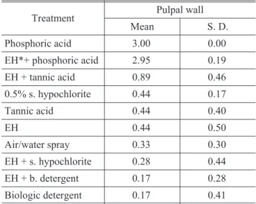
TABLE 1 -Mean scores and standard deviation for effici-ency degree of smear layer removal on pulpal walls.
Treatment Pulpal wall
Mean S. D.
Phosphoric acid 3.00 0.00
EH*+ phosphoric acid 2.95 0.19
EH + tannic acid 0.89 0.46
0.5% s. hypochlorite 0.44 0.17
Tannic acid 0.44 0.40
EH 0.44 0.50
Air/water spray 0.33 0.30
EH + s. hypochlorite 0.28 0.44
EH + b. detergent 0.17 0.28
Biologic detergent 0.17 0.41
Analysis of variance and Kruskall-Wallis analysis7of the results revealed different effects of
non or slightly demineralizing treatments accord-ing to the cavity walls in dentin (pulpal walls = 0.0584; lateral walls = 0.0073), and equiva-lent effect on the enamel of gingival walls (p = 0.8467). Student-Newman-Keuls technique14
revealed that the treatment with enamel hatchet and 5% tannic acid was more efficient than both biologic detergent treatments on pulpal walls, and
that the biologic detergent treatment was more ef-ficient than the one with the enamel hatchet and 0.5% sodium hyphoclorite or 5% tannic acid, on lateral walls (Figures 1, 2 and 3).
DISCUSSION
This study showed that all non or slightly de-mineralizing treatments of cavity walls produced some removal of the smear layer, although differ-ences between these treatments were quite subtle.
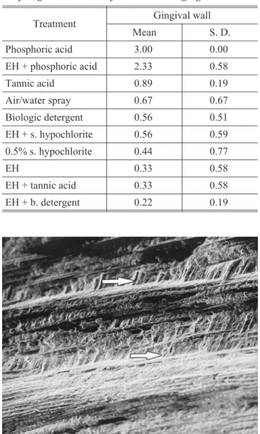
TABLE 3 -Mean scores and standard deviation for effici-ency degree of smear layer removal on gingival walls.
Treatment Gingival wall
Mean S. D.
Phosphoric acid 3.00 0.00
EH + phosphoric acid 2.33 0.58
Tannic acid 0.89 0.19
Air/water spray 0.67 0.67
Biologic detergent 0.56 0.51
EH + s. hypochlorite 0.56 0.59
0.5% s. hypochlorite 0.44 0.77
EH 0.33 0.58
EH + tannic acid 0.33 0.58
EH + b. detergent 0.22 0.19 TABLE 2 -Mean scores and standard deviation for
effici-ency degree of smear layer removal on lateral walls.
Treatment Lateral wall
Mean S. D.
Phosphoric acid 3.00 0.00
EH + phosphoric acid 2.66 0.35
Biologic detergent 0.78 0.17
Air/water spray 0.56 0.54
Tannic acid 0.56 0.46
0.5% s. hypochlorite 0.28 0.39
EH + b. detergent 0.28 0.33
EH 0.22 0.34
EH + s. hypochlorite 0.11 0.27
EH + tannic acid 0.00 0.00
FIGURE 1 -Enamel hatchet associated with 5% tannic acid (mean score 0.89 ± 0.19). Electron micrograph sho-wing the smear layer on a pulpal wall with a smooth ap-pearance; arrows indicate some evidences of transver-sally cut dentinal tubules (2000 X).
As expected, the performance of phosphoric acid exceeded that of the others, completely removing the smear layer. The application methods were carried out based on the literature: cotton pellets are the instruments more frequently employed to take the substances into the cavities; acids, in gen-eral, were applied passively without rubbing, and organic solvents or detergents, with rubbing2,3,9,21.
Moreover, the study of the treatments on MOD cavities allowed to observe their effects on different cavity walls, which would not be possible if just one surface of dental tissue had been employed. The enamel hatchet was used alone because it is difficult to apply this instrument with uniformity on all of the walls during a shorter period of time. Meanwhile, in association with the chemical sub-stances, the period of application was the same (15 s). We attempted to use clinical situations adapted to experimental conditions. The statistical analyses compared each one of the treatments to each other.
The treatment of cavity walls with phosphoric acid has been applied in different concentrations for the removal of the smear layer, specially in adhesive restorative procedures. As the hazard-ous effects of phosphoric acid on dentin were demonstrated both in dentinal structure and in dentinal permeability, its use in small concentra-tion for a short time has been suggested to mini-mize these effects and achieve the adhesion reactions1,5,8,9,18,20.
This study demonstrated diverse performances of non or slightly demineralizing agents, according to the cavity walls in dentin. We have to consider the possible effect of mechanical procedures changing the initial result of chemical treatments. Thus, the act of scrubing with cotton pellets, or the use of enamel hatchet may be considered respon-sible for the final results on cavity walls. The sim-ple friction of cotton pellets with water may result in some removal of the smear layer5,9. When
phos-phoric acid was associated with enamel hatchet, the smear layer was removed but some occluded tubules were detected. This fact may be explained by the greater compaction of the smear layer parti-cles on the aperture of the tubules, which occurs during manual instrumentation17.
The effect of a given treatment was diverse ac-cording to the kind of cavity wall. When tannic acid was applied alone, similar results on lateral and pulpal dentinal walls were observed. However, when tannic acid was associated with enamel hatchet, better results were observed on pulpal walls. The use of the enamel hatchet may be easier on pulpal walls and it may increase the effect of the chemical treatment. On the other hand, the use of biologic detergent applied with cotton pellets with rubbing produced better results on the dentin of lateral walls. The rubbing of cotton pellets might have been more effective on lateral walls.
We consider that treatment of the smear layer of enamel margins on gingival walls is important to prevent microleakage. Actually, we could not ob-serve a uniform effect produced by any of the treat-ments on the enamel of gingival walls. These find-ings can justify the greater standard deviation of the statistical analysis, except for the tannic acid and biologic detergent associated with enamel hatchet, which produced the highest and the smallest mean score value, respectively. Exam-ining the removal of the smear layer on the enamel surfaces of gingival walls, tannic acid had the higher mean score value among the treatments. This may be explained by its acidic nature21which
imputes to it a slight demineralizing effect. In addi-tion, the composition of the enamel smear layer is different from that of dentin, as the former tissue has greater mineral concentration.
Air/water spray could remove some smear par-ticles, as shown by the use of the high speed handpiece with water cooling during cavity preparation19,25. Air/water spray treatment came in
second place among the treatments on the lateral and gingival walls, although there was no statisti-FIGURE 3 -5% tannic acid (mean score 0.89 ± 0.17).
cally significant difference between this treatment and the others.
CONCLUSIONS
1. Phosphoric acid (37%) received the highest score due to complete removal of the smear layer; 2. non or slightly demineralizing agents presented
diverse performances according to the cavity walls in dentin;
3. enamel hatchet with 5% tannic acid was more efficient on pulpal walls, while the biologic de-tergent was more efficient on lateral walls; 4. non or slightly demineralizing agents presented
equivalent effect on the enamel of gingival walls.
LUZ, M. A. A. de C.; GARONE NETTO, N.; ARANA-CHAVEZ, V. E.; SOBRAL, M. A. P.; SINGER, J. da M. Avaliação dos tratamentos químicos e/ou mecânicos da camada de esfregaço conforme observado por microscopia eletrônica de varredura – estudo cego comparativo.Pesq Odont Bras, v. 14, n. 2, p. 101-106, abr./jun. 2000.
Um estudo comparativo cego de tratamentos químicos e/ou mecânicos da camada de esfregaço, de acordo com ima-gens da microscopia eletrônica de varredura, foi realizado. Analisou-se o efeito dos tratamentos sobre a camada de es-fregaço de paredes de cavidades mésio-ocluso-distais, preparadasin vitro, em terceiros molares humanos. Os agentes empregados foram spray ar/água, ácido fosfórico a 37%, ácido tânico a 5%, detergente biológico, hipoclorito de sódio a 0,5% e machado para esmalte isoladamente ou associado a estas substâncias. Eletromicrografias foram avaliadas por três profissionais de acordo com o grau de visualização da dentina ou esmalte subjacentes. O ácido fosfórico recebeu as maiores graduações devido à remoção completa da camada de esfregaço. Análises estatísticas revelaram perfor-mances diversas entre tratamentos não/ou levemente desmineralizantes de acordo com as paredes cavitárias, em dentina e efeitos equivalentes entre si, em paredes de esmalte.
UNITERMOS: Camada de esfregaço; Dentina; Microscopia eletrônica de varredura.
BIBLIOGRAPHIC REFERENCES
1. ARAÚJO, M. A. J.; RODE, S. M.; VILLELA, L. C. et al.
Avaliação qualitativa do efeito de agentes de limpeza na camada de lama dentinária: estudo ultra-estrutural em microscopia eletrônica de varredura.Rev Odontol Univ São Paulo, v. 12, n. 2, abr/jun, 1998.
2. BITTER, N. C. The effect of 25% tannic acid on prepared dentin: a scanning electron microscope methylene blue dye study.J Prosthet Dent, v. 64, n. 1, p. 12-16, July 1990.
3. BITTER, N. C. Tannic acid for smear removal: pilot study with scanning electron microscope.J Prosthet Dent, v. 61, n. 4, p. 503-507, Apr. 1989.
4. BRÄNNSTRÖM, M.Dentin and pulp in restorative den-tistry. London : Wolfe, 1982. 125 p.
5. BRÄNNSTRÖM, M.; JOHNSON, G. Effect of various condi-tioners and cleaning agents on prepared dentin surfa-ces: a scanning electron microscopic investigation. J Prosthet Dent, v. 31, n. 4, p. 422-430, Apr. 1974. 6. BRÄNNSTRÖM, M.; GLANTZ, P. O.; NORDENVALL, K. J.
The effect of some cleaning solutions on the morphology of dentin prepared in different ways: anin vivostudy.J Dent Child, v. 46, n. 3, p. 19-23, July/Aug. 1979. 7. BROWN Jr, B. W.; HOLLANDER, M.Statistics: a
biomedi-cal introduction. New York : John Wiley & Sons, 1977. 456 p.
8. CHARLTON, D. G. Dentin bonding: past and present. Ge-neral Dent, v. 44, n. 6, p. 498-507, Nov./Dec. 1996. 9. FRANCISCHONE, C. E.; CANDIDO, M. S. M.; BERBERT; A.
et al. Efeito de alguns agentes de limpeza sobre a dentina, observado através da microscopia eletrônica de
varredura. Estomatol Cult (Bauru), v. 14, n. 1/2, p. 49-56, jan./dez. 1984.
10. JODAIKIN, A.; AUSTIN, J.C. The effects of cavity smear la-yer removal on experimental marginal leakage around amalgam restorations. J Dent Res, v. 60, n. 11, p. 1861-1866, Nov. 1981.
11. KAQUELER, J. C.; DECOMBAS, M.; LE MAY, O. Structu-res bucco-dentaiStructu-res en microscopie électronique a balayage. Paris : Masson, 1989. 204 p.
12. NAKABAYASHI, N. Bonding of restorative materials to den-tine: the present status in Japan.Int Dent J, v. 35, n. 2, p. 145-154, 1985.
13. NAKABAYASHI, N.; SAIMI, Y. Bonding to intact dentin.J Dent Res, v. 75, n. 9, p. 1706-1715, Sept. 1996. 14. NETER, J.; WASSERMAN, W.; KUTNER, M. H.Applied
li-near statistical methods. 3. ed. Homewwood: Richard D. Irwin, 1990. 1127 p.
15. PASHLEY, D. H.; CARVALHO, R. M. Dentine permeability and dentine adhesion.J Dent, v. 25, n. 5, p. 355-372, Sept. 1997.
16. PASHLEY, D. H.; DEPEW, D. D. Effects of the smear layer, copalite and oxalate on microleakage. Operat Dent, v. 11, n. 3, p. 95-102, 1986.
17. PASHLEY, D. H.; LEIBACH, J. G.; HORNER, J. A. The ef-fects of burnishing NaF/Kaolin/Glycerin paste on den-tin permeability.J Periodontol, v. 58, n. 1, p. 19-23, Jan. 1987.
18. PERDIGÃO, J.; LAMBRECHTS, P.; VAN MEERBEEK, B.; VANHERLE, G.; LOPES, A. L. B. Adesão aos tecidos dentários: o estado da arte II.Rev Port Estomatol Cir Maxilofac, v. 35, n. 4, p. 141-150, 1994.
microscope study.Brit Dent J, v. 150, n. 9, p. 243-247, May 1981.
20. SWIFT, E. J.; PERDIGÃO, J.; HEYMANN, H. O. Bonding to enamel and dentin: a brief history and state of the art, 1995.Quintessence Int, v. 26, n. 2, p. 95-110, Feb. 1995.
21. TAKAHASHI, H.; OKAMOTO, Y.; FUJINAKA, S.et al.Pilot study of exposure of the smear layer to tannic acid solu-tions. J Prosth Dent, v. 70, n. 3, p. 261-263, Sept. 1993.
22. VAN MEERBEEK, B.; PERDIGÃO, J.; LAMBRECHTS, P.et
al.The clinical performance of adhesives.J Dent, v. 26, n. 1, p. 1-20, Jan. 1998.
23. VAN MEERBEEK, B.; DHEM, A.; GORET-NICAISE, M.et al.Comparative SEM and TEM examination of
ultras-tructure of resin-dentin interdifusion zone.J Dent Res, v. 72, n. 2, p. 495-501, Feb. 1993.
24. WHITE, G. J.; BEECH, D. R.; TYAS, M. J. Dentin smear la-yer: an asset or a liability for bonding?Dent Mater, v. 5, n. 6, p. 379-383, Nov. 1989.
25. ZAIMOGLU, A.; AYDIN, K. A. An evaluation of smear layer with various desensitizing agents after tooth preparati-on.J Prosth Dent, v. 68, n. 3, p. 450-457, Sept. 1990.
Recebido para publicação em 12/01/00 Enviado para reformulação em 17/02/00 Aceito para publicação em 24/04/00
Errata
Effect of professional dental prophylaxis with sodium bicarbonate
jet on the cariogenic microbiota
Efeito da profilaxia profissional com jato de bicarbonato de sódio
sobre a microbiota cariogênica
Célia Regina Moreira LANZA José Eduardo de Oliveira LIMA Sergio Aparecido TORRES
Maria Aparecida de Andrade Moreira MACHADO