Progression of Lung Carcinoma
Jing Jin1, Binqing Fu1,2, Xinyu Mei3, Ting Yue1, Rui Sun1,2, Zhigang Tian1,2*, Haiming Wei1,2*
1Institute of Immunology, School of Life Sciences, University of Science and Technology of China, Hefei, Anhui, China,2Hefei National Laboratory for Physical Sciences at Microscale, University of Science and Technology of China, Hefei, Anhui, China,3Anhui Provincial Hospital, Hefei, Anhui, China
Abstract
NK cells are a major component of the antitumour immune response that limits tumour progression. However, it has been reported that tumour-infiltrating NK (TINK) cells from patients with non-small-cell lung carcinoma (NSCLC) exhibit profound defects in degranulation and IFN-cproduction. In support of this notion, we report a novel mechanism associated with tumour escape from NK cell-mediated antitumour immunity in lung carcinoma. In this study, we investigated the phenotypic profile of TINK cells based on the expression of the NK-cell maturation markers CD11b and CD27. Interestingly, we found a substantial CD11b2CD272(DN) NK-cell population harboured within the tumour tissues. The presence of this
CD11b2CD272 NK subset indicated that the TINK cells were of an immature and inactive phenotype. Remarkably, we
determined that the presence of DN NK cells had an impact on the clinical outcomes of patients with NSCLC, as the frequency of tumour-infiltrating DN NK cells was positively correlated with the tumour stage and tumour size. We further used a murine Lewis lung cancer (LLC) model to confirm the correlation between the frequency of tumour-infiltrating DN NK cells and the progression of lung carcinoma. Together, our findings demonstrate that the tumour microenvironment may render TINK cells less tumouricidal and thereby contribute to cancer progression.
Citation:Jin J, Fu B, Mei X, Yue T, Sun R, et al. (2013) CD11b2CD272NK Cells Are Associated with the Progression of Lung Carcinoma. PLoS ONE 8(4): e61024. doi:10.1371/journal.pone.0061024
Editor:Xi Yang, University of Manitoba, Canada
ReceivedSeptember 19, 2012;AcceptedMarch 5, 2013;PublishedApril 2, 2013
Copyright:ß2013 Jin et al. This is an open-access article distributed under the terms of the Creative Commons Attribution License, which permits unrestricted use, distribution, and reproduction in any medium, provided the original author and source are credited.
Funding:This work was supported by the Natural Science Foundation of China (31021061, 30730084) and Ministry of Science and Technology of China (973 Basic Science Project 2009CB522403, 2012CB519004). The funders had no role in study design, data collection and analysis, decision to publish, or preparation of the manuscript.
Competing Interests:The authors have declared that no competing interests exist.
* E-mail: [email protected] (HW); [email protected] (ZT)
Introduction
Lung cancer is the leading cause of cancer-related deaths worldwide [1]. Innovative and efficacious therapeutic strategies, including immunotherapies, are therefore urgently needed. A thorough understanding of anti-tumour immune responses and especially of the role of NK cells in this process is necessary [2–6]. NK cells are innate lymphocytes that have a natural ability to recognise and kill aberrant cells, including cancer cells [7–10]. However, elegant studies have provided evidence that NSCLC-infiltrating NK cells exhibit profound defects in degranulation and IFN-c production [11]. These defects are associated with coordinated changes in the NK-cell receptor repertoire, suggesting a local tumour-induced impairment of NK-cell function. These findings suggest that it is not the quantity but the quality of intratumoural NK cells that accounts for their dysfunction in NSCLC. Notably, that study also revealed that intratumoural NK cells expressed dramatically lower levels of killer-cell immuno-globulin-like receptor (KIR) than did peripheral blood NK (pNK) cells from the same patients. Similarly, another study demonstrat-ed the loss of KIR expression on intratumoural NK cells when compared with NK cells from the peritumoural lung tissue [12]. Interestingly, the lack of KIR expression on tumour-infiltrating NK (TINK) cells is also observed in human breast tumours [13], whereas KIR is present on NK cells infiltrating healthy mammary tissue (Mt-NK cells). This finding suggests that TINK cells, like non-educated cells, have no cytotoxic capacity [14,15]. This study
also indicated that the phenotype of TINK cells was characteristic of immature and nonfunctional NK cells [13]. In addition, the terminal differentiation of NK cells is characterised by the appearance of KIRs and decreased NKG2A expression [16,17]. Accordingly, the study describing the loss of KIR expression on TINK cells from patients with breast cancer suggests that a strong inhibitory environment, such as the tumour microenvironment, can reorient or reverse the transcriptional program of NK cell maturation toward a nonreactive self-tolerant profile [13]. In support of this hypothesis, several recent studies have shown that the NK-cell developmental program is not entirely fixed and that mature NK cells can be re-educated by their environment [18– 20]. Hence, it is likely that the tumour microenvironment may have a negative impact on NK-cell maturation.
28]. Despite the knowledge of these maturation markers, the functional differences between these populations remain poorly understood. Thus, much effort has recently been devoted toward subdividing human NK cells into functionally distinct subpopula-tions. Recent studies have shown that CD27, a member of the tumour necrosis factor receptor superfamily, is an important marker that can be used to define NK-cell subsets [29,30]. The
surface density of CD27 and CD11b can be used to divide murine NK cells into 4 subsets that define different levels of maturation [31,32]. Based on the findings in previous studies, we hypothesised that the expression of CD11b and CD27 may similarly define distinct populations of human NK cells [33]. Despite advances in understanding NK-cell development, the specific NK-cell subsets harboured within tumours and the mechanisms that might
account for the NK-cell infiltration of tumours have not yet been defined.
A recent study revealed that mature CD27highNK cells are the predominant NK-cell subpopulation that accumulates within the tumour microenvironment. However, this work was performed in an early-developing murine MCA205 fibrosarcoma tumour model [34]. The impact of mouse studies on the clinical monitoring of human NK-cell function and on the design of improved therapeutics for cancer has been limited. Hence, we aimed to identify whether the same NK-cell subset was present in the human tumour microenvironment and to precisely characterise NK-cell differentiation within human tumours. To this end, we investigated the expression of CD11b and CD27 on NK cells isolated from primary human NSCLC tumour specimens and peripheral blood from the same patients as well as peripheral blood from healthy control subjects. In agreement with the findings of previous studies, nearly all of the pNK cells from NSCLC patients and healthy control subjects were CD11b+CD272 (CD11b+SP) NK cells. However, TINK cells
isolated from patients with NSCLC contained CD11b2CD272 (DN) NK cells in addition to CD11b+SP NK cells. Furthermore,
we demonstrated that the presence of the CD11b2CD272 population was associated with clinical outcomes in patients with NSCLC, as the frequency of CD11b2CD272 cells within the tumour positively correlated with the tumour stage and tumour size. We further verified the correlation between the frequency of CD11b2CD272TINK cells and tumour progression in a murine Lewis lung cancer (LLC) model. Taken together, our findings provide direct evidence that CD11b2CD272 NK cells, which have been defined as immature NK cells in previous studies, accumulate within tumours, suggesting that the tumour microen-vironment may render TINK cells less tumouricidal and thereby contribute to cancer progression. Importantly, our results reveal a novel mechanism underlying NK-cell dysfunction during antitu-mour immune responses.
Results
DN NK cells are present within the TINK-cell population To determine the maturation status of TINK cells, we compared the expression of CD11b and CD27 on TINK cells with that on pNK cells from autologous patients and healthy control subjects. Approximately 10% of TINK cells were CD27+,
whereas less than 5% of pNK cells from autologous patients and healthy control subjects were CD27+. Interestingly, TINK cells
exhibited a significant reduction in CD11b expression when compared with the other two types of NK cells (Fig. 1A). Furthermore, we detected a DN (CD11b2CD272) NK-cell subset that accounted for approximately 35% of the TINK population (Fig. 1B). By contrast, the proportion of CD11b+SP
(CD11b+CD272) NK cells was lower within the TINK population than within the pNK population (Fig. 1C). These data demon-strate, for the first time, that a large population of DN NK cells exists within the TINK-cell population in human tumours, suggesting an immature phenotype of TINK cells.
TINK cells display an immature and inactive phenotype To determine whether TINK cells have an immature pheno-type, we investigated the expression of NK-cell receptors associated with maturity on CD56+CD32 TINK cells and pNK cells from autologous patients and healthy control subjects. Downregulation of CD57, which is expressed on highly mature NK cells [35], was observed on TINK cells, whereas no change in CD57 expression was observed on pNK cells from autologous
patients and healthy control subjects (Fig. 2A and B). Conversely, CD127 and CD117, receptors that are virtually absent on traditional mature NK cells [21], were clearly expressed on TINK cells (Fig. 2A and B). Moreover, NKG2A, an inhibitory receptor reported to be highly expressed by immature NK cells, was slightly upregulated on TINK cells when compared with the other two types of NK cells (Fig. 2A and B). Collectively, these results demonstrate that TINK cells display an immature phenotype.
We next examined the expression of NK-cell receptors that have been reported to be involved in NK-cell activation [36,37]. Previous reports have demonstrated that NK cells infiltrating human non-small-cell lung cancer are enriched in CD162 cells[11,12]. Similarly, we found that the expression of CD16 (Fccreceptor III) was dramatically reduced on TINK cells from NSCLC patients as compared with pNK cells from autologous patients and healthy control subjects (Fig. 3A and B). CD16 facilitates antibody-dependent cellular cytotoxicity and is used as an NK-cell maturation marker [38]. Hence, reduced expression of CD16 on TINK cells further confirms that TINK cells are phenotypically immature. In addition, CD226 and NKp30 were expressed at significantly lower levels on TINK cells than on pNK cells (Fig. 3A and B). Together, these data confirm that TINK cells exhibit an inactive phenotype, supporting previous reports of their poor cytotoxic capacity [11,12]. Based on these results, we hypothesise that TINK-cell dysfunction is associated with their immature phenotype, as the TINK-cell population contains a large subset of DN NK cells.
DN NK cells account for the immature characteristics of TINK cells
To assess the impact of the DN NK cells on TINK-cell maturation, we evaluated the phenotypes of both the DN and the CD11b+SP NK-cell subsets within the TINK-cell population. The
proportions of the DN NK cells expressing CD16 and CD57 were significantly lower than those of the CD11b+SP NK cells.
Moreover, CD127 (the IL-7 receptora-chain) and CD117 (also known as KIT), two key markers of immature NK cells, were expressed at higher levels on DN NK cells than on CD11b+SP NK
cells (Fig. 4A and B). These findings suggest that DN NK cells within the TINK-cell population display an immature phenotype. To further dissect the roles of the DN and CD11b+SP NK-cell
subsets of TINK cells, we assessed the expression of activating NK receptors on cells within the two subsets. We found that fewer DN NK cells were CD226+ or NKp30+ when compared with
CD11b+SP NK cells (Fig. 4A and B). Hence, the DN NK-cell
subset within human tumours exhibits an inactive phenotype, which is consistent with the immature developmental status of human DN NK cells reported in recent studies.
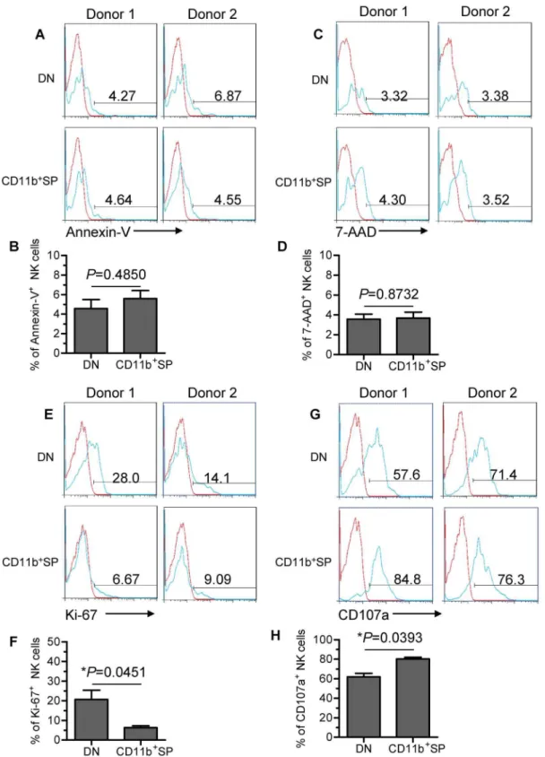
DN NK cells have proliferative capacity and poor cytotoxic capacity
To investigate the potential developmental capability of DN and CD11b+SP subsets, we first evaluated the viability of the two
subsets using Annexin-V and 7-Amino-Actinomycin D (7-AAD) staining. Minor proportions of the two subsets (,10%) were Annexin-V+or 7-AAD+, indicating that both subsets were viable
and not undergoing apoptosis (Fig. 5A–D). Furthermore, we found that the DN subset proliferated significantly more than the CD11b+SP subset as determined by higher Ki-67 expression
(Fig. 5E and F), indicating that the DN NK-cell subset has developmental potential.
functional marker that identifies NK cell-mediated lysis of target cells. In addition to the observed reduction in CD16 expression (Fig. 4A and B), the DN subset had lower CD107a expression than the CD11b+SP subset, suggesting reduced cytotoxicity of the DN
subset (Fig. 5G and H). This observation is consistent with published report showing that CD11b can mediate cytotoxic priming by beta-glucan [39]. Hence, the DN NK-cell subset has a poor cytotoxic capacity. We also measured the cytotoxic capacity of TINK cells compared with pNK cells from autologous patients by analysing the expression of CD107a. Diminished CD107a staining was observed on TINK cells (Fig. S1), suggesting reduced cytotoxicity for these cells, which is consistent with the previous study [13].
The appearance of DN NK cells is associated with tumour progression in humans
To determine the functional role of DN NK cells in tumours, we evaluated the influence of DN NK cell presence on clinical outcomes. The NSCLC patients included in this study were classified into 3 groups (Ia/b, IIa/b and IIIa/b) based on tumour
node metastasis (TNM). The clinical characteristics of the patients are summarised in Table S1. We first determined that a negative correlation exists between the absolute counts of total TINK cells and tumour size and progression stage (Fig. S2). Furthermore, a representative phenotypic analysis of CD11b and CD27 expres-sion in TINK cells isolated from tumours of 3 different stages is depicted in Figure 6A. Remarkably, the frequency of DN (CD11b2CD272) NK cells within the TINK-cell population increased as tumours progressed (Fig. 6B). We next examined the relationship between the frequency of DN NK cells and the size of the tumours. Notably, the frequency of DN NK cells within the TINK-cell population positively correlated with the maximum diameter of the resected tumours (Fig. 6C). These observations indicate that DN NK cells play an important role in tumour progression. We further investigated overall survival (OS) curve by Kaplan-Meier method. The patients included were classified into 3 groups (Low, Median and High DN NK%) based on the frequency of DN NK cells. Analysis of Kaplan-Meier survival curve showed a significant difference between the groups with low
Figure 2. TINK cells display an immature phenotype.(A) Representative flow cytometry analysis of the expression of NK-cell maturation receptors (CD57, CD127, CD117 and NKG2A) on gated CD56+CD32TINK cells as compared with that on pNK cells from autologous patients and healthy control subjects. Quadrants depicted were set on isotype controls. (B) The frequency of CD57+, CD127+, CD117+and NKG2A+NK cells within the above-mentioned three NK-cell populations (n = 15; mean6SEM).
DN NK% and high DN NK% (Fig. S3), suggesting that the frequency of DN NK cells is associated with clinical outcome.
The kinetics of DN NK-cell accumulation in lung tissue is associated with tumour progression in vivo
To further confirm the correlation between the frequency of tumour-infiltrating DN NK cells and the progression of lung carcinoma, we examined the expression of CD11b and CD27 on TINK cells from lung tissue in an intrapleurally implanted murine Lewis lung cancer (LLC) model [40]. Consistent with previous reports, the frequency of TINK cells in the lung tissue gradually decreased over time following LLC injection (Fig. 7A and C). Analysis of the frequency of each NK-cell subset within the TINK-cell population at different time-points revealed several findings. We observed striking differences in the frequency of each subset within the lung TINK-cell population at each time-point (here depicted as 0 d, 10 d and 20 d) (Fig. 7B). The injection of LLC induced a reduction in the proportion of CD11b+SP NK cells
within the lung tissue and a concomitant increase in the proportion of DN NK cells over time (Fig. 7D–F). Collectively, our findings suggest that the frequency of DN NK cells within the TINK-cell population is associated with the progression of lung carcinoma.
To determine whether the tumour microenvironment was responsible for the accumulation of tumour-infiltrating DN NK cells, we measured the expression of ICAM-1, a ligand for
CD11b/CD18 (also referred to as Mac-1 and CR3), on tumour cell lines. Human adenocarcinoma (ADC)-derived A549 cells and mouse LLC cells expressed minimal surface ICAM-1 (Fig. S4A). In contrast, high concentrations of soluble ICAM-1 (sICAM-1) were detected in malignant pleural fluid samples and A549 supernatants compared with plasma from NSCLC patients and healthy controls (Fig. S4B), implying that the level of sICAM-1 is high in the tumour microenvironment. Thus, we hypothesise that the secretion of sICAM-1 by human tumours may hinder the interaction between ICAM-1 and CD11b/CD18, leading to the downregulation of CD11b expression on NK cells and thereby resulting in the DN NK-cell accumulation.
Discussion
Analogous to adaptive immunity, NK cell-mediated immunity can be divided into distinct developmental and effector phases. Although recent studies have described the developmental stages of NK cells [29,30,33], the relationship between NK-cell development and function, especially in the context of disease, remains poorly understood. In this study, we present several lines of evidence indicating that tumour-infiltrating NK cells have a unique distribution of NK-cell subsets, due to the effects exerted by the tumour microenvironment.
Our prior studies have shown that CD11b and CD27 expression defines NK-cell maturation status, reflecting a
devel-Figure 3. TINK cells display an inactive phenotype.(A) Representative flow cytometry analysis of the expression of NK-cell activation receptors (CD16, CD226 and NKp30) on gated CD56+CD32 TINK cells as compared with that on pNK cells from autologous patients and healthy control subjects. Quadrants depicted were set on isotype controls. (B) The frequency of CD16+, CD226+and NKp30+NK cells within the above-mentioned three NK-cell populations (n = 15; mean6SEM).
opmental continuity of human NK cells [33]. By investigating the expression of CD11b and CD27 on TINK cells from NSCLC patients, we characterised the maturation status of tumour-infiltrating NK-cell subpopulations. Interestingly, we found a substantial CD11b2CD272NK-cell subset within the TINK-cell population but not within the pNK-cell population from the same patients or from healthy control subjects. However, the mecha-nism by which such a large DN NK-cell subset accumulates within a tumour remains unsolved. One possibility is a higher rate of active death for the mature NK cells (CD11b+SP NK subset) than
for the immature NK cells (DN NK subset) within the tumour, leading to a relative accumulation of tumour-infiltrating DN NK cells. However, we showed that DN and CD11b+SP subsets
display equal viability, thus ruling out this possibility (Fig. 5A–D). It is also possible that the tumour microenvironment could induce NK cells of an immature phenotype, just as a previous study indicated that the phenotypic alterations of TINK cell could be induced by the breast tumour microenvironment [13]. However, the mechanism by which the tumour microenvironment influences TINK-cell maturation remains unclear. Although little is known about NSCLC-infiltrating NK cells, in situ NK cells have been investigated in well-conducted studies [11–13]. NK cells are recruited to the tumour environment, where they are localised mainly to the tumour stroma rather than the tumour nest, implying that TINK cells are not in direct contact with cancer cells. The lack of physical contact between TINK cells and tumour cells suggests that tumour cells may produce soluble factors that influence TINK-cell development [41]. TGF-b is a likely candidate [13,42] because it is known to down-regulate GATA-3, and GATA-3-deficient bone marrow NK cells display a CD11blow phenotype [43]. Thus, we assessed TGF-b levels in malignant pleural fluid samples and supernatants from NSCLC-derived A549 cells by ELISA. However, the levels of TGF-b detected in these samples were not significantly different from those detected in plasma samples from NSCLC patients and healthy control subjects (data not shown). We thus searched for another soluble factor that could affect TINK-cell maturation. Previous studies have shown that the expression of ICAM-1, a ligand for CD11b/CD18 (also referred to as Mac-1 and CR3), is frequently reduced or absent in NSCLC with tumour cell dissemination [44,45]. Accordingly, A549 and LLC cells expressed minimal surface ICAM-1. However, high concentrations of soluble ICAM-1 (sICAM-1) were detected in malignant pleural fluid samples and A549 supernatants, implying that the level of sICAM-1 is high in the tumour microenvironment. The interac-tion between ICAM-1 and CD11b/CD18 integrin plays a critical role in leukocyte trafficking, immune synapse formation and co-stimulation [46,47]. Moreover, expression of CD11b is associated with NK-cell effector function [39]. Analogous to the effect of sMICA on NK-cell function [48,49], it is possible that the secretion of sICAM-1 by human tumours may hinder the interaction between ICAM-1 and CD11b/CD18, leading to the downregulation of CD11b expression on NK cells and thereby affecting the TINK-cell functional maturation. However, elevated levels of sICAM-1 only partially explain the presence of immature NK cells within the tumour. Therefore, we cannot rule out the possibility that other soluble factors produced by tumour cells contribute to this process. Our findings demonstrate that the tumour microenvironment may render TINK cells less tumour-icidal and thereby contribute to cancer progression, as CD11b downregulation was detected on TINK cells but not on pNK cells from the NSCLC patients. An alternate mechanism that could account for our findings would be that immature NK might be attracted to infiltrate tumour tissue, as opposed to an active role of tumour tissue in stimulating the loss or reversal of NK maturity. Future studies are required to elaborate on the mechanism by which the tumour microenvironment affects the functional maturation of TINK cells.
Numerous studies have examined the relationship between tumour-infiltrating NK cells and clinical outcomes. One study demonstrates that the frequency of NK cells within NSCLC tumours directly correlates with the size of the resected tumours [12]. Strikingly, our results highlight the important role of DN NK cells in tumour progression, not only because the frequency of DN
Figure 4. The DN NK subset in TINK cells accounts for the immature phenotype.(A) Representative flow cytometry analysis of the expression of various surface molecules (CD16, CD57, CD127, CD117, CD226 and NKp30) on the DN NK subset versus the CD11b+SP NK subset in TINK cells. Dot plots were gated on live NK cells using a lymphocyte gate based on forward scatter versus side scatter and an NK-cell gate identifying CD56+CD32cells. DN NK cells were identified based on gating for CD56+CD32CD11b2CD272, while CD11b+SP NK cells were identified based on a CD56+CD32CD11b+CD272 gate. Quadrants depicted were set on isotype controls. (B) The major phenotypic differences detected in the two subsets (DN and CD11b+SP NK) are summarised. Data shown represent the findings from 15 patients (mean6SD).
Figure 5. DN NK cells have proliferative capacity and poor cytotoxic capacity.(A and C) Two representative flow cytometry analyses of the expression of Annexin-V and 7-AAD (green graphs) relative to isotype-matched controls (red graphs) on the gated DN NK subset versus the CD11b+SP NK subset in TINK cells. (B and D) The frequency of Annexin-V+and 7-AAD+NK cells on two gated NK-cell subsets (n = 6; mean
6SD). (E) Two representative flow cytometry analyses of Ki-67 expression (green graphs) relative to isotype-matched controls (red graphs) on the gated DN NK subset versus the CD11b+SP NK subset within TINK cells. (F) The frequency of Ki-67+NK cells on two gated NK-cell subsets (n = 6; mean
6SD). (G) Two representative flow cytometry analyses of CD107a expression (green graphs) relative to isotype-matched controls (red graphs) on the gated DN NK subset versus the CD11b+SP NK subset in TINK cells. (H) The frequency of CD107a+NK cells on two gated NK-cell subsets (n = 6; mean
(CD11b2CD272) NK cells within the TINK-cell population is clearly correlated with tumour stage and tumour size, but also because DN NK cells have developmental potential.
In conclusion, our study has provided new insights into NK-cell maturation within human tumours. To the best of our knowledge, this is the first report of a significant immature CD11b2CD272 NK-cell subset within the tumour-infiltrating NK-cell population and of a correlation between DN NK-cell frequency and tumour progression. More importantly, our data provide evidence that the tumour microenvironment may render TINK cells less tumour-icidal and thereby contribute to cancer progression.
Materials and Methods
Ethics Statement
The study was approved by the Institutional Review Board of the University of Science and Technology of China. Human primary lung tumour tissue was obtained during surgery from 38 treatment-naive patients with NSCLC. Peripheral blood samples were collected from the same patients prior to surgery. In accordance with the Declaration of Helsinki and the Institutional Review Board of the University of Science and Technology of China, all participants provided written informed consent, which was obtained before enrolment in the study.
The study was carried out in strict accordance with the recommendations in the Guide for the Care and Use of Laboratory Animals. The animal experiment facilities were approved by the Anhui Provincial Department of Science and Technology, the approval ID is SYXK (Anhui) 2005-004. All protocols involving animal work were approved by the Ethics Committee of Animal Experiments of the University of Science
and Technology of China (Permit Number: USTCA-CUC1201051). All surgery was performed under anaesthesia, and all efforts were made to minimize animal suffering.
Samples from patients and healthy control subjects Peripheral blood and tumour tissue were acquired from the Department of Chest Surgery, Anhui Provincial Hospital (Hefei, China). Detailed patient characteristics are shown in Table S1. Peripheral blood samples were obtained from healthy donors at the Blood Centre of Anhui Province (Hefei, China) and used as controls. All of the samples were collected after obtaining written informed consent from the donors and following approval by the Institutional Review Board of the University of Science and Technology of China.
Mice and in vivo tumour challenge
A total of 54 female C57BL/6 mice were purchased from Shanghai Experimental Animal Centre, Chinese Science Acade-my (Shanghai, China), and were maintained in a specific pathogen-free (SPF) facility under barrier conditions at Laboratory Animal Centre of the University of Science and Technology of China. All mice were fed a regular mouse chow and housed in normal night-day conditions under standard temperature and humidity. All animal studies were carried out at the biosafety laboratory of the Laboratory Animal Centre, University of Science and Technology of China. The experiment complied with the Animal Management Rule of the Ministry of Public Health, People’s Republic of China (document No. 55, 2001), and the experiment protocol was approved by the Ethics Committee of Animal Experiments of the University of Science and Technology of China (Permit Number: USTCACUC1201051). All mice were
Figure 6. The frequency of tumour-infiltrating DN NK cells is highly associated with clinical outcome.(A) Representative flow cytometry analysis of the expression of CD27 and CD11b on TINK cells from NSCLC patients with tumours of distinct stage classifications based on tumour node metastasis (TNM). Dot plots were gated on live NK cells using a lymphocyte gate based on forward scatter and side scatter and an NK-cell gate (CD56+CD32). DN NK cells were analysed by gating on CD56+CD32CD11b2CD272cells. Quadrants depicted were set on isotype controls. (B) The frequency of tumour-infiltrating DN NK cells is correlated with the malignant progression of lung carcinoma (n = 20; mean6SEM). (C) The frequency of DN NK cells within tumours is directly correlated with tumour size. They-axis represents the maximum diameter of the resected tumours.
used at 8–10 weeks of age [50], and were randomly divided into 3 groups (n = 6 each). One group of mice with no treatment were marked as control group (here depicted as 0 d group). To establish tumours [40], the other two groups of mice were intrapleurally injected with 56105live Lewis lung cancer (LLC) cells in a volume of 0.1 mL phosphate buffered saline (PBS). The mice were examined daily after LLC injection. To assess tumour progression in the lung, one group of tumour cell-inoculated mice were sacrificed on 10 day following LLC injection, and the remaining group of tumour cell-inoculated mice were sacrificed on 20 day following LLC injection. Mice were sacrificed using CO2. This
study was performed three times.
Isolation of lymphocytes from samples
Primary lung tumours were washed extensively with phosphate-buffered saline (PBS) to remove peripheral blood lymphocytes and were processed by mincing the tissue with operative scissors. Mechanically disrupted tumour tissue was digested with 0.1% collagenase type IV (Sigma-Aldrich, St. Louis, MO) in serum-free RPMI 1640 for 1 hour at 37uC under agitation. After enzymatic digestion, single-cell suspensions were filtered and washed, and mononuclear cells (MNCs) were purified using Ficoll-Hypaque density gradient centrifugation.
Similarly, peripheral blood mononuclear cells (PBMCs) were isolated from blood samples by Ficoll centrifugation.
To isolate mononuclear cells (MNCs), tumour-bearing mice were euthanised, and the lung tissues were collected. The tissues
Figure 7. The kinetics of DN NK-cell accumulation in tumours is associated with tumour progression in vivo.(A) To establish tumours, C57BL/6 mice were intrapleurally injected with 56105live Lewis lung cancer (LLC) cells. Representative flow cytometry analysis of lung TINK cells at different time-points after LLC injection. Dot plots were gated on live NK cells using a lymphocyte gate based on forward scatter versus side scatter and an NK-cell gate using NK1.1+CD32cells. Quadrants depicted were set on isotype controls. (B) Representative flow cytometry analysis of CD27/ CD11b expression in lung TINK cells at different time-points after LLC injection. Quadrants depicted were set on isotype controls. (C) The frequency of TINK cells in lung tissue isolated from C57BL/6 mice at each time-point (n = 6 each). (D–F) The frequency of each subset of lung TINK cells isolated from C57BL/6 mice at each time-point (n = 6 each). All experiments were performed three times with similar results.
were mechanically disrupted and then digested with collagenase I (Sigma-Aldrich, St. Louis, MO) to create single-cell suspensions. After washing, the cell pellets were resuspended in 40% Percoll (GE Healthcare, Uppsala, Sweden), gently overlaid onto 70% Percoll, and centrifuged at room temperature. MNCs were isolated from the Percoll interface.
Flow cytometric analysis of NK-cell phenotypes
Lymphocytes from human blood/tumour samples were incu-bated for 30 minutes at 4uC with the following mouse anti-human monoclonal antibodies: CD3, CD56, CD27, CD11b, CD16, CD57, CD127, CD117, anti-NKG2A, anti-NKp30 and anti-CD226 (all from BD Bioscience, San Jose, CA). Mouse serum was used to block non-specific Fc-receptor (FcR) binding, and isotype-matched IgGs were used as negative control antibodies. FACS staining was performed according to the manufacturer’s instructions. The FACS gating strategy is shown in Figure S5.
Mouse lung lymphocytes were prepared and stained with mAbs. FcRs were blocked with normal rat serum. Abs specific for the following Ags were used to stain murine lung lymphocytes: NK1.1, CD3, CD11b and CD27 (all from BD Bioscience, San Jose, CA). The samples were analysed on a FACSCalibur flow cytometer (BD Biosciences). Flow cytometry data were analysed using FlowJo software (Tree Star, Inc., Ashland, OR).
Apoptosis detection by Annexin-V and 7-AAD staining Lymphocytes from human tumour samples were incubated with the following mouse human monoclonal antibodies: anti-CD3, anti-CD56, anti-CD27 and anti-CD11b, as described above. After two washes in cold binding buffer (10 mM HEPES/NaOH [pH 7.4], 140 mM NaCl, 2.5 mM CaCl2), cells were stained with
Annexin-V and 7-AAD (BD PharMingen) for 15 min at room temperature in the dark and analysed by flow cytometry.
Ki-67 and CD107a detection
Lymphocytes from human tumour samples were incubated with the following mouse human monoclonal antibodies: anti-CD3, anti-CD56, anti-CD27, anti-CD11b and anti-CD107a for 30 minutes at 4uC. After fixation and permeabilisation, cells were further stained with anti-CD107a Ab and anti-Ki-67 Ab for 1 h at 4uC, washed twice with permeabilisation buffer, and analysed by flow cytometry. Appropriate isotype Abs were used as controls for intracellular staining.
Cell lines
Human adenocarcinoma (ADC)-derived A549 cells, obtained from the Chinese Academy of Sciences Cell Bank (Shanghai, China), were cultured in RPMI-1640 supplemented with 10% foetal calf serum (FCS) at a density of 107cells/ml. After incubation for 72 h, the culture supernatants were collected and assayed for cytokine levels by ELISA.
LLC cells derived from C57BL/6 mice with Lewis lung carcinoma, obtained from the Chinese Academy of Sciences Cell Bank (Shanghai, China), were cultured in DMEM supplemented with 10% FCS. The cells were cultured for less than 6 months after resuscitation. Early-passage cells were used for all experi-ments and were not reauthenticated.
Cytokine detection by ELISA
To measure cytokine levels in NSCLC patients, plasma samples were collected from all patients and healthy control subjects and stored at 270uC until use. Likewise, malignant pleural fluid
samples and A549 supernatants were also appropriately collected and stored before utilisation. TGF-band sICAM-1 were assayed using standard sandwich ELISA kits (CUSABIO BIOTECH CO., LTD.) according to the manufacturer’s instructions.
Statistical analyses
We used two-tailed paired Student’s t-tests (difference between two groups) or two-tailed unpaired Student’s t-tests to determine the statistical significance (*P#0.05; **P#0.01; ***P#0.001). Overall survival (OS) curve was estimated by Kaplan-Meier method and differences between the groups of patients were evaluated using the log-rank test at minimalPvalue.
Supporting Information
Figure S1 TINK cells had poor cytotoxic potential compared with paired pNK cells. (A) Two representative flow cytometry analyses for CD107a expression (green graphs) relative to isotype-matched controls (red graphs) on TINK cells as compared with that on pNK cells from autologous patients. (B) The frequency of CD107a+NK cells within the above-mentioned
two NK-cell populations (n = 6; mean6SD). (TIF)
Figure S2 The absolute counts of TINK cells are highly associated with tumour progression.(A) Absolute counts of TINK cells are negatively correlated with the malignant progression of lung carcinoma (n = 20; mean6SEM). (B) Absolute counts of TINK cells are negatively correlated with tumour size. The y-axis represents the maximum diameter of the resected tumours.
(TIF)
Figure S3 Kaplan-Meier curve of overall survival (OS). The patients included were classified into 3 groups (Low, Median and High DN NK%) based on the frequency of DN NK cells. The group with low DN NK% (black curves), median DN NK% (green curves) and high DN NK% (red curves) are shown.
(TIF)
Figure S4 sICAM-1 levels are elevated in malignant pleural fluid samples and A549 supernatants.(A) Repre-sentative flow cytometry analysis of ICAM-1 expression (green graphs) relative to isotype-matched controls (red graphs) on the surface of A549 and LLC cells. (B) Levels of sICAM-1 in plasma samples from healthy donors and NSCLC patients, malignant pleural fluid samples and A549 supernatants were quantified by sandwich ELISA.
(TIF)
Figure S5 FACS gating strategy. (A) The FACS gating strategy for excluding dead and irrelevant cells. (B) The FACS gating strategy for isolating total CD3-CD56+TINK cells within
the lymphocyte gate. (TIF)
Table S1 Clinical characteristics of 38 patients with NSCLC. NOTE: Pathologic staging and histologic types of lung cancer were determined according to the TNM staging system and the histologic classification of the World Health Organization, respectively.
(TIF)
Acknowledgments
Author Contributions
Obtained permission for the use of clinical samples: XM. Conceived and designed the experiments: HW ZT JJ. Performed the experiments: JJ TY.
Analyzed the data: HW JJ ZT BF. Contributed reagents/materials/ analysis tools: RS XM. Wrote the paper: JJ HW BF.
References
1. Siegel R, Ward E, Brawley O, Jemal A (2011) Cancer statistics, 2011: the impact of eliminating socioeconomic and racial disparities on premature cancer deaths. CA Cancer J Clin 61: 212–236.
2. Smyth MJ, Hayakawa Y, Takeda K, Yagita H (2002) New aspects of natural-killer-cell surveillance and therapy of cancer. Nat Rev Cancer 2: 850–861. 3. Swann JB, Smyth MJ (2007) Immune surveillance of tumors. J Clin Invest 117:
1137–1146.
4. Ljunggren HG, Malmberg KJ (2007) Prospects for the use of NK cells in immunotherapy of human cancer. Nature Reviews Immunology 7: 329–339. 5. Chan CJ, Andrews DM, Smyth MJ (2008) Can NK cells be a therapeutic target
in human cancer? Eur J Immunol 38: 2964–2968.
6. Cerwenka A, Falk CS, Watzl C (2007) NK cells-from basic research to cancer therapy. Eur J Immunol 37: 1161–1164.
7. Lanier LL (2005) Nk Cell Recognition. Annual Review of Immunology 23: 225– 274.
8. Vivier E, Raulet DH, Moretta A, Caligiuri MA, Zitvogel L, et al. (2011) Innate or Adaptive Immunity? The Example of Natural Killer Cells. Science 331: 44– 49.
9. Vivier E, Tomasello E, Baratin M, Walzer T, Ugolini S (2008) Functions of natural killer cells. Nature Immunology 9: 503–510.
10. Sun JC, Lanier LL (2009) Natural killer cells remember: an evolutionary bridge between innate and adaptive immunity? Eur J Immunol 39: 2059–2064. 11. Platonova S, Cherfils-Vicini J, Damotte D, Crozet L, Vieillard V, et al. (2011)
Profound Coordinated Alterations of Intratumoral NK Cell Phenotype and Function in Lung Carcinoma. Cancer Research 71: 5412–5422.
12. Carrega P, Morandi B, Costa R, Frumento G, Forte G, et al. (2008) Natural killer cells infiltrating human nonsmall-cell lung cancer are enriched in CD56brightCD16– cells and display an impaired capability to kill tumor cells. Cancer 112: 863–875.
13. Mamessier E, Sylvain A, Thibult ML, Houvenaeghel G, Jacquemier J, et al. (2011) Human breast cancer cells enhance self tolerance by promoting evasion from NK cell antitumor immunity. Journal of Clinical Investigation 121: 3609– 3622.
14. Jonsson AH, Yokoyama WM (2009) Natural killer cell tolerance licensing and other mechanisms. Adv Immunol 101: 27–79.
15. Kim S, Poursine-Laurent J, Truscott SM, Lybarger L, Song YJ, et al. (2005) Licensing of natural killer cells by host major histocompatibility complex class I molecules. Nature 436: 709–713.
16. Bjorkstrom NK, Riese P, Heuts F, Andersson S, Fauriat C, et al. (2010) Expression patterns of NKG2A, KIR, and CD57 define a process of CD56dim NK-cell differentiation uncoupled from NK-cell education. Blood 116: 3853– 3864.
17. Zimmer J, Be´ziat V, Descours B, Parizot C, Debre´ P, et al. (2010) NK Cell Terminal Differentiation: Correlated Stepwise Decrease of NKG2A and Acquisition of KIRs. PLoS ONE 5: e11966.
18. Brodin P, Ka¨rre K, Ho¨glund P (2009) NK cell education: not an on-off switch but a tunable rheostat. Trends in Immunology 30: 143–149.
19. Elliott JM, Wahle JA, Yokoyama WM (2010) MHC class I-deficient natural killer cells acquire a licensed phenotype after transfer into an MHC class I-sufficient environment. Journal of Experimental Medicine 207: 2073–2079. 20. Joncker NT, Shifrin N, Delebecque F, Raulet DH (2010) Mature natural killer
cells reset their responsiveness when exposed to an altered MHC environment. Journal of Experimental Medicine 207: 2065–2072.
21. Huntington ND, Vosshenrich CAJ, Di Santo JP (2007) Developmental pathways that generate natural-killer-cell diversity in mice and humans. Nature Reviews Immunology 7: 703–714.
22. Di Santo JP (2008) Functionally distinct NK-cell subsets: developmental origins and biological implications. Eur J Immunol 38: 2948–2951.
23. Freud AG (2006) Evidence for discrete stages of human natural killer cell differentiation in vivo. Journal of Experimental Medicine 203: 1033–1043. 24. Cooper MA, Fehniger TA, Caligiuri MA (2001) The biology of human natural
killer-cell subsets. Trends Immunol 22: 633–640.
25. Caligiuri MA (2008) Human natural killer cells. Blood 112: 461–469. 26. Colucci F, Caligiuri MA, Di Santo JP (2003) What does it take to make a natural
killer? Nature Reviews Immunology 3: 413–425.
27. Fehniger TA (2002) CD56bright natural killer cells are present in human lymph nodes and are activated by T cell-derived IL-2: a potential new link between adaptive and innate immunity. Blood 101: 3052–3057.
28. Romagnani C, Juelke K, Falco M, Morandi B, D’Agostino A, et al. (2007) CD56brightCD16- killer Ig-like receptor- NK cells display longer telomeres and
acquire features of CD56dim NK cells upon activation. J Immunol 178: 4947– 4955.
29. Hayakawa Y, Smyth MJ (2006) CD27 dissects mature NK cells into two subsets with distinct responsiveness and migratory capacity. Journal of Immunology 176: 1517–1524.
30. Vossen MT, Matmati M, Hertoghs KM, Baars PA, Gent MR, et al. (2008) CD27 defines phenotypically and functionally different human NK cell subsets. J Immunol 180: 3739–3745.
31. Chiossone L, Chaix J, Fuseri N, Roth C, Vivier E, et al. (2009) Maturation of mouse NK cells is a 4-stage developmental program. Blood 113: 5488–5496. 32. Hayakawa Y, Huntington ND, Nutt SL, Smyth MJ (2006) Functional subsets of
mouse natural killer cells. Immunol Rev 214: 47–55.
33. Fu B, Wang F, Sun R, Ling B, Tian Z, et al. (2011) CD11b and CD27 reflect distinct population and functional specialization in human natural killer cells. Immunology 133: 350–359.
34. Hayakawa Y, Sato-Matsushita M, Takeda K, Iwakura Y, Tahara H, et al. (2011) Early activation and interferon-c production of tumor-infiltrating mature CD27high natural killer cells. Cancer Science 102: 1967–1971.
35. Lopez-Verges S, Milush JM, Pandey S, York VA, Arakawa-Hoyt J, et al. (2010) CD57 defines a functionally distinct population of mature NK cells in the human CD56dimCD16+NK-cell subset. Blood 116: 3865–3874.
36. Lakshmikanth T, Burke S, Ali TH, Kimpfler S, Ursini F, et al. (2009) NCRs and DNAM-1 mediate NK cell recognition and lysis of human and mouse melanoma cell lines in vitro and in vivo. Journal of Clinical Investigation 119: 1251–1263. 37. Lee SH, Biron CA (2010) Here today--not gone tomorrow: roles for activating receptors in sustaining NK cells during viral infections. Eur J Immunol 40: 923– 932.
38. Vivier E, Morin P, O’Brien C, Druker B, Schlossman SF, et al. (1991) Tyrosine phosphorylation of the Fc gamma RIII(CD16): zeta complex in human natural killer cells. Induction by antibody-dependent cytotoxicity but not by natural killing. J Immunol 146: 206–210.
39. Xia Y, Borland G, Huang J, Mizukami IF, Petty HR, et al. (2002) Function of the lectin domain of Mac-1/complement receptor type 3 (CD11b/CD18) in regulating neutrophil adhesion. J Immunol 169: 6417–6426.
40. Chen LL, He ZX, Qin L, Li QY, Shi XB, et al. (2011) Antitumor Effect of Malaria Parasite Infection in a Murine Lewis Lung Cancer Model through Induction of Innate and Adaptive Immunity. PLoS ONE 6.
41. Richards JO, Chang X, Blaser BW, Caligiuri MA, Zheng P, et al. (2006) Tumor growth impedes natural-killer-cell maturation in the bone marrow. Blood 108: 246–252.
42. Allan DS, Rybalov B, Awong G, Zuniga-Pflucker JC, Kopcow HD, et al. (2010) TGF-beta affects development and differentiation of human natural killer cell subsets. Eur J Immunol 40: 2289–2295.
43. Samson SI, Richard O, Tavian M, Ranson T, Vosshenrich CAJ, et al. (2003) GATA-3 promotes maturation, IFN-gamma production, and liver-specific homing of NK cells. Immunity 19: 701–711.
44. Le Maux Chansac B, Moretta A, Vergnon I, Opolon P, Lecluse Y, et al. (2005) NK cells infiltrating a MHC class I-deficient lung adenocarcinoma display impaired cytotoxic activity toward autologous tumor cells associated with altered NK cell-triggering receptors. J Immunol 175: 5790–5798.
45. Passlick B, Pantel K, Kubuschok B, Angstwurm M, Neher A, et al. (1996) Expression of MHC molecules and ICAM-1 on non-small cell lung carcinomas: association with early lymphatic spread of tumour cells. Eur J Cancer 32A: 141– 145.
46. Orange JS, Harris KE, Andzelm MM, Valter MM, Geha RS, et al. (2003) The mature activating natural killer cell immunologic synapse is formed in distinct stages. Proceedings of the National Academy of Sciences of the United States of America 100: 14151–14156.
47. Crozat K, Eidenschenk C, Jaeger BN, Krebs P, Guia S, et al. (2011) Impact of beta2 integrin deficiency on mouse natural killer cell development and function. Blood 117: 2874–2882.
48. Groh V, Wu J, Yee C, Spies T (2002) Tumour-derived soluble MIC ligands impair expression of NKG2D and T-cell activation. Nature 419: 734–738. 49. Salih HR, Rammensee HG, Steinle A (2002) Cutting edge: down-regulation of