U minho | 20 13 Catherine F err eira Thiol-disulphide o xidoreduct
ases: production, purification and s
tructural anal
ysis of a cold adap
ted DsbA
.
Universidade do Minho
Escola de Ciências
Catherine Oliveira Ferreira
Abril de 2013
Thiol-disulphide oxidoreductases:
production, purification and
structural analysis of a cold
adapted DsbA.
UNIVERSIDADE DO MINHO
Escola de Ciências
CATHERINE OLIVEIRA FERREIRA
Thiol-disulphide oxidoreductases:
production, purification and structural
analysis of a cold adapted DsbA.
Master Thesis
Master in Molecular Genetics
Work done under supervision of: Doutor Tony Collins
Professora Doutora Margarida Casal
i DECLARAÇÃO
Nome
Catherine Oliveira Ferreira
Endereço electrónico: catherine.o.ferreira@gmail.com Número do Cartão de Cidadão: 13845225
Título da tese: Thiol-disulphide oxidoreductases: production, purification and structural analysis of a cold adapted DsbA.
Orientadores:
Doutor James Anthony Collins
Professora Doutora Margarida Paula Pedra Amorim Casal Ano de conclusão: 2013
Designação do Mestrado: Genética Molecular
É AUTORIZADA A REPRODUÇÃO PARCIAL DESTA TESE/TRABALHO (indicar, caso tal seja necessário, nº máximo de páginas, ilustrações, gráficos, etc.), APENAS PARA EFEITOS DE INVESTIGAÇÃO, MEDIANTE DECLARAÇÃO ESCRITA DO INTERESSADO, QUE A TAL SE COMPROMETE;
Universidade do Minho, ___/___/______
ii
A
CKNOWLEDGEMENTS
/
AGRADECIMENTOS
‘Serei prosa serei verso? Instrumento útil neste Universo. Guiada num controverso caminhar,
tendo em missão seu guião findar. Dia de hoje, que será sempre um só, Produto palpável dos sonhos passados. Desses, tantos se desmoronaram num pó,
e a tantos outros permanecem meus quereres abraçados.’
Catherine Ferreira De um jeito poético começo por descrever a forma como encaro cada desafio. Tendo em mente que tudo se resume à descoberta incansável do nosso caminho, do nosso lugar. Este caminho de linhas tortas que se constrói das nossas vontades, das nossas opções mas que nunca é resultado de um só ‘eu’. Nesta viagem beneficiamos da ajuda, dos conselhos e da força daqueles com que a vida nos quis cruzar. Começo assim por agradecer a quem permitiu que este passo fosse possível, a quem tão bem me recebeu e me integrou. Agradeço imenso aos meus orientadores pela indispensável presença que tanto me auxiliou neste percurso. À Professora Doutora Margarida Casal pelas palavras amigas que sempre reservou para mim, e pelo contagiante sorriso com que sempre me recebeu. Não dispenso valorizar-lhe a boa disposição, a serenidade e os certeiros conselhos que tanto me auxiliaram. Ao Professor Doutor Tony Collins pela incansável paciência, pela constante disponibilidade e pelo esforço acrescido que sempre demonstrou em contornar todas as situações inesperadas tão comuns de qualquer percurso. Agradeço também a todos que floriram os meus dias no laboratório. Assim, agradeço do fundo do coração aos que me arrancaram sorrisos em dias mais difíceis, aos que me presentearam com a sua presença e cuja personalidade de cada um fez daquele laboratório um lugar tão bom e tão melhor. À Joana Sá Pessoa, ao Raul Machado, ao João Silva, ao André Costa, ao Pedro Castro e ao Fernando Branca. De uma forma geral agradeço a forma tão especial como fui recebida, integrada e tratada. Enchem-me de alegria, antecipada saudade e da certeza de que todos os momentos estarão sempre carinhosamente guardados na minha memória.
Agradeço com um especial carinho a todas as pessoas que de uma forma ou de outra contribuíram no sentido de me ajudar neste percurso. Á Joana Tulha, à Filipa Pereira, à Andreia Pacheco, à Dulce Cunha, à Sara Alves, ao Flávio Azevedo, à
iii Helena Pereira e à Catarina Carneiro. Sem esquecer todos os técnicos que fizeram parte desta jornada, reconhecendo-lhes o tão importante trabalho que sempre tiveram e com o qual nada disto seria possível.
À Professora Doutora Maria João Sousa pela recepção no Mestrado em Genética Molecular.
‘Quando o sonho abraça a fé, a coragem é véstia mais bela. Atributo invejado por quem vive só da razão. De quem vê e não entende, de quem toca e não sente, de quem passa sem viver. Pois se o sonho é meu e a fé minha loucura… a coragem enaltece o brilho de quem tem morada em mim. Dos que me vêm de verdade, dos que me sentem no infinito e me entendem sem perguntar. Nesse olhar, nesse abraço, nessas palavras plenas de silêncio. Na promessa de alinhar na primeira fila de cada batalha vossa, sei que vos terei em cada luta minha. Pois é desse brilho que vivo. E quanto mais invejado e incompreendido mais intenso e desejado será o seu brilho. Selvagens almas, misteriosos seres… sem vocês não seria mais eu.’
Catherine Ferreira
Agradeço o que as palavras não conseguem descrever, à minha família, especialmente aos meus pais, pois o seu apoio a todos os níveis foi sem dúvida essencial. Aos meus irmãos pois não teria o mesmo sentido sem eles. Aos meus avós que sempre me apoiaram também. Dedico todo este esforço a eles e em especial à minha avó e à minha bisavó, esperando que se orgulhem desta batalha.
Aos meus amigos que em todos os momentos estiveram presentes, que foram incansáveis na ajuda, que albergaram a maior paciência e mesmo assim se mantiveram firmes ao meu lado. Por todos os momentos bons e menos bons. Agradeço assim à Rita Cunha, ao Pedro Castro, ao António Rego e à Sara Peixoto.
Um agradecimento especial ao Pedro Martinez que no momento certo surgiu, que do seu jeito único me estendeu a mão e que desde então se mantém fielmente ao meu lado. Para o que der e vier sei que estarás sempre lá.
Ao João digo que são poucas as palavras capazes de descrever a minha gratidão. Que nada seria possível se nos dias mais difíceis eu não encaminhasse a minha mente para um futuro melhor. E acredito tanto nele. É nesse futuro que descanso e vejo a paz e a felicidade de que preciso.
iv
A
BSTRACT
Thiol-disulphide oxidoreductases (DsbAs) are bacterial extra-cytoplasmic enzymes which catalyse oxidative disulphide bond formation (S-S) between the thiol sulphurs (-SH) of cysteine side chains in newly synthesised proteins (Shouldice, et al., 2011). In medicine, the key role of DsbA in catalysing the correct folding of many essential proteins that enable pathogenesis has led to suggestions for this enzyme as a potential antimicrobial drug target (Heras, et al., 2009). DsbA catalyses the correct folding of virulence factors associated with cell adhesion, bacterial mobility and host cell manipulation and hence its inhibition would reduce or impede bacterial pathogenesis. Indeed bacterial infections are a major cause of death in the world and this, in addition to current high levels of antibiotic resistance in many pathogenic bacteria, highlights the urgent need for new validated targets and for the design of new antibacterial agents against these targets. Due to its role in pathogenesis DsbA offers such a target for a new therapeutic approach and a better understanding of this enzyme and its function is of much importance.
In the present study a cold adapted DsbA from Pseudoalteromonas haloplanktis TAC125 was studied with the long term aim of better understanding its structure and function relationship. Previous studies of this enzyme made use of non-optimised production and purification procedures and production levels were found to be poor (approximately 50 mg/L) with large losses being noted during purification. Therefore the present study was focused on optimising the shake-flask batch production in E. coli and simplifying and improving the purification protocol for this protein. Furthermore, as an initial step in our quest for a better understanding of this enzyme, a comparative structural analysis (with homologous enzymes) was carried out to identify structural factors which may be important for the low temperature activity of cold-adapted DsbAs. Mutants were then designed and prepared in an attempt to investigate the roles of the observed structural differences.
We have shown that the rich medium Terrific broth (TB) with induction during the stationary phase of growth allowed for optimum DsbA production. Interestingly, high production levels were attained even in the absence of induction with IPTG. Optimisation of the purification protocol allowed for the development of a simplified procedure yielding 250 mg of purified protein per litre of production culture (a 5-fold increase on that previously reported) with a reduced DsbA loss during the process. Structural comparisons allowed for the identification of two loop insertions in the cold-adapted enzyme as compared to homologs adapted to higher temperatures and four deletion mutants investigating these insertions have been prepared.
v
R
ESUMO
As tiol-dissulfito oxidorredutases (DsbAs) são enzimas bacterianas extra citoplasmáticas que catalisam a formação oxidativa de pontes dissulfito (S-S) entre os grupos tióilicos (-SH) das cadeias laterais das cisteínas em proteínas recentemente sintetizadas (Shouldice, et al., 2011). Na medicina, o papel chave da DsbA prende-se com a catálise do correcto rearranjo de muitas proteínas essenciais na patogénese, têm assim surgido sugestões de que esta enzima possa ser um alvo de potenciais drogas antimicrobianas (Heras, et al., 2009). A DsbA é catalisadora do correcto rearranjo de factores de virulência associados à adesão celular, mobilidade bacteriana e manipulação das células hospedeiras. Na verdade, as infecções bacterianas são já a maior causa de morte no mundo e este facto, em junção com o actual alto nível de resistência a antibióticos por parte de várias bactérias patogénicas, enaltece a urgente necessidade tanto de validar novos alvos como de objectivar o desenho de novos agentes antibacterianos que actuem nesses alvos. Devido ao seu papel na patogénese, a DsbA, sendo um possível alvo apresenta-se assim uma nova abordagem terapêutica destacando-se a elevada importância de um melhor entendimento desta enzima e da sua função.
No presente estudo, foi estudada uma DsbA adaptada ao frio proveniente da bactéria Pseudoalteromonas haloplanktis TAC125 objectivando-se a longo prazo um melhor entendimento da relação entre a sua estrutura e função. Estudos anteriores centrados nesta enzima têm feito uso de processos de produção e purificação não optimizados tendo resultado em baixos níveis de produção (aproximadamente 50 mg/L), com grandes perdas observadas no processo de purificação. Assim sendo, o actual estudo centrou-se tanto na optimização da produção em batch em E. coli como em simplificar e melhorar o protocolo de purificação para esta proteína. Além disso, como parte inicial da nossa investigação focada na obtenção de um melhor entendimento desta enzima, uma comparativa análise estrutural (com enzimas homólogas) foi levada a cabo de modo a identificar os factores estruturais que possam ser importantes na actividade a baixas temperaturas desta DsbA naturalmente adaptada ao frio. Os mutantes foram então desenhados na tentativa de investigar o papel das diferenças estruturais observadas.
Demonstrou-se neste estudo que a junção do meio rico Terrific Broth (TB) com indução na fase estacionária de crescimento permitiu um nível óptimo de produção da DsbA. Interessantemente, elevados níveis de produção foram alcançados inclusive na ausência de indução. No entanto, altos níveis de produção foram também observados
vi aquando da indução com 1 mM de IPTG na fase de declínio exponencial. A optimização do protocolo de purificação permitiu o desenvolvimento de um simplificado procedimento, rendendo 250 mg de proteína purificada por litro de cultura d produção (5 vezes mais) com a reduzida perda de proteína ao longo do processo. Comparações estruturais permitiram a identificação de duas inserções em loop’s da enzima adaptada ao frio quando comparada com os homólogos adaptados a temperaturas mais elevadas. Neste sentido, quatro mutantes centrados na investigação desses locais foram concebidos.
vii
A
BBREVIATIONS AND SYMBOLS
TRX – Thioredoxin
-SH – Thiol group of cysteine
S-S – Disulphide bond between cystines
E. coli – Escherichia coli
Cys – Cysteine Val – Valine Lys – Lysine Leu – Leucine Ala – Alanine Ser – Serine His – Histidine Asp – Aspargine
pKa – Dissociation constant
NMR – Nuclear Magnetic Resonance
PhDsbA – DsbA protein from Pseudoalteromonas haloplanktis PhDsbB - DsbB protein from Pseudoalteromonas haloplanktis
Tcp – Toxin co-regulated pilus
B. pertussis – Bordetella pertussis
kcat – enzymatic reaction rate
A - Frequency factor related to the frequency of collision of the reactants and to the probability of the reactants being in the appropriate orientation to react
T – Temperature
R - Universal gas constant
Ea - Activation energy necessary for the reaction IPTG - isopropyl β-D-1-thiogalactopyranoside
viii
kDa – Atomic mass unit
LB – Lysogeny broth
TB – Terrific broth
SB – Super broth
HPLC – High-performance liquid chromatography FPLC – Fast protein liquid chromatography DTNB - 5,5’–dithiobis–2–nitrobenzoic acid
TNB – 2-nitro-5-thiobenzoic acid
NaCl – Sodium chloride
pI – Isoelectric point
HIC – Hydrophobic interaction chromatography
IEX – Ion exchange chromatography
WT – Wild type
K2HPO4.3H2O – dipotassium phosphate trihydrate
KH2PO4 – potassium dihydrogen phosphate
UV – Ultra violet
Vis – Visible
EDTA - Ethylenediamine tetraacetic acid
DTT – Dithiothreitol
MOPS - 3-(N-morpholino)propanesulfonic acid
BSA – Bovine serum albumin
DNA – deoxyribonucleic acid
RPM – Rotations per minute
CH3COOK – Potassium acetate CaCl2 – Calcium chloride
RbCl2– Rubidium chloride
ix
PCR – Polymerase chain reaction
MnCl2 – Magnesium chloride G – Guanidine C – Cytosine H2O – Water Tris-HCl – Tris-hydrochloride DNTP’s – deoxyribonucleotides Tm – Melting temperature
MgSO4– Magnesium sulphate
GSSG – Oxidised glutathione
GSH – Reduced glutathione
DSC – Differential Scanning Calorimetry
x
T
ABLE OF CONTENTS
Chapter 1: State of the art ... 1
THIOL-DISULPHIDE OXIDOREDUCTASE (EC 1.8.4.-) ... 2
1. 1. 1. 1. 1. Why study DsbA? ... 5
1. 1. 2. A comparative study: understanding DsbAs and cold-adaptation ... 7
1. 1. 3. Understanding life in cold environments ... 8
1. 1. 4. THE pET22b(+)/E. coli BL21(DE3) EXPRESSION SYSTEM ... 11
1. 1. 5. MEASUREMENT OF DsbA ACTIVITY ... 14
1. 1. 6. PSYCHROPHILIC DsbA PURIFICATION ... 14
1. 1. 7. OBJECTIVES ... 15
Chapter 2: Materials and methods ... 17
2. 1. BIOLOGICAL MATERIAL ... 18
2. 1. 1. Escherichia coli strains ... 18
2. 1. 2. DsbA Production: optimisation of medium, aeration and induction (time, period) 19 2. 1. 3. SDS-PAGE analysis... 21
2. 1. 4. DsbA PURIFICATION ... 23
2. 1. 5. DsbA reducing activity assay ... 25
2. 1. 6. Sugar detection assay ... 26
2. 1. 7. Bradford assay for protein quantification ... 27
2. 1. 8. MUTANT CONSTRUCTION ... 27
Chapter 3: results and discussion ... 35
3.1. PRODUCTIONOPTIMIZATION ... 36
3.2. PHDSBAPURIFICATIONOPTIMIZATION ... 42
3.3. MUTANTCONSTRUCTION ... 44
3.4. ACTIVITYASSAY ... 48
Chapter 4: Final remarks and future perspectives ... 50
1
2
THIOL-DISULPHIDE OXIDOREDUCTASE (EC 1.8.4.-)
1. 1. Thiol-disulphide oxidoreductases (DsbAs) are bacterial extra-cytoplasmic
enzymes which catalyse disulphide bond formation in newly synthesised proteins. Of small size (typically around 21 kDa), they belong to the thioredoxin (TRX) superfamily of structurally related proteins (Collet & Bardwell, 2002) and have been mainly isolated from Gram-negative bacteria, with those from Escherichia coli and Vibrio cholerae being the most studied (Shouldice, et al., 2011; Ruddock, et al., 1996). DsbA homologs have also been identified in Gram-positive organisms, but, in contrast, these have been poorly studied.
The first report of DsbA was made when the enzyme’s gene sequence was
identified in E. coli and its function determined through the analysis of dsbA- mutants
(Bardwell, et al., 1991). These mutants showed a defect in the construction of disulphide bonds in newly synthesized periplasmic proteins and hence the function of DsbA was correlated to the oxidative formation of disulphide bonds, i.e. covalent chemical bond formation between the thiol sulphurs of cysteine side chains (Figure 1) (Shouldice, et al., 2011; Fabianek, et al., 2000). Here, the reduced thiols (-SH) of a proteins cysteine residues are oxidized to give the disulphide derivative cystines (S-S). This is a key step in the folding and stability of many secreted proteins and forms part of a complex cycle involving numerous other intervenient enzymes (Madonna, et al., 2006). Indeed DsbA activity plays a key role in cell survival as the activity, stability (chemical and thermal) and resistance to proteases of many essential proteins are dependent on correct disulphide bond formation (Dutton, et al., 2010).
3 DsbA forms part of the disulphide bond formation system (DSB), with the first oxidative event of this system being the oxidized form of DsbA interacting with reduced substrates (nascent proteins translocated to the periplasm) to catalyze the oxidation of the cysteine residues and form disulphide bonds (Heras, et al., 2009; Fabianek, et al., 2000; Madonna, et al., 2006). During the reaction, DsbA becomes reduced on receiving two electrons from the substrate protein and is thereafter reactivated via re-oxidation by a membrane bound partner known as DsbB (Figure 2). This latter then transfers the two electrons from DsbA to membrane-bound quinones (Dutton, et al., 2010; Horne, et al., 2007). Further enzymes are involved in the DSB system, including DsbC, a disulphide isomerase that proofreads and reshuffles incorrectly formed disulphides and DsbD a partner of DsbC that maintains this in its active reduced form (Heras, et al., 2009; Horne, et al., 2007). Indeed all proteins of the DSB system are essential and act together in ensuring correct disulphide bond formation, DsbAs directly act on the substrate cysteines, DsbBs are essential for re-oxidation of DsbA (Figure 2) and DsbC and DsbD are essential for correcting improperly formed disulphide bonds (Fabianek & Thöny-Meyer, 2000). In the present study we will focus only on DsbA and in particular on a DsbA isolated from the Gram negative cold adapted bacterium Pseudoalteromonas haloplanktis TAC125.
Figure 2: Schematic representation of the disulphide bond formation cycle in Escherichia coli. In: (Collet & Bardwell, 2002)
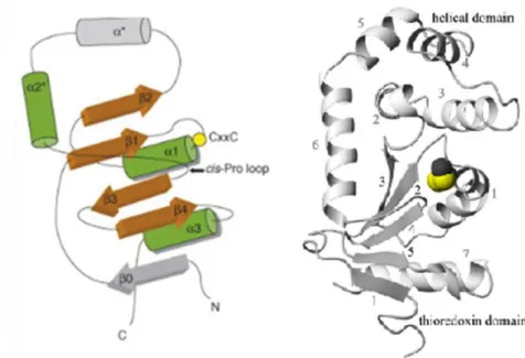
All proteins of the TRX superfamily share the structural characteristic of an α-helical domain juxtaposed with β-strands and with a pair of redox active cysteines
4 (Cys-X-X-Cys) located at the N-terminal end of the first helix at the active site of the enzyme (Figure 3) (Heras, et al., 2009; Guddat, et al., 1997; Ruddock, et al., 1996).
Figure 3: Common structural organization of DsbA fold. At left: Helices are illustrated in green and β
sheets in brown. The locations of the catalytic CxxC motif (shown by a yellow sphere) and the cis-Pro loop (arrow) are also specified. Data from: (Gruber, et al., 2006). On the right: Crystal structure representation of oxidized Vibrio cholerae DsbA. The elements of secondary structure are sequentially numbered from the N terminus. Helices are presented with black numbers and the grey numbers denote strands. The active site is presented in a CPK representation. In: (Horne, et al., 2007).
DsbAs are the most oxidizing proteins known, probably as a result of the CXXC
motif structure and, more precisely, due to an unusually low pKa of the most N-terminal
cysteine in the active site (Collet & Bardwell, 2002). The high reactivity of this Cys is
due to an electrostatic interaction with a nearby His which stabilizes the Cys in its
thiolate anion form (Guddat, et al., 1997). The redox potential of the enzyme also depends on the type of residues, XX, flanked by the two cysteines of the general motif (Ito & Inaba, 2008). Indeed the canonical sequence of the active site motif for TRX enzymes is C-P-H-C (Paxman, et al., 2009; Madonna, et al., 2006) but variations in the third residue (i.e the histidine) have been observed. In fact, Guddat and collaborators showed in 1997 that a mutation of the histidine residue of the active site motif leads to a significant decrease in the redox potential of these mutants (Guddat, et al., 1997).
As mentioned above, generally disulphide bonded proteins are more stable than their non-disulphide bonded forms yet it has been reported that the disulphide bond of DsbA is very unstable and that the stability of this protein is increased on disulphide
5 bond reduction (Zapun, et al., 1993). Furthermore, a previous study showed that oxidised DsbA from E. coli is more rapidly cleaved by proteases than its reduced form. These results suggest a lower stability and higher flexibility of the oxidized form of the enzyme which may have importance in its disulphide bond donation activity and in the the accommodation of substrate (Horne, et al., 2007).
1. 1. 1. WHY STUDY DsbA?
DsbAs have been the focus of much study over recent years, not only because of their fundamental interest, but also because of their potential applied importance. From a fundamental point of view, a better understanding of the implications of this enzyme in protein folding is of obvious importance. In relation to its applied interest, its potential for developments in the fields of biotechnology and medicine has led to a drastic increase in interest in this enzyme.
In biotechnology, the use of DsbA in catalysing the correct folding of disulphide bond containing proteins has led to suggestions for co-expression of this enzyme during recombinant protein production, for its use as an additive in cell free protein production systems (Kuroita, et al., 2007) and even for the refolding of misfolded proteins (Antonio-Pérez, et al., 2012). Indeed studies have shown that the use of this in cell free systems leads to higher production levels (approximately two fold higher) of active properly folded disulphide containing proteins (Kuroita, et al., 2007). Obviously, such increases in production levels warrants the use of DsbA as a tool in protein production procedures and the development of further more cheaply produced and more highly active DsbAs is called for.
In medicine, the key role of DsbA in catalysing the correct folding of many essential proteins, and in particular those that enable pathogenesis (i.e. the virulence factors), in pathogenic organisms has led to suggestions for this enzyme as a potential antimicrobial drug target (Lasica & Jagusztyn-Krynicka, 2007); Heras, et al., 2009; Shouldice, et al., 2011). DsbA has an essential role in pathogenesis as it catalyses the correct folding of virulence factors associated with adhesion (e.g. fimbriae, intimin), bacterial mobility (flagella) and host cell manipulation (e.g. toxins, such as the cholera and pertussis toxins) (see below for a more in-depth discussion) (Heras, et al., 2009; Shouldice, et al., 2011). Blocking DsbA activity would therefore interfere with the functioning of these virulence factors and hence impede the pathogens ability to cause disease. Indeed previous studies have demonstrated that hosts with defective DsbA
6 display reduced virulence in animal infection models (Bardwell, et al., 1991; Lin, et al., 2008; Heras, et al., 2009; Shouldice, et al., 2011). Therefore, better understanding DsbA and its inhibition opens up exciting new possibilities for novel antibacterial agents (Früh, et al., 2010). Indeed bacterial infections are a major cause of death in the world and this, in addition to current high levels of antibiotic resistance in many pathogenic bacteria, highlights the urgent need for new validated targets and for the design of new antibacterial agents against these targets. Due to its role in pathogenesis DsbA offers such a target for a new therapeutic approach. Nevertheless, it is important to note that drugs acting against DSB may not necessarily kill pathogens, but instead would impede or reduce bacterial pathogenesis by interfering with multiple essential virulence factors encoded by the pathogens (Heras, et al., 2009). However, this may not be a disadvantage as it may even result in less evolutionary pressure for bacteria to develop resistance (Heras, et al., 2009).
Some examples of virulence factors, how they intervene in the disease causing process and documented examples of the role of DsbA in their correct functioning will now be discussed.
1. 1. 2. 1. DsbA in cell adhesion
For a large number of bacterial pathogens the first and possibly the most important step is adhesion to the host cell. This process is essential for host colonisation and in establishing the disease. Adhesion is initially mediated by pili or fimbriae which are hair like structures typically made up of multiple protein subunits that propagate from the surface of the bacterium (Heras, et al., 2009). E. coli DsbA is reported to be important in the formation of disulphide bonds in the P fimbrial adhesion subunit protein PapG that recognises and binds to carbohydrates in the urinary tract surface (Heras, et al., 2009). DsbA has also been shown to be important in fimbriae construction in another urinary tract pathogen, Proteus mirabilis and plays a critical role in functional pili assembly for Vibrio cholerae colonisation mediated by the toxin co-regulated pilus (Tcp).
1. 1. 2. 2. DsbA in host cell manipulation
Following adhesion the success of bacterial colonisation is mainly dependent on the capacity to manipulate the hosts. Here, mass cell damage and destruction induced by secreted toxins and proteases occurs. Indeed, numerous secreted virulence factors and the secretion systems required for their discharge necessitates the DSB system to
7 catalyse their correct folding and function. This necessity shows their important role in this more advanced stage of bacterial pathogenesis.
Pathogens dispose of six different methods for secreting the toxins required for cell damage and DsbA activity is reported to be important in both type II and type III secretion systems (Lasica & Jagusztyn-Krynicka, 2007; Durand, et al., 2009). Type II secretion systems export proteins from the periplasm via a multimeric complex (Durand, et al., 2009) while type III secretion uses a multi-subunit molecular syringe like structure that directly injects the virulence proteins into the cytosol of eukaryotic cells (Heras, et al., 2009). In both of these cases DsbA is essential for correct folding of protein subunits which themselves are essential for the establishment of the correct structural conformation and function of the secretion apparatuses (Heras, et al., 2009). Furthermore, DsbA also acts as a catalyst in the structural assembly of many of the actual toxins to be secreted. Examples include functional assembly of: the cholera toxin of Vibrio cholera, the heat-labile enterotoxin assembly in E. coli and functional disulphide bond formation in almost the entire complex structure of the pertussis toxin of B. pertussis. Here, DsbA plays an important role in the structural assembly of a complex structure that includes six subunits and eleven intramolecular disulphide bonds (Heras, et al., 2009).
1. 1. 2. 3. DsbA in cellular spread and survival
Attachment through fimbriae permits bacteria to establish infections in cells. However mobility, which is the opposite phenotype, is also very important for virulence and bacterial fitness because it enables the bacteria to spread across the host cells (Heras, et al., 2009). Studies in several bacteria show that mutation of dsbA impedes functional flagella production and hence also bacterial mobility. As an example, in E. coli, DsbA is reported to be required for catalysing the formation of disulphide bonds in the FlgI protein that acts as the flagellar P-ring motor, for cell mobility (Dailey & Berg, 1993).
1. 1. 2. A COMPARATIVE STUDY: UNDERSTANDING DsbAs AND COLD-ADAPTATION
As part of a long term goal of obtaining a better understanding of DsbAs so as to enable the development of their use in protein production and, more importantly, in the design of novel antibacterial agents, a comparative study of a cold adapted and
8 mesophilic homolog has been initiated (Collins et al, 2010). To attain the objectives, the structures-stabilities-functions and also dynamics of both the oxidised and reduced states of the two DsbA homologs, adapted to low (~5°C) and moderate temperatures (~37°C), will be compared. The results obtained will give clues on Nature’s strategies for modifying proteins to attain a desired catalytic rate within the environmental constraints and will help show how evolution optimises and balances dynamics, stability and activity (Tomatis, et al., 2008). Mutagenesis studies will then be carried out to better investigate these observations. DsbA is a well studied enzyme, both at the structural and biochemical levels (Heras, et al., 2009; Horne, et al., 2007; Schirra, et al., 1998) but most previous studies have focused on individual enzymes under specific conditions and we believe that an in-depth comparative approach should offer a more ‘complete picture’ and better pinpoint those regions important for function and stability. This, in turn, should aid in identifying the most appropriate regions for targeting by inhibitor.
The comparative studies should also allow for a better comprehension of life in the extremes and in particular of enzyme adaptation to various temperatures. More specifically, the study of a cold adapted DsbA should enable a better understanding of the molecular determinants of low temperature adaptation in enzymes. Furthermore, the expected high activity of the cold-adapted enzyme could allow for the development of a novel highly active tool for cell free protein synthesis of disulphide bond containing proteins.
A more in-depth discussion of the state of the art in cold-adaptation will now be presented.
1. 1. 3. UNDERSTANDING LIFE IN COLD ENVIRONMENTS
Life on Earth is ubiquitous, it is not restricted to those regions which we, as humans, classify as being normal but it is also found in those ‘extreme regions’ on Earth such as the deep seas, the polar regions, the volcanic regions or/and the saline
pools. On the one hand, these regions constitute the major portion of the Earth’s
surface and are far from being sterile (Lonhienne, et al., 2000), but on the other hand, to survive these various extremes, these organisms had to adapt at all levels of organization; from structural to physiological adaptation. The present study is focused on adaptation to low temperatures and how organisms are able to not only tolerate, but to grow and maintain high enzyme activities in this permanently extreme condition.
9 Cold-adapted microorganisms capable of growing at 0ºC were identified by Forster as early as 1887 when he isolated them from fish (Zecchinon, et al., 2001). In fact, for some cold adapted organisms, low temperatures are not only optimal, but mandatory, for continued cell proliferation, with moderate to high temperatures (e.g., >12 °C (Xu, et al., 2003)) being inhibitory. These unique organisms, called psychrophiles, have effectively colonized cold environments thanks to successful adjustments which counteract the negative effects of low temperatures. These negative effects include a reduction of reaction rates, alterations in enzyme-substrate interaction strength, increase in solvent viscosity and a modified solubility of proteins, gases and salts and finally also, protein cold-denaturation (Georlette, et al., 2004). Psychrophiles have overcome all these challenges and reveal metabolic fluxes at low temperatures more or less comparable to those shown by mesophilic species living at moderate temperatures
(Zecchinon, et al., 2001). Indeed, bacterial cell densities as high as 107 ml-1 have been
found in the Antarctic oceans, similar to the densities of temperate waters (Gerday, et al., 2000).
The enzymes produced by psychrophilic organisms have adapted to temperatures close to the freezing point of water and typically display high catalytic rates and low stability as compared to their higher temperature adapted homologs i.e enzymes from mesophiles and thermophiles. Indeed many enzymes are incapable of carrying out their function under these low temperature conditions due to the reduced kinetic energy available at low temperatures, this effectively ‘freezing’ enzymatic motion. Currently it is hypothesised that psychrophilic enzymes have evolved an increased flexibility to overcome this (Collins, et al., 2003), thereby allowing for a high activity but also leading to the observed reduced stability. A reduced number or strength of intramolecular interactions are frequently reported for these enzymes as compared to their higher temperature adapted homologs (Gerday, et al., 1997) and it has ben hypothesized that these reduced interactions allow for the proposed flexibility of these enzymes and hence the enhanced activity at low temperatures. The actual molecular basis for the adaptation is enzyme specific however it still completely understood and direct evidence of the proposed increased flexibility is scant, with previous attempts to demonstrate this leading to conflicting results.
1. 1. 4. 1. The psychrophilic – mesophilic pair
This study is centred on a cold adapted thiol-disulphide oxidoreductase from a Gram negative psychrophilic bacterium (Pseudoalteromonas haloplanktis TAC125)
10 which has been isolated from an Antarctic coastal sea water sample collected in the vicinity of the French Antarctic station at Dumont d’Urville in Terre Adélie, Antarctica (66º 40’ S; 140º 01’ E) (Médigue, et al., 2005; Collins, et al., 2003). The gene encoding the cold-adapted enzyme has been cloned and the protein overexpressed using the pET22b(+)/E. coli BL21(DE3) expression system and purified from the periplasmic
extracts. Production was carried out with Terrific Broth medium, using isopropyl β-D
-1-thiogalactopyranoside (IPTG) for induction, and purification involved a combination of hydrophobic interaction chromatography and anion exchange chromatography. The production and purification procedures were not optimised and production levels were approximately 50 mg/L with large losses during purification being noted. Furthermore, precipitation of the protein, and in particular of the oxidised form, led to large losses of protein over time and no biochemical, dynamics or activity studies were carried out on
this protein. Backbone and side-chain 1H, 15N and 13C NMR assignments for the
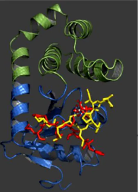
reduced form were however reported (Collins, et al., 2010a) and the NMR structure has been recently determined (Figure 4).
Figure 4: Structure of the psychrophilic PhDsbA revealing the thioredoxin domain in blue and in green the
α-helical domain. Peptide substrate (yellow) and the oxidising loop of PhDsbB (red) were overlayed onto the structure of PshDsbAp by aligning it with the E.coli DsbA structural complexes DsbA-peptide and DsbA-DsbB. In: (Collins, et al., 2010a)
This cold adapted protein was found to be very similar to previously reported homologous mesophilic DsbAs with the 4 α-helices of the helical domain inserted into a thioredoxin like fold composed of a central 5 stranded β-sheet flanked by 3 α-helices.
11 In addition, this contains the consensus DsbA active site sequence (i.e. Cys-Pro-His-Cys) at a break in the first α-helix in the thioredoxin-like domain.
In contrast to the psychrophilic DsbA, a large number of mesophilic DsbAs have been studied in detail, both at the structural and biochemical levels (Heras, et al., 2009; Horne, et al., 2007; Schirra, et al., 1998). Examples include DsbAs from Vibrio cholera (Horne, et al., 2007), Escherichia. coli (Mössner, et al., 1998; Fabianek, et al., 2000), Neisseria meningitidis (Vivian, et al., 2009) Salmonella enterica serovar Typhimurium (Heras, et al., 2010) and Staphylococcus aureus (Williams, et al., 2010). Of these, that from Vibrio cholera is one of the best understood, with both NMR and crystallographic structures of both the oxidised and reduced states being reported as well as investigations of activity, stability and dynamics (by NMR) (Horne, et al., 2007). This mesophilic Vibrio cholerae DsbA is to be used for comparison in this project, it has already been cloned, successfully overexpressed in E. coli and purified, and protocols for these have already been optimised and reported (Horne, et al., 2007).
The availability of in-depth information for mesophilic enzymes homologous to the cold adapted protein of the present study and in particular for the Vibrio cholera DsbA should allow for a more comprehensive comparative analysis of the activity, stability, structure and dynamics of the enzymes. This should enable a better understanding of structure and function relationships in DsbAs as well as of cold adaptation in this enzyme.
1. 1. 4. THE pET22B(+)/E. COLI BL21(DE3) EXPRESSION SYSTEM
The first report of the Gram-negative, rod-shaped bacterium, Escherichia coli, was made in 1885 by Theodor Escherich. Escherichia coli is an abundant inhabitant of the mammalian colon and is one of the most thoroughly studied organisms known (Jeong, et al., 2009). It is well understood, easy to manipulate, grows rapidly on relatively cheap media (Khow & Suntrarachun, 2012) and is described as one of the most efficient vehicles for over-expression of both eukaryotic and prokaryotic proteins (Miroux & Walker, 1996). The current term ‘over-expression’ is here mentioned to define the capacity to produce target proteins at levels much higher than those of its own repertoire of proteins. Studies reveal that monomeric proteins that contain few cysteines and have an average size smaller than 60 kDa will give good production in an E. coli expression host (Bell, 2001). Indeed, in some cases up to 60 % of the total protein produced can be constituted by the recombinant protein.
12 The pET22b(+)/E. coli BL21(DE3) expression system based on the bacteriophage T7 promoter expression system is one of the most widely used laboratory systems for recombinant protein expression in E. coli. This is based on an inducible machinery that permits control of target gene expression. It consists of a lac operator sequence directly downstream of the T7 promoter, and the gene for the lac repressor (termed lacI) all encoded on the expression vector. The E.coli BL21 expression host used with this system contains a chromosomal copy of the T7 DE3 lysogen which comprises the T7 polymerase gene under control of the E. coli lacUV5 promoter as well as a chromosomal copy of the lacI repressor gene. In DE3 lysogens the lac repressor acts not only at the lacUV5 promoter in the host chromosome and thereby repressing T7 RNA polymerase gene transcription by the host polymerase, but also at the vector at the T7lac promoter, blocking the transcription of the target gene. The lacUV5 and T7 lac promoters are inducible with IPTG or lactose, the addition of which to the growth medium ‘inactivates’ the lac I repressor and induces the production of the T7 RNA polymerase whereupon binding to the T7 lac promoter transcribes the target DNA (Figure 5).
Figure 5: Representative scheme of IPTG induction in the pET/E. coli BL21(DE3) expression system. In:
(Novagen, 2003)
Production with this system can make use of batch or fed-batch approaches, with batch production in shake flasks being the most common at a laboratory scale. Typically, the most frequently used shake flask production approach uses lysogeny
13 broth (LB) with IPTG induction at the mid-exponential phase of growth (Teulé, et al., 2009) but the use of richer media such as terrific broth (TB) or super broth (SB) has recently become common place. While being a highly used and efficient method for recombinant protein production in E. coli, this system does however sometimes suffer from low yields of protein product (mg/L) (Teulé, et al., 2009), with yields being dependent on the actual system used, the target protein, induction conditions and environmental factors. In fact, process optimisation to maximise productivity is an essential first step in the production of any recombinant protein.
In the case of the cold-adapted DsbA of the present study, the specific expression system used is the pET22b(+)/E. coli BL21(DE3) system already described above. Here, the use of the pET22b(+) expression vector allows for expression of unmodified and untagged DsbA in the host periplasm (Novagen, 2003). The wild-type signal sequence of DsbA which targets the produced protein to the periplasmic space and is removed during the translocation process is used.
14 1. 1. 5. MEASUREMENT OF DsbA ACTIVITY
Several assays have previously been described for measuring DsbA activity. These assays are centred on the observation and/or quantification of the conformational and/or chemical differences between the two states of the protein, more precisely the reduced and oxidised states. The oxidised state displays a disulphide bond in the active site of DsbA and can oxidise any substrate that exhibits two free cysteines, itself becoming reduced in the process. Several strategies to measure DsbA activity have been reported, these include: an insulin activity assay, Ellmans assay, HPLC analysis, a fluorimetric assay, an SDS-PAGE based detection method and an assay using a synthetic fluorescent peptide. In this study the insulin assay will be used to monitor the reducing activity of the protein studied.
1. 1. 6. PSYCHROPHILIC DsbA PURIFICATION
Chromatography, which separates compounds on the basis of their differential partitioning between two phases (i.e. a mobile phase and a stationary phase), will be used to purify the cold adapted DsbA from the E. coli endogenous proteins. A variety of chromatographic approaches can be used for protein purification, including gel filtration
chromatography, ion-exchange chromatography, hydrophobic interaction
chromatography and affinity chromatography. Of these, hydrophobic interaction and ion-exchange chromatographies have been previously investigated for purification of the cold adapted homologs DsbA and will be encountered in this study.
Hydrophobic interaction chromatography (HIC): This technique makes use of subtle differences in protein surface hydrophobicity for separation. Here a reversible interaction occurs between exposed hydrophobic patches on the protein and hydrophobic ligands (e.g. phenyl, octyl, butyl, isopropyl etc.) on the column matrix. It is very similar to reverse phase chromatography but the ligands used are much less hydrophobic and hence less extreme elution conditions are required, thereby avoiding the denaturing conditions often used in reverse phase chromatography. Hydrophobic binding in HIC is often facilitated by use of neutral salts effective in ‘salting out’ (e.g. ammonium sulphate, NaCl), with these reducing protein solvation and leading to higher exposure of protein hydrophobic groups and thus improving binding. Reduction of the salt concentration is used in protein elution and these are eluted in order of increasing hydrophobicity (Amersham Pharmacia Biotech, 2000).
15 Ion exchange chromatography (IEX): This technique is based on the reversible interaction between a charged protein and an oppositely charged chromatographic medium. Proteins carry many ionisable groups such as the basic groups on the side chains of lysine, arginine and histidine as well as the acidic groups on the side chains of aspartic acid and glutamic acid residues. The charge of these side chains is
influenced by the dissociation constant (pKa) of the side chains, the environment of
these side chains (i.e. their neighbouring residues in space) as well as the pH of the solution. In turn, the charge, number and structural positioning of these ionisable groups determines the net charge of the protein in a particular condition. Indeed, knowledge of the pI of a protein, this latter being defined as the pH at which the protein carries a net zero charge i.e. equal number of positive and negative charges, and control of the pH of the solution can be used in deciding the type of ion exchange approach to use. Namely, at a pH above the pI proteins carry a net negative charge and will bind to an anion exchanger (positively charged matrix) whereas at a pH below the pI a net positive charge is displayed and a cation exchanger (negatively charged matrix) should be used (Amersham Biosciences, 2002).
1. 1. 7. OBJECTIVES
The present study will be focused on the cold-adapted DsbA termed here as PhDsbA (Ph represents Pseudoalteromonas haloplanktis). Within the long term objectives of obtaining a better understanding of the structure-function relationship of DsbAs as well as of cold-adaptation in this enzyme, here we will focus on the following multiple objectives:
Figure 7: Anion and cation exchange chromatography. In anion exchange chromatography (shown
at left) the matrix carries a positive charge and attracts negatively charged molecules. On the right cation exchange chromatography is illustred, this is based on the attraction of positively charged molecules by a negatively charged matrix. In: (Amersham Biosciences, 2002)
16
Optimisation of the batch production in shake flask of PhDsbA with the pET/E. coli BL21(DE3) expression system
Current production levels are approximately 50 mg/L and here culture medium, culture aeration (via medium volume to flask volume ratio), induction time and induction period will be investigated in an attempt to improve production levels
Development of a simplified purification protocol for PhDsbA
An initial attempt to purify this protein involved periplasmic extraction, HIC, dialysis and IEX, but this was found to result in large losses of PhDsbA. Here we will attempt to develop a simplified efficient purification protocol for this study.
Identify determinants of cold-adaptation.
A comparative structural analysis of PhDsbA with its mesophilic homologs and in particular with that from Vibrio cholera will be carried out so as to identify mutations or alterations which may be important in adaptation to low temperatures. Primary, secondary and tertiary structures will be compared.
Construction of mutants
Mutants identified in the structural comparison will be constructed so as to allow for identification of their role in the protein.
17
18 2. 1. BIOLOGICAL MATERIAL
2. 1. 1. Escherichia coli strains
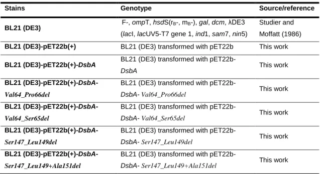
In this study, the principal working strain used was E. coli BL21 (DE3), a descendent strain from the native E. coli strain B (Daegelen, et al., 2009). Transformants of this working strain that were constructed and used in this study to produce wild-type and mutant DsbA are described in Table 1.
Table 1: Strains used in this work
Stains Genotype Source/reference
BL21 (DE3) F-, ompT, hsdS(rB-, mB-), gal, dcm, λDE3
(lacI, lacUV5-T7 gene 1, ind1, sam7, nin5)
Studier and Moffatt (1986)
BL21 (DE3)-pET22b(+) BL21 (DE3) transformed with pET22b This work
BL21 (DE3)-pET22b(+)-DsbA BL21 (DE3) transformed with
pET22b-DsbA This work
BL21
(DE3)-pET22b(+)-DsbA-Val64_Pro66del
BL21 (DE3) transformed with
pET22b-DsbA- Val64_Pro66del This work
BL21
(DE3)-pET22b(+)-DsbA-Val64_Ser65del
BL21 (DE3) transformed with
pET22b-DsbA- Val64_Ser65del This work
BL21
(DE3)-pET22b(+)-DsbA-Ser147_Leu149del
BL21 (DE3) transformed with
pET22b-DsbA- Ser147_Leu149del This work
BL21
(DE3)-pET22b(+)-DsbA-Ser147_Leu149+Ala151del
BL21 (DE3) transformed with
pET22b-DsbA- Ser147_Leu149+Ala151del This work
All plasmids used in this work are listed and detailed in Table 2 with the respective characteristics and sources. The pET22b(+)-dsbA construct provided for this study contains the dsbA gene inserted in the pET22b(+) multiple cloning site between the NdeI and EcoRI restriction sites. The inserted sequence contains the wild–type N-terminal signal sequence for periplasmic expression.
19 Table 2: Plasmids used in this work
Stains Characteristics Source/reference
pET22b(+) amp
R
, T7lac, optional C-terminal His.Tag® sequence,
signal sequence for potential periplasmic localization Novagen
pET22b(+)-dsbA pET 22b, dsbA Tony Collins
collection
pET22b(+)-DsbA-Val64_Pro66del pET22b(+)-DsbA-Val64_Pro66del
This work
pET22b(+)-DsbA-Val64_Ser65del pET22b(+)-DsbA-Val64_Ser65del This work
pET22b(+)-DsbA-Ser147_Leu149del pET22b(+)-DsbA-Ser147_Leu149del This work
pET22b(+)-DsbA-Ser147_Leu149+Ala151del pET22b(+)-DsbA-Ser147_Leu149+Ala151del This work
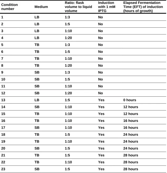
2. 1. 2. DsbA Production: optimisation of medium, aeration and induction (time, period)
In an attempt to optimise production levels of the cold adapted DsbA we investigated various media (Table 3) and various production conditions for induced and non-induced cultures (Table 4).
Table 3: Composition of the media used in this work
The rich media TB, SB and LB were investigated for both induced and non-induced cultures. Aeration was investigated by varying the medium volume to flask volume ratio from 1:3 to 1:20, with the lower medium volume (i.e. higher ratio) allowing for better culture mixing and hence better aeration. Elapsed fermentation times (EFT) before induction with 1 mM IPTG of 12, 16, 24 and 28 hours, corresponding Medium Composition
LB Bacto tryptone (1% w/v), yeast extract (0.5% w/v), and sodium chloride (0.5% w/v)
TB Bacto tryptone (1.2% w/v), yeast extract (2.4% w/v), glycerol 99.5 % (0.4% w/v), 70 mM
K2HPO4.3H2O and 20 mM KH2PO4
20 respectively, to the early, mid, declining exponential and stationary phases of growth were also examined and compared to non-induced cultures. Induction periods of 2, 4, 6
and 12 hours after induction with 1 mM isopropyl β-D-1-thiogalactopyranoside (IPTG)
were investigated. Cultivations in the absence of induction were carried out to
determine growth curves. Biomass (OD600nm) and DsbA production levels (SDS-PAGE)
were compared for all production optimisation conditions. PROTOCOL:
Plate out glycerol cultures or fresh transformants of the producing strains on
LB-agar plates containing 100 µg/mL of ampicillin as the selection marker and incubate overnight at 37 ºC.
Inoculate 100 mL LB+ampicillin preculture in a 500 ml erlenmeyer with a cfu of
the plate culture. Incubate for approximately 15 hours at 25 ºC and 200 rpm.
Inoculate production cultures (in 500 mL erlenmeyers) to an initial OD600nm of
0.1. Incubate at 20 ºC and 200 rpm (25 mm orbital).
Induce when required with 1 mM IPTG. Collect 0.5 mL cell pellet samples at 2,
4, 6 and 12 hours after induction for determination of production levels by SDS-PAGE. Monitor biomass levels throughout the productions by measuring OD600nm.
21 Table 4: Production conditions examined
Condition number Medium Ratio: flask volume to liquid volume Induction with 1 mM IPTG Elapsed Fermentation Time (EFT) of induction (hours of growth) 1 LB 1:3 No 2 LB 1:5 No 3 LB 1:10 No 4 LB 1:20 No 5 TB 1:3 No 6 TB 1:5 No 7 TB 1:10 No 8 TB 1:20 No 9 SB 1:3 No 10 SB 1:5 No 11 SB 1:10 No 12 SB 1:20 No 13 LB 1:5 Yes 0 hours 14 SB 1:10 Yes 12 hours 15 TB 1:10 Yes 12 hours 16 TB 1:10 Yes 16 hours 17 SB 1:10 Yes 16 hours 18 TB 1:5 Yes 24 hours 19 TB 1:10 Yes 24 hours 20 SB 1:5 Yes 24 hours 21 TB 1:5 Yes 28 hours 22 TB 1:10 Yes 28 hours 23 SB 1:5 Yes 28 hours 2. 1. 3. SDS-PAGE analysis
12 % SDS-PAGE was used for analysis of protein production levels as well as for monitoring the purification. Here proteins are linearized and imparted with a negative charge by SDS and DTT pre-treatment before being separated on the basis of differences in their size on an acrylamide-bis-acrylamide gel (see gel components in Table 5). Separated protein bands are then visualised by staining with Coomassie Blue which interacts ionically and hydrophobically with proteins.
22 Table 5: SDS-PAGE stacking and running gel composition.
Solution Stacking gel Running gel (12%)
Acrylamide 40% 216 µL 1.62 mL Bis-acrylamide 2% 117 µL 960 µL 0.25 M Tris-HCl (pH 6.8), 2%SDS 1.1 mL - 0.75 M Tris-HCl (pH 8.8), 2%SDS - 2.8 mL TEMED 3 µL 4.5 µL APS (10%) 12.5 µL 30 µL H2O 750 µL 170 µL Final volume 2.2 mL 5.6 mL
PROTOCOL (for preparation of production sample for SDS-PAGE):
Collect cells from 500 µL production samples by centrifuging at maximum
speed for 5 minutes and discard supernatant.
Add 100 µL of a 50 mM Tris, 1 mM EDTA solution at pH 8 and mix well.
Add 25 µL of SDS-PAGE loading solution (10 % SDS, 10 mM
β-mercaptoethanol, 20 % glycerol, 0.2 M Tris at pH 6.8 and bromophenol blue) and vortex.
Centrifuge at max speed for 25 minutes.
Run 4 µL of supernatant on a 12 % SDS-PAGE gel at a constant current flow of
10 amps.
PROTOCOL (for preparation of purification samples for SDS-PAGE):
Gently mix samples by inversion.
To 20 µL of sample add 5 µL of SDS-PAGE loading solution.
Mix to homogenize and run 20 µL in a 12 % SDS-PAGE gel at a constant
current flow of 10 amps.
The Coomassie Blue staining solution is composed of 10 % acetic acid in deionised water with Coomassie Brilliant Blue R-250 addition until the solution attains a strong blue colour. The de-staining solution has the same composition with the exception of the Coomassie Brilliant Blue R-250 component.
23 ImageJ was employed for quantification of Coomassie Blue stained protein bands.
2. 1. 4. DsbA PURIFICATION
A previously used purification protocol was optimised in this project. This involved the following steps: DsbA periplasmic extraction, hydrophobic interaction chromatography, dialysis for buffer exchange and ion exchange chromatography.
A Pharmacia Biotech FPLC system composed of a LCC-501 Plus LKB controller, two P500 pumps, a UV-M II optical unit, a FRAC 100 fraction collector and a Rec 102 chart recorder was used for all chromatographic steps.
2. 1. 4. 1. DsbA periplasmic extraction
An osmotic shock when transferring cells from a high sucrose concentration to a
dilute MgSO4 solution in conjunction with a thermal shock by rapidly decreasing the
temperature from approximately 25 ºC (room temperature) to 4 ºC allows for liberation of periplasmic proteins.
PROTOCOL:
Collect cells by centrifugation at 7000 rpm for 5 minutes at 4 ºC.
Add 1/40th the volume of the initial culture volume of 30 mM Tris-HCl at pH 8
and gently ressuspend pellet, on ice, with a Pasteur pipette.
Add 1/40th the volume of initial culture volume of 2×PEB and mix by gentle
inversion. (2×PEB is composed of 30 mM Tris-HCl at pH8, 40 % of sucrose and 2 mM EDTA)
Transfer to 40 mL centrifuge tubes and leave for 20 minutes at room
temperature.
Centrifuge at 14000 rpm for 30 minutes at 20 ºC and discard supernatant.
Immediately add 1/20th the volume of initial culture volume of cold 5 mM MgSO4
and mix well.
Leave tubes for 20 minutes on ice.
Centrifuge tubes at 14000 rpm for 40 minutes at 4 ºC.
24
Add 0.1 mM calcium and 5 -10 units of DNase (Fermentas®) and incubate for 10
minutes at room temperature.
Store at 4 ºC.
2. 1. 4. 2. Hydrophobic interaction chromatography (HIC)
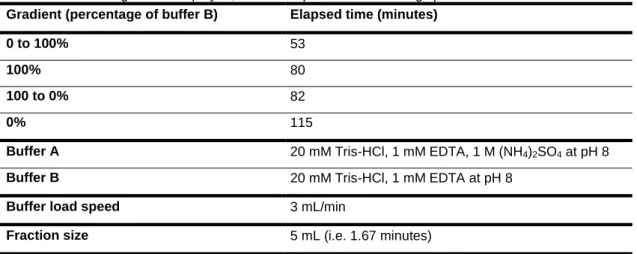
HIC is based on the reversible binding of proteins with exposed hydrophobic groups. A 1.6 cm x 20 cm, 40 mL Phenyl Sepharose High Performance (Pharmacia) column was used. This contains a hydrophobic phenyl group covalently coupled to a highly porous cross-linked 4 % agarose matrix. The sample solution is filtered through a 0.45 µm filter and 1 M ammonium sulphate added before loading to the column. The ammonium sulphate is added to enhance protein hydrophobicity and hence column binding and a gradient of decreasing ammonium sulphate (buffer B) is used for elution. See table 9 for details of the chromatographic conditions used.
Table 6: Details of gradient employed, buffer composition and loading speed used in HIC. Gradient (percentage of buffer B) Elapsed time (minutes)
0 to 100% 53
100% 80
100 to 0% 82
0% 115
Buffer A 20 mM Tris-HCl, 1 mM EDTA, 1 M (NH4)2SO4 at pH 8
Buffer B 20 mM Tris-HCl, 1 mM EDTA at pH 8
Buffer load speed 3 mL/min
Fraction size 5 mL (i.e. 1.67 minutes)
2. 1. 4. 3. Dialysis
This is based on the diffusion of solutes across a semi-permeable membrane from a region of high concentration to a region of low concentration. It is used for the exchange of buffers.
PROTOCOL:
Load sample into a pre-wetted 12000-14000 kDa MWCO dialysis tubing and dialyse, with constant mixing, overnight in 4 to 5 litres of the appropriate buffer (i.e. buffer A for the IEX).
25 2. 1. 4. 4. Ion exchange chromatography (IEX)
Ion-exchange chromatography separates proteins based on their charge. A 1.6 cm x 20 cm, 40 mL DEAE Fast Flow Sepharose (Pharmacia) column was used.
This anion exchanger contains the positively charged reactive group
diethylaminoethanol (DEAE) covalently linked to a sepharose (a polysaccharide polymer) matrix. A gradient of increasing NaCl concentration (buffer B) is used for protein elution.
In the present study the pH of the equilibration buffer used was optimised to maximise DsbA binding: pH 7.2, 7.5 and 8.0 were investigated.
Table 7: Details of the gradient employed, buffer composition and running speed used in IEX. Gradient (percentage of buffer B) Elapsed time (minutes)
0 to 50% 56
50 to 100% 59
100% 79
100 to 0% 81
0% 105
Buffer A 10 mM MOPS, 1 mM EDTA tested at pH 7.2; 7.5 and 8.
Buffer B 10 mM MOPS, 1 mM EDTA, 1 M NaCl tested at pH 7.2;
7.5 and 8.
Buffer load speed 5 mL/min
Fraction size 5 mL (i.e. 1 minute)
2. 1. 5. DsbA reducing activity assay
DsbA reducing activity was confirmed according to a procedure described by Arne Holmgren in 1979. This assay is based on the reduction of insulin disulphide bonds by DsbA under the conditions used and measuring the resultant insulin precipitation by monitoring the increase in absorbance at 650 nm. DTT is used to recycle oxidised DsbA following catalysis.
The assay was used to monitor the purification process as well as to determine the activity of wild-type and mutant DsbAs.
26 PROTOCOL (for insulin solution preparation at 10 mg/mL):
Add 50 mg of insulin to 4 mL of 0.05 M Tris-HCl at pH 8.
Add 1M HCl to adjust pH between a range of 2 or 3.
Immediately add 1M NaOH to adjust pH to 8.
Bring the volume to 5 mL with deionized water.
PROTOCOL:
Add a known concentration of DsbA to a 1 mL cuvette and add 2 mM EDTA,
0.75 mg/mL insulin and 20 mM MOPS at pH 7 to bring the final volume to 1 mL.
Mix gently by inversion and blank the spectrophotometer at 650 nm with this
solution
Add 0.33 mM DTT, mix by inversion.
Immediately monitor the change in A650nm over time. A Genesys 20 (R)
spectrophotometer from Thermo Spectronic (R) was used.
A negative control with all components except DsbA should also be monitored.
2. 1. 6. Sugar detection assay
Sugar detection was performed according to a protocol first described in 1956 by Dubois and collaborators (Dubois, et al., 1956). This is a quantitative and sensitive colorimetric test where sulphuric acid in the presence of phenol is used for sugar detection. The sugars are converted to hydroxymethylfufurals in the hot acidic conditions and form a green product on interaction with phenol. Even small quantities of sugars can be detected and quantified based on the direct relationship between the
enhancement of colour (A490nm) and the sugar quantity.
PROTOCOL:
To 1 mL of sample solution (in water) add 1 mL of a 5 % phenol solution.
Add 5 mL of 96 % sulphuric acid to each tube and mix well.
Leave 10 minutes at room temperature, mix and place in a water bath at 25 to
30 ºC for 20 minutes.
Blank spectrophotometer with water.
27
Calculate the amount of total carbohydrate using a standard curve prepared
using 0.02 – 0.1 mg/mL glucose.
All reactions were carried out in glass tubes in a laminar flow hood.
2. 1. 7. Bradford assay for protein quantification
The Bradford assay was used to calculate protein concentration spectrophotometrically at 595 nm. This method is based on the reaction between the
NH3+ and possibly also the aromatic groups of amino-acids with the coomassie blue
reagent (Kruger, 2002). For this purpose a standard curve with known concentrations of BSA was used.
Bradford Reagent: Dissolve 100 mg Coomassie Blue G-250 in 50 ml 95% ethanol, add 100 ml 85% (w/v) phosphoric acid to this solution and dilute the mixture with 850 mL of water. Leave to agitate overnight and filter twice.
PROTOCOL:
Place 5 to 20 µL of sample solution in a 1 mL cuvette and bring to a final
volume of 100 µL with distilled water.
Add 1 mL of Bradford solution and mix immediately.
Leave at room temperature to allow reaction to develop for 10 minutes.
Blank spectrophotometer with the mixture without enzyme sample solution.
Read absorbance at 595 nm and calculate concentration from a standard curve
prepared with BSA concentrations of 5 to 500 µg/ml.
2. 1. 8. MUTANT CONSTRUCTION
In an attempt to better understand adaptation to temperature, structural differences between DsbAs adapted to various temperatures were identified and a number of mutants designed and prepared so as to investigate these differences. This involved a number of steps.
Mutant selection: sequence and structure comparisons so as to identify
structural differences in DsbAs adapted to various temperatures.
28
Introduction of selected mutations into cold-adapted DsbA gene sequence by
inverse PCR, recircularisation of vector by ligation, and transformation to an expression host (Scheme 1).
Plasmid isolation, confirmation of mutation by restriction digestion analysis and
sequencing.
Scheme 1: Representation of process used for deletion of amino acids in the PhDsbA sequence.
2. 1. 9. 1. Mutant selection: structure comparisons
Here, the primary, secondary and tertiary structures of the cold-adapted enzyme were compared to homologous DsbAs. Major differences in the cold-adapted DsbA primary sequence (and in any other cold-adapted DsbA sequence available in the UniProtKB databank) were first identified by sequence comparisons of DsbAs available at the UniProtKB databank. Mutations identified in the primary sequences were then investigated in more detail by comparisons of the tertiary structures of the cold-adapted (reduced state) DsbA with the E. coli and V. cholera DsbAs.
DsbA sequences for primary structure analysis were identified and retrieved
from the uniprotkb database by a similarity search with the PhDsbA amino acid sequence (e-value) and by manually retrieving sequences based on a bibliography