Imaging in idiopathic pulmonary fibrosis: diagnosis
and mimics
Bruno Hochhegger,IEdson Marchiori,IIMatheus Zanon ,I,* Adalberto Sperb Rubin,IIIRenata Fragomeni,IV
Stephan Altmayer,ICarlos Roberto Ribeiro Carvalho,VBruno Guedes BaldiV
IDepartamento de Radiologia, Laboratorio de Pesquisas em Imagens Medicas (LABIMED), Irmandade Santa Casa de Misericordia de Porto Alegre,
Universidade Federal de Ciencias da Saude de Porto Alegre, Porto Alegre, RS, BR.IIDepartamento de Radiologia, Universidade Federal do Rio de Janeiro,
Rio de Janeiro, RJ, BR.IIIDepartamento de Pneumologia, Irmandade Santa Casa de Misericordia de Porto Alegre, Universidade Federal de Ciencias
da Saude de Porto Alegre, Porto Alegre, RS, BR.IVDepartamento de Patologia, Universidade Federal de Ciencias da Saude de Porto Alegre, Porto
Alegre, RS, BR.VDivisao Pulmonar, Instituto do Coracao (InCor), Hospital das Clinicas HCFMUSP, Faculdade de Medicina, Universidade de Sao Paulo,
Sao Paulo, SP, BR.
Hochhegger B, Marchiori E, Zanon M, Rubin AS, Fragomeni R, Altmayer S, et al. Imaging in idiopathic pulmonary fibrosis: diagnosis and mimics. Clinics. 2019;74:e225
*Corresponding author. E-mail: mhgzanon@hotmail.com
Idiopathic pulmonary fibrosis is a chronic disease of unknown etiology that usually has a progressive course and is commonly associated with a poor prognosis. The main symptoms of idiopathic pulmonary fibrosis, including progressive dyspnea and dry cough, are often nonspecific. Chest high-resolution computed tomography is the primary modality used in the initial assessment of patients with suspected idiopathic pulmonary fibrosis and may have considerable influence on subsequent management decisions. The main role of computed tomo-graphy is to distinguish chronic fibrosing lung diseases with a usual interstitial pneumonia pattern from those presenting with a non-usual interstitial pneumonia pattern, suggesting an alternative diagnosis when possible. A usual interstitial pneumonia pattern on chest tomography is characterized by the presence subpleural and basal predominance, reticular abnormality honeycombing with or without traction bronchiectasis, and the absence of features suggestive of an alternative diagnosis. Idiopathic pulmonary fibrosis can be diagnosed according to clinical and radiological criteria in approximately 66.6% of cases. Confirmation of an idiopathic pulmonary fibrosis diagnosis is challenging, requiring the exclusion of pulmonary fibroses with known causes, such as asbestosis, connective tissue diseases, drug exposure, chronic hypersensitivity pneumonitis, and other forms of idiopathic interstitial pneumonitis. The histopathological hallmark of usual interstitial pneumonia is a heterogeneous appearance, characterized by areas of fibrosis with scarring and honeycombing alternating with areas of less affected or normal parenchyma. The aim of this article was to review the clinical, radiological, and pathological features of idiopathic pulmonary fibrosis and of diseases that might mimic idiopathic pulmonary fibrosis presentation.
KEYWORDS: Differential Diagnosis; Idiopathic Pulmonary Fibrosis; Radiology; Pathology; Interstitial Lung Diseases.
’ INTRODUCTION AND HISTORY
Idiopathic pulmonary fibrosis (IPF) is a chronic disease of unknown etiology that usually has a progressive course, and IPF is commonly associated with a poor prognosis. Although IPF has been described extensively in the litera-ture, some controversy exists regarding its history. The first description of IPF as a new clinical and pathological entity is usually attributed to a 1933 report by Hamman & Rich (1). Nevertheless, many reports in German have previously
published descriptions of autopsy findings that were consis-tent with IPF (2). Patients presented with progressively wors-ening cough and dyspnea, and autopsies usually revealed hypertrophied right ventricles, small rigid lungs with wid-ened and thickwid-ened bronchioli, and an increased quantity of interstitial tissue without pleural adhesions (2). In these German reports, different terms were proposed to describe these interstitial changes, namely, cirrhosis cystica pulmonum andlymphangitis reticularis pulmonum(2).
Clinical and laboratory assessment
A detailed clinical assessment is essential for the diagnosis of patients with interstitial lung diseases (ILDs) and for the diagnostic confirmation of IPF. A detailed investigation of exposure to external agents, such as mold, birds, and drugs, should be performed. Evidence of extrapulmonary manifes-tations, such as arthralgia, Raynaud phenomenon, dry mouth and eyes, and skin lesions, are essential to the approach for ILDs as these factors can be helpful in establishing
DOI:10.6061/clinics/2019/e225
Copyright&2019CLINICS–This is an Open Access article distributed under the terms of the Creative Commons License (http://creativecommons.org/licenses/by/ 4.0/) which permits unrestricted use, distribution, and reproduction in any medium or format, provided the original work is properly cited.
No potential conflict of interest was reported.
Received for publication onJuly 3, 2018.Accepted for publication on
the diagnosis of connective tissue diseases (CTDs), which can also present a usual interstitial pneumonia (UIP) pattern. An investigation of the family history of lung disorders is also recommended because CTDs and hereditary diseases are potential etiologies of ILDs (3-5).
IPF mainly affects patients in their sixth and seventh decades of life, with a higher prevalence in males and smokers or former smokers, and IPF affects the lungs exclusively (3). Gastroesophageal reflux is a common association (3,6).
The main symptoms of IPF, including progressive dyspnea and dry cough, are often nonspecific (6). Frequent signs on physical examination include the presence of bilateral inspi-ratory crackles (Velcro-like) predominantly in the lower lung zones, and digital clubbing (3,5). Pulmonary function tests (PFTs) in IPF are characterized by a restrictive pattern com-bined with a decreased diffusing capacity. Diminished exer-cise performance and hypoxemia at rest or during exerexer-cise may be found (7).
Serological analyses, including tests for rheumatoid factor (RF), cyclic citrullinated peptide, and nuclear anti-body (ANA), are helpful in the differential diagnosis as the UIP pattern can also be found in CTDs (7,8). How-ever, mildly positive ANA and/or RF serology can be found in IPF (5).
Computed tomography signs (definition, accuracy, interobserver agreement and differential diagnosis)
A ground-glass opacity (GGO), a reticular pattern, traction bronchiectasis, and honeycombing are among the most com-mon features of ILDs on high-resolution computed tomogra-phy (HRCT), and tomogra-physicians should be familiar with the definitions, accuracies, and differential diagnoses of these features for the diagnostic work-up.
Ground-glass opacity
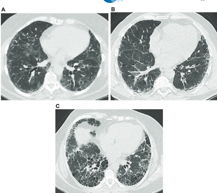
On computed tomography (CT) imaging, GGO presents as a dense area of increased opacity within the lungs that conserves bronchial and vascular margins (Figure 1A) (9). GGO is less hazy than consolidation, in which bronchovas-cular margins cannot be distinguished. GGO can be due to the partial filling of airspaces, interstitial thickening (as a result of fluid, cells, and/or fibrosis), the partial collapse of alveoli, an increased capillary blood volume, or a combina-tion of these, whereas all are related to the common partial displacement of air (9). Good interobserver agreement has been reported in the detection of GGO (kappa value, 0.78-0.90) (10).
Reticular pattern
A reticular pattern is defined as a collection of several small linear opacities that resemble a net-like aspect (Figure 1B) (9). The components of a reticular pattern are clearly observed on thin-section CT, and they can represent interlobular septal thickening, intralobular lines, or the cyst walls of honeycomb-ing. This finding is usually associated with ILD, but conges-tion and infecconges-tions (e.g., viral) are also important differential diagnoses (9).
Honeycombing
Honeycombing is defined as clustered cystic airspaces that are usually subpleural with well-defined walls and diameters ranging from 0.3-1 cm, reaching 2.5 cm is rare cases (Figure 1C) (9). Commonly considered specific for IPF,
honeycombing is an essential criterion for UIP diagnosis, and the terminology should be used carefully as it may directly influence patient management (11). Centrilobular emphy-sema, traction bronchiectasis, and cystic lung disease should be included in the differential diagnoses. Interobserver agre-ement for honeycombing is moderate (kappa=0.59±0.12). In a study by Watadani et al., there was disagreement on the identification of honeycombing in 29% of cases due to the co-existence of traction bronchiectasis, large cysts, and overlapping pulmonary emphysema (12).
Traction bronchiectasis
Traction bronchiectasis and bronchiolectasis represent nonuniform bronchial and bronchiolar dilatation, respec-tively (Figure 1A) (9). Dilated airways can also present as cysts (bronchi) or microcysts (bronchioles in the lung periphery). In IPF, traction bronchiectasis is better explained as a result of bronchiolar proliferation instead of utter mechanical traction (13). Recent studies have suggested that traction bronchiectasis and honeycombing are parts of a spectrum of the presentation of a singular and continuous mechanism of bronchiolar dysplastic proliferation in IPF (13,14). On the other hand, in nonspecific interstitial pneu-monia (NSIP), bronchocentricity is predominant, and trac-tion bronchiectasis is exclusively surrounded by fibrotic tissue, characteristics that suggest the mechanical traction as the main component in the development of traction bron-chiectasis in NSIP (13,14). Interobserver agreement for trac-tion bronchiectasis is moderate, with kappa values ranging from 0.24 to 0.42 (11).
Idiopathic pulmonary fibrosis
Computed tomography diagnostic criteria (usual inter-stitial pneumonia). Chest HRCT plays an essential role in the initial assessment of suspected IPF, considerably influen-cing subsequent management decisions. The primary role of HRCT is to distinguish chronic fibrosing lung diseases with an UIP pattern from those presenting a non-UIP pattern, sug-gesting an alternative diagnosis when possible. The most common HRCT protocol used to evaluate diffuse lung diseases is a volumetric acquisition of thin sections (generally
o1.5 mm), combined with a high spatial frequency
recon-struction algorithm (15). The 2018 ATS/ERS/JRS/ALAT guidelines for diagnosis of IPF (6) suggested four diagnostic entities: (a) UIP, (b) probable UIP, (c) indeterminate for UIP, and (d) alternative diagnosis (Table 1). The HRCT criteria for a UIP diagnosis include the presence of honeycombing with or without peripheral traction bronchiectasis or bronchiolectasis and a basal and subpleural distribution (6). Although GGO is common in UIP, it must be less extensive than the reticulation and be superimposed on a fine reticular pattern to be characterized as an UIP pattern (6). Another common feature on HRCT is mediastinal and hilar lymph node enlargement, which is present in up to 70% to 86% of patients with a UIP pattern (typicallyo15 mm) (16).
Figure 1 -Common features on high-resolution computed tomography in interstitial lung diseases.(a)Images from a 63-year-old female presenting a nonspecific interstitial pneumonia pattern. There are predominant areas of ground-glass opacities, with some traction bronchiectasis and cortical interlobular septal thickening. (b) Images from a 61-year-old male with idiopathic pulmonary fibrosis. There are diffuse areas of interlobular septal thickening, predominantly in the cortical lung zones.(c)Images from a 56-year-old female with idiopathic pulmonary fibrosis. There are extensive areas of honeycombing, with some interlobular septal thickening.
Table 1-HRCT Criteria for the UIP Pattern.
UIP Probable UIP Indeterminate for UIP Alternative diagnosis
- Predominantly subpleural and basal distribution - Reticular abnormality - Honeycombing with or
without traction bronchiectasis
- Predominantly subpleural and basal distribution - Reticular abnormality with
peripheral traction bronchiectasis - May have mild GGO
- Predominantly subpleural and basal distribution
- Mild reticulation, mild GGO or distortion
- CT features that do not suggest any specific etiology
- Distribution other than subpleural/basal - Extensive GGO (extension4reticular
abnormality)
- Profuse micronodules (bilateral, predominantly upper lobes)
- Discrete cysts (multiple, bilateral, apart from areas of honeycombing)
- Diffuse mosaic attenuation/air-trapping (bilateral, in three or more lobes)
- Consolidation in bronchopulmonary segment(s)/ lobe(s)
appropriate clinical settings at specialized centers (19). A confident diagnosis of UIP based on HRCT is accurate in 80% to 95% of cases using pathology as a reference (20). In multiple studies assessing the CT accuracy for an IPF dia-gnosis, pathology was considered a gold standard. However, there is substantial interobserver variation among patholo-gists in the assessment of non-neoplastic lung disease (21). An interobserver variation over 50% has been reported for the pathological diagnosis of NSIP and its distinction from UIP (21).
Histopathology diagnostic criteria (usual interstitial pneumonia). The main histopathologic feature and diag-nostic criterion of UIP is a patchy presentation comprising affected zones of fibrosis with scarring and honeycombing alternating with zones of less affected or healthy tissue (Table 2) (Figure 2C) (22). These findings usually prevail in the subpleural and paraseptal parenchyma. Inflammation
is commonly absent or mild, consisting of an irregular interstitial infiltrate of lymphocytes and plasma cells asso-ciated with the hyperplasia of type 2 pneumocytes and the bronchiolar epithelium. Although the fibrotic areas consist primarily of dense collagen, the diffuse convex subepithelial foci of proliferating fibroblasts and myofibroblasts are typical findings (fibroblast foci). These should be differentiated from fibroblastic alveolar plugs of organizing pneumonia. While the former is always within the interstitial spaces, the latter is associated with polypoid intrusions into the alveolar space. Zones of honeycombing are formed by cystic fibrotic airspaces that are commonly lined by a bronchiolar epithelium and filled with mucus and inflammatory cells (22).
Pathogenesis. The pathogenesis of IPF has been dis-cussed for several years, and one of the most accepted views is that the initial inflammatory events might initiate a dys-regulated fibroblast-mediated wound-healing response that
can be sustained by several factors, leading to lung fibrosis (23). One of the most accepted models is the‘‘alveolar stem cell exhaustion’’model, which suggests that an accelerated parenchymal senescence that is determined by predisposing factors, such as genetic mutations or variations and telome-rase dysfunction, together with environmental exposures, such as tobacco smoke, can severely compromise the regen-erative potential of parenchymal epithelial stem cells (13,24,25). This epithelial damage to the airway wall can induce alveolar collapse, what can trigger a fibroproliferation process and promote the destruction of airways, eventually leading to severe and irreversible functional impairment (24-26).
Mai et al. 2016 recently used micro-computed tomogra-phy to analyze the geometric progression of lung changes caused by IPF, associating the analysis concomitantly with histopathological and CT findings, and the authors encoun-tered evidence suggesting that the disease started preferen-tially at the peripheries of the secondary pulmonary lobules, gradually extended toward the centrilobular region, and ultimately replaced the entire lobule (26). The sequence of lung changes on CT in patients with IPF starts with areas of GGO that turn into areas of reticulation and end with cystic changes (26,27). The so-called‘‘microscopic honeycombing’’
represents the formation of these cystic spaces. These spaces should be differentiated from the honeycombing visualized in CT scans; these spaces are present in most cases and are not necessarily specific to a UIP diagnosis, while honeycom-bing is highly specific to UIP and is associated with a poor prognosis (26,28).
When is biopsy necessary? Surgical lung biopsy (SLB) is the diagnostic method of choice for patients whose imag-ing findimag-ings are not definitive for UIP, includimag-ing findimag-ings of probable UIP, indeterminate for UIP and alternative diagnosis. Hence, an integrated multidisciplinary approach assessing both imaging and pathological features is essential to establish a final diagnosis (3,6).
Tissue samples should be obtained from two to three lobes due to the high variability in the distribution and morphology of abnormalities. Careful evaluation of the pre-sence of potential risks before performing SLB is essential as the risks associated with the procedure can outweigh the benefits of determining a diagnosis, especially in older patients, in those with severe impairment in PFTs or those with co-existing comorbidities, such as pulmonary hyperten-sion and severe heart failure (6,29). Recent evidence suggests that cryobiopsy is a promising method that is less invasive
than SLB and can be used in place of SLB to obtain lung samples to confirm an IPF diagnosis (30).
Mimics of idiopathic pulmonary fibrosis
Clinical mimics. Confirmation of an IPF diagnosis is
challenging, requiring the exclusion of pulmonary fibrosis with known causes, such as asbestosis, CTDs, drug expo-sure, chronic hypersensitivity pneumonitis (HP), and other forms of idiopathic interstitial pneumonitis (3,5,6). Although these entities have similarities with IPF, such as progressive dyspnea, dry cough, and a restrictive pattern with a decreased diffusing capacity in the PFT, they are associated with better prognosis and an improved responsiveness to immunosup-pressive drugs (7).
Connective tissue diseases. These disorders often affect
young women, and rheumatoid arthritis and systemic sclerosis are the most common CTDs associated with the UIP pattern. Extrapulmonary manifestations, such as arthralgia, Sicca symptoms, Raynaud phenomenon, and esophageal manifesta-tions, are commonly identified in ILD associated with CTD. However, interstitial lung changes can be the sole manifestation of CTD. The presence of serological abnormalities is an important feature of CTDs (7). ILD is also associated with interstitial pneumonia with autoimmune features, which should be included in the differential diagnosis of IPF (31,32).
Hypersensitivity pneumonitis. HP is an inflammatory
syndrome associated with an exposure history to suspected antigens, such as mold, birds and drugs. Hence, potential exposures should be extensively investigated in patients with
pulmonary fibrosis. Fatigue, fever, and weight loss may be
found in acute and subacute HP and rarely in those with chronic forms. Progressive dyspnea is the main symptom in chronic HP, and inspiratory squeaks on pulmonary auscultation and digital clubbing may be identified. If the responsible inhaled antigen can be identified, complete avoidance is the most efficient manage-ment (5,29). Acute disease is commonly self-limited, resolving without any specific therapy. Corticosteroid therapy might be necessary for an acute relief of symptoms and can hasten initial recovery in severe cases. However, long-term efficacy has not been demonstrated in prospective clinical trials (33).
Idiopathic nonspecific interstitial pneumonia. Compared
with IPF, idiopathic NSIP occurs predominantly in young women and can have associated systemic manifestations, such as fever, fatigue, and weight loss, and features suggestive of CTD (4,7,34).
Asbestosis. The main feature for distinguishing asbestosis
from IPF is a history of occupational exposure to asbestos, Table 2-Histopathological Criteria for the UIP Pattern.
UIP Probable UIP Indeterminate for UIP Alternative diagnosis
- Evidence of marked fibrosis / architectural distortion,± honey-combing in a pre-dominantly subpleural / paraseptal distribution - Patchy involvement of lung
parenchyma by fibrosis - Fibroblast foci
- Some characteristics of column 1 but not sufficient to corroborate the diagnosis of UIP - Exclusively honeycombing
changes
- Absence of features to suggest an alternative diagnosis
- Some histologic features of column 1 associated with features that suggest another diagnosis or UIP secondary to another cause (granulomas, hyaline membranes, cellular inflammatory infiltrate in areas away from honeycombing, prominent lymphoid hyperplasia, bronchiolocentric distribution, exuberant chronic fibrous pleuritis and organizing pneumonia)
- Hyaline membranes - Organizing pneumonia - Granulomas
- Marked interstitial inflammatory cell infiltrate away from honeycombing - Predominant airway-centered
changes
- Other features suggestive of an alternate diagnosis
such as asbestos mining, shipbuilding, and welding. Deter-mining a temporal association between the exposure and the occurrence of symptoms is essential (7).
Drug-induced lung diseases. Investigations of exposures to
drugs that can induce ILD, such as bleomycin, amiodarone, nitrofurantoin, methotrexate, and cyclophosphamide, is essen-tial in the assessment of patients with a suspicion of IPF. Determining a temporal relationship between the exposure to the potential drug and the occurrence of respiratory manifesta-tions is also necessary (5).
Radiological mimics. NSIP accounts for most of the
disagreements among observers for idiopathic interstitial pneumonias. HRCT findings that favor NSIP rather than UIP include the presence of GGOs, which are found in most cases. Subpleural sparing is highly suggestive of NSIP and is present in 64% of cases compared to 11% in chronic HP and 4% in IPF (34). The features that best differentiate chronic HP from IPF and NSIP are lobular zones with decreased attenuation, the presence of centrilobular nodules, and a lack of a lower zone predominance of abnormalities. The former has been reported in 80% of patients with chronic HP, 43% of patients with IPF, and 34% of patients with NSIP (34). The UIP pattern on HRCT in patients with CTDs or fibrotic HP is similar to the pattern found in idiopathic UIP, and this should be distinguished on a clinical basis. However, radiologists may contribute to such differentiation based on additional findings, such as esophageal dilatation, and on pleural or pericardial effusion or thickening. Asbestosis typically has a histologic and HRCT pattern of UIP, and pleural plaques usually aid in the diagnosis. Fibrotic sarco-idosis can be distinguished from UIP by the upper lobe, a predominant peribronchial distribution and other HRCT findings, including pulmonary nodules, lymph node enlar-gement and calcifications.
Histopathological mimics. During an examination of an
ILD patient biopsy, the goal of the pathologist is to differ-entiate UIP from other diseases that may simulate this pattern, such as CTDs, chronic HP, pneumoconiosis, and NSIP (7,35).
Connective tissue diseases. CTDs are a heterogeneous
group of diseases, with different patterns on biopsy. Although the most common pattern is an NSIP pattern, UIP is frequently observed. The presence of an exuberant lymphocytic infiltrate that forms germinal centers and of follicular bronchiolitis can be helpful features for CTD diagnosis in these situations.
In addition, the presence of pleuralfibrosis is more characteristic
of CTDs (7,35).
Chronic hypersensitivity pneumonitis. In chronic HP,fibrosis
is predominantly airway centered but can occasionally manifest as UIP. Features that might suggest a diagnosis of chronic HP include a predominance of upper lobes, the presence of interstitial granulomas, which are mainly peribronchiolar, and bronchiolitis with peribronchiolar metaplasia (7,35). Recent studies focusing on genetic factors associated with chronic HP have suggested a common pathobiology with IPF. A MUC5B rs35705950 single-nucleotide polymorphism and a short telomere length, as seen in sporadic and familial forms of IPF, can
pre-dispose patients with HP to lung remodeling andfibrosis (36).
Asbestosis. Asbestosis is histopathologically characterized
by the presence of predominantly subpleural interstitial fibrosis,
which is similar to UIP. However, in early cases,fibrosis can be
more prominent around bronchioles. Other helpful features in
the differential diagnosis with UIP are collagenfibrosis with the
absence offibroblastic foci and a mild inflammatory infiltrate.
To confirm the histological diagnosis, asbestos bodies must be present (37).
Nonspecific interstitial pneumonia. NSIP is a type of
chronic interstitial pneumonia that is characterized by a relative spatial homogeneity in the involvement of lung parenchyma, which is different from the typical patchy pattern of UIP where regions of the normal lung are interspersed with the affected parenchyma. In addition, NSIP is characterized by a temporal
homogeneity of lesions (inflammation and/or fibrosis), which
is different from the typical temporal heterogeneity of UIP (7,35,38-41). Other features of UIP, such as smooth muscle
hyperplasia, fibroblastic foci, and an aggregation of elastic
fibers, are not observed in most cases of NSIP (39). In addition, peripheral accentuation is absent or inconspicuous (41).
Although the UIP pattern may be observed in several ILDs, there are relevant clinical, tomographic, and histopathologi-cal features that contribute to differentiating them from IPF. The diagnosis of IPF is challenging, and a multidisciplinary discussion with ILD experts, including physicians, radio-logists, and pathoradio-logists, improves the accuracy of the diagnosis, which is essential to establishing the appropriate management strategy.
’ AUTHOR CONTRIBUTIONS
Hochhegger B is the guarantor of the integrity of the entire study. Hochhegger B, Marchiori E, Rubin AS, Carvalho CR and Baldi BG were responsible for the study concepts and design. Hochhegger B, Zanon M, Fragomeni R and Altmayer S were responsible for the literature research. Hochhegger B, Marchiori E, Zanon M, Rubin AS, Fragomeni R, Altmayer S, Carvalho CR and Baldi BG were responsible for the manuscript preparation and editing.
’ REFERENCES
1. Hamman L, Rich AR. Clinical pathologic conference. Int Clin. 1933;1:196-231. 2. Homolka J. Idiopathic pulmonary fibrosis: a historical review. CMAJ.
1987;137(11):1003-5.
3. Baddini-Martinez J, Baldi BG, Costa CH, Jezler S, Lima MS, Rufino R. Update on diagnosis and treatment of idiopathic pulmonary fibrosis. J Bras Pneumol. 2015;41(5):454-66, http://dx.doi.org/10.1590/S1806-37132015000000152.
4. Travis WD, Costabel U, Hansell DM, King TE Jr, Lynch DA, Nicholson AG, et al. An official American Thoracic Society/European Respiratory Society statement: Update of the international multidisciplinary classifi-cation of the idiopathic interstitial pneumonias. Am J Respir Crit Care Med. 2013;188(6):733-48, http://dx.doi.org/10.1164/rccm.201308-1483ST. 5. Wuyts WA, Cavazza A, Rossi G, Bonella F, Sverzellati N, Spagnolo P. Differential diagnosis of usual interstitial pneumonia: when is it truly idiopathic? Eur Respir Rev. 2014;23(133):308-19, http://dx.doi.org/ 10.1183/09059180.00004914.
6. Raghu G, Remy-Jardin M, Myers JL, Richeldi L, Ryerson CJ, Lederer DJ, et al. Diagnosis of Idiopathic Pulmonary Fibrosis. An Official ATS/ERS/ JRS/ALAT Clinical Practice Guideline. Am J Respir Crit Care Med. 2018;198(5):e44-e68, http://dx.doi.org/10.1164/rccm.201807-1255ST. 7. King TE Jr. Idiopathic pulmonary fibrosis. In: Schwarz MI, King TE Jr
(eds). Interstitial Lung Disease, 5thed. Shelton: People
’s Medical Pub-lishing House-USA; 2011. p 895.
8. Moua T, Maldonado F, Decker PA, Daniels CE, Ryu JH. Frequency and implication of autoimmune serologies in idiopathic pulmonary fibrosis. Mayo Clin Proc. 2014;89(3):319-26, http://dx.doi.org/10.1016/j.mayocp. 2013.11.018.
9. Hansell DM, Bankier AA, MacMahon H, McLoud TC, Müller NL, Remy J. Fleischner Society: glossary of terms for thoracic imaging. Radiology. 2008;246(3):697-722, http://dx.doi.org/10.1148/radiol.2462070712. 10. Sergiacomi G, Cicciò C, Boi L, Velari L, Crusco S, Orlacchio AN, et al.
Ground-glass opacity: high-resolution computed tomography and 64-multi-slice computed tomography findings comparison. Eur J Radiol. 2010;74(3): 479-83, http://dx.doi.org/10.1016/j.ejrad.2009.03.034.
12. Watadani T, Sakai F, Johkoh T, Noma S, Akira M, Fujimoto K, et al. Interobserver variability in the CT assessment of honeycombing in the lungs. Radiology. 2013;266(3):936-44, http://dx.doi.org/10.1148/radiol. 12112516.
13. Piciucchi S, Tomassetti S, Ravaglia C, Gurioli C, Gurioli C, Dubini A, et al. From ‘‘traction bronchiectasis’’ to honeycombing in idiopathic pulmonary fibrosis: A spectrum of bronchiolar remodeling also in radi-ology? BMC Pulm Med. 2016;16(1):87, http://dx.doi.org/10.1186/s12890-016-0245-x.
14. Walsh SL, Wells AU, Sverzellati N, Devaraj A, von der Thüsen J, Yousem SA, et al. Relationship between fibroblastic foci profusion and high resolution CT morphology in fibrotic lung disease. BMC Med. 2015;13: 241, http://dx.doi.org/10.1186/s12916-015-0479-0.
15. Kusmirek JE, Martin MD, Kanne JP. Imaging of Idiopathic Pulmonary Fibrosis. Radiol Clin North Am. 2016;54(6):997-1014, http://dx.doi.org/ 10.1016/j.rcl.2016.05.004.
16. Jung JI, Kim HH, Jung YJ, Park SH, Lee JM, Hahn ST. Mediastinal lym-phadenopathy in pulmonary fibrosis: correlation with disease severity. J Comput Assist Tomogr. 2000;24(5):706-10, http://dx.doi.org/10.1097/ 00004728-200009000-00007.
17. Raghu G, Mageto YN, Lockhart D, Schmidt RA, Wood DE, Godwin JD. The accuracy of the clinical diagnosis of new-onset idiopathic pul-monary fibrosis and other interstitial lung disease: a prospective study. Chest. 1999;116(5):1168-74, http://dx.doi.org/10.1378/chest.116. 5.1168.
18. Kaarteenaho R. The current position of surgical lung biopsy in the diag-nosis of idiopathic pulmonary fibrosis. Respir Res. 2013;14:43, http://dx. doi.org/10.1186/1465-9921-14-43.
19. Raghu G, Lynch D, Godwin JD, Webb R, Colby TV, Leslie KO, et al. Diagnosis of idiopathic pulmonary fibrosis with high-resolution CT in patients with little or no radiological evidence of honeycombing: secondary analysis of a randomised, controlled trial. Lancet Respir Med. 2014;2(4):277-84, http://dx.doi.org/10.1016/S2213-2600(14)70011-6. 20. Nicholson AG, Addis BJ, Bharucha H, Clelland CA, Corrin B, Gibbs AR,
et al. Inter-observer variation between pathologists in diffuse parench-ymal lung disease. Thorax. 2004;59(6):500-5, http://dx.doi.org/10.1136/ thx.2003.011734.
21. Flaherty KR, King TE Jr, Raghu G, Lynch JP 3rd, Colby TV, Travis WD, et al. Idiopathic interstitial pneumonia: what is the effect of a multidis-ciplinary approach to diagnosis? Am J Respir Crit Care Med. 2004;170(8): 904-10, http://dx.doi.org/10.1164/rccm.200402-147OC.
22. Martinez FJ, Chisholm A, Collard HR, Flaherty KR, Myers J, Raghu G, et al. The diagnosis of idiopathic pulmonary fibrosis: current and future approaches. Lancet Respir Med. 2017;5(1):61-71, http://dx.doi.org/ 10.1016/S2213-2600(16)30325-3.
23. Wilson MS, Wynn TA. Pulmonary fibrosis: pathogenesis, etiology and regulation. Mucosal Immunol. 2009;2(2):103-21, http://dx.doi.org/10.1038/ mi.2008.85.
24. Wolters PJ, Collard HR, Jones KD. Pathogenesis of idiopathic pulmonary fibrosis. Annu Rev Pathol. 2014;9:157-79, http://dx.doi.org/10.1146/ annurev-pathol-012513-104706.
25. Chilosi M, Doglioni C, Murer B, Poletti V. Epithelial stem cell exhaustion in the pathogenesis of idiopathic pulmonary fibrosis. Sarcoidosis Vasc Diffuse Lung Dis. 2010;27(1):7-18.
26. Mai C, Verleden SE, McDonough JE, Willems S, De Wever W, Coolen J, et al. Thin-Section CT Features of Idiopathic Pulmonary Fibrosis Corre-lated with Micro-CT and Histologic Analysis. Radiology. 2017;283(1): 252-63, http://dx.doi.org/10.1148/radiol.2016152362.
27. Akira M, Sakatani M, Ueda E. Idiopathic pulmonary fibrosis: progression of honeycombing at thin-section CT. Radiology. 1993;189(3):687-91, http://dx.doi.org/10.1148/radiology.189.3.8080483.
28. Chung JH, Chawla A, Peljto AL, Cool CD, Groshong SD, Talbert JL, et al. CT scan findings of probable usual interstitial pneumonitis have a high predictive value for histologic usual interstitial pneumonitis. Chest. 2015; 147(2):450-9, http://dx.doi.org/10.1378/chest.14-0976.
29. Hodnett PA, Naidich DP. Fibrosing interstitial lung disease. A practical high-resolution computed tomography-based approach to diagnosis and management and a review of the literature. Am J Respir Crit Care Med. 2013;188(2):141-9, http://dx.doi.org/10.1164/rccm.201208-1544CI. 30. Tomassetti S, Wells AU, Costabel U, Cavazza A, Colby TV, Rossi G, et al.
Bronchoscopic lung cryobiopsy increases diagnostic confidence in the multidisciplinary diagnosis of idiopathic pulmonary fibrosis. Am J Respir Crit Care Med. 2016;193(7):745-52, http://dx.doi.org/10.1164/rccm.201504-0711OC.
31. Fischer A, Antoniou KM, Brown KK, Cadranel J, Corte TJ, du Bois RM, et al. An official European Respiratory Society/American Thoracic Society research statement: interstitial pneumonia with autoimmune features. Eur Respir J. 2015;46(4):976-87, http://dx.doi.org/10.1183/13993003.00150-2015. 32. Pereira DA, Dias OM, Almeida GE, Araujo MS, Kawano-Dourado LB, Baldi BG, et al. Lung-dominant connective tissue disease among patients with interstitial lung disease: prevalence, functional stability, and com-mon extrathoracic features. J Bras Pneumol. 2015;41(2):151-60, http://dx. doi.org/10.1590/S1806-37132015000004443.
33. Selman M, Pardo A, King TE Jr. Hypersensitivity pneumonitis: insights in diagnosis and pathobiology. Am J Respir Crit Care Med. 2012;186(4): 314-24, http://dx.doi.org/10.1164/rccm.201203-0513CI.
34. Silva CI, Müller NL, Lynch DA, Curran-Everett D, Brown KK, Lee KS, et al. Chronic hypersensitivity pneumonitis: differentiation from idio-pathic pulmonary fibrosis and nonspecific interstitial pneumonia by using thin-section CT. Radiology. 2008;246(1):288-97, http://dx.doi.org/10.1148/ radiol.2453061881.
35. Smith M, Dalurzo M, Panse P, Parish J, Leslie K. Usual interstitial pneu-monia-pattern fibrosis in surgical lung biopsies. Clinical, radiological and histopathological clues to aetiology. J Clin Pathol. 2013;66(10):896-903, http://dx.doi.org/10.1136/jclinpath-2013-201442.
36. Ley B, Newton CA, Arnould I, Elicker BM, Henry TS, Vittinghoff E, et al. The MUC5B promoter polymorphism and telomere length in patients with chronic hypersensitivity pneumonitis: an observational cohort-control study. Lancet Respir Med. 2017;5(8):639-47, http://dx.doi.org/ 10.1016/S2213-2600(17)30216-3.
37. Roggli VL, Gibbs AR, Attanoos R, Churg A, Popper H, Cagle P, et al. Pathology of asbestosis - An update of the diagnostic criteria: Report of the asbestosis committee of the college of American pathologists and pulmonary pathology society. Arch Pathol Lab Med. 2010;134(3):462-80. 38. Larsen BT, Colby TV. Update for pathologists on idiopathic interstitial
pneumonias. Arch Pathol Lab Med. 2012;136(10):1234-41, http://dx.doi. org/10.5858/arpa.2012-0225-RA.
39. Hashisako M, Fukuoka J. Pathology of Idiopathic Interstitial Pneumonias. Clin Med Insights Circ Respir Pulm Med. 2016;9(Suppl 1):123-33. 40. Visscher DW, Myers JL. Histologic spectrum of idiopathic interstitial
pneumonias. Proc Am Thorac Soc. 2006;3(4):322-9, http://dx.doi.org/ 10.1513/pats.200602-019TK.