Analysis of 39 cases of idiopathic chronic interstitial
pneumonia*
ROGÉRIO RUFINO1, LEONARDO RIZZO2, CLÁUDIA HENRIQUE DA COSTA3, ROBERTO JOSÉ DE LIMA4, KALIL MADI5
*Study carried out at the Universidade do Estado do Rio de Janeiro (UERJ, Rio de Janeiro State University), Rio de Janeiro, Brazil.
1. Postdoctoral degree from the National Heart and Lung Institute - Imperial College/London. Adjunct Professor of Pulmonology and Phthisiology at the Universidade do Estado do Rio de Janeiro (UERJ, Rio de Janeiro State University) School of Medical Sciences, Rio de Janeiro, Brazil
2. Resident in Pathology at the Instituto Nacional do Câncer (INCA, Cancer National Institute), Rio de Janeiro, Brazil. 3. Doctorate in Pulmonology from the Universidade Federal do Rio de Janeiro (UFRJ, Federal University of Rio de Janeiro), Rio de Janeiro, Brazil. Assistant professor of Pulmonology and Phthisiology at the Universidade do Estado do Rio de Janeiro (UERJ, Rio de Janeiro State University) School of Medical Sciences, Rio de Janeiro, Brazil
4. Assistant Professor in the Department of Pathology of the Universidade Federal do Rio de Janeiro (UFRJ, Federal University of Rio de Janeiro) School of Medicine, Rio de Janeiro, Brazil.
5. Full Professor in the Department of Pathology of the Universidade Federal do Rio de Janeiro (UFRJ, Federal University of Rio de Janeiro) School of Medicine, Rio de Janeiro, Brazil.
Correspondence to: Rogério Rufino. Rua Mário Pederneiras, 10/121, Humaitá - CEP: 22041-020, Rio de Janeiro, RJ, Brasil. Tel: 55 21 2527-6871. Email:[email protected]
Submitted: 3 March 2006. Approved, after review: 5 April 2006.
ABSTRACT
Objective: To make a retrospective analysis of lung biopsy samples obtained from patients diagnosed with chronic
idiopathic interstitial pneumonia, as defined in the American Thoracic Society/European Respiratory Society classification system made public in 2000. Methods: Samples from 252 open-lung biopsies of patients with interstitial lung disease, all performed between 1977 and 1999, were reviewed, and 39 cases of idiopathic interstitial lung disease were selected and re-evaluated by two pathologists in accordance with the American Thoracic Society/European Respiratory Society classification system. Results: Among those 39 cases, the diagnoses were maintained in 28 (71.8%). A new pathologic entity, nonspecific interstitial pneumonia, was included in the reclassification, and overlapping patterns were observed in 6 cases. Of the 28 cases in which the diagnosis of chronic idiopathic interstitial pneumonia remained unchanged, idiopathic pulmonary fibrosis was accompanied by cryptogenic organizing pneumonia in 4, cryptogenic organizing pneumonia was accompanied by nonspecific interstitial pneumonia in 1, and desquamative interstitial pneumonia was accompanied by nonspecific interstitial pneumonia in 1. All cases of idiopathic pulmonary fibrosis were confirmed, although 3 of those were found to be accompanied by cryptogenic organizing pneumonia. Virtually all prior diagnoses were maintained in the review of the biopsy samples (p > 0,05). Conclusion: The American Thoracic Society/European Respiratory Society system of classifying interstitial lung disease is a useful tool for pathologists who deal with lung biopsies.
INTRODUCTION
Most idiopathic diffuse interstitial lung diseases are chronic inflammatory processes that can result in fibrosis and deformation of the parenchyma, representing an heterogeneous group of entities classified by clinical, functional, radiological and histopathologic criteria.(1-2) In
many cases, the histopathologic diagnosis contributes decisively to defining the disease, especially when there is a positive correlation with the radiological aspects and clinical evolution. In a smaller number of cases, difficulties arise due to the superimposition of morphological patterns and the low representativeness of the specimen when compared to the lung as a whole. Another problem found in the final decades of the twentieth century is related to the conceptual aspect, a number of diagnostic labels having been created, thereby making it more difficult to come t o a c l e a r u n d e r s t a n d i n g o f t h e v a r i o u s morbidities linked to this special lung ailment.(3-4)
T h e A m e r i c a n T h o r a c i c S o c i e t y / E u r o p e a n Respiratory Society (ATS/ERS) classification, published in 2000,(1) is a multidisciplinary
consensus and defines seven types of idiopathic interstitial pneumonias. This consensus describes morphological patterns that are not specific to one disease, such as 'usual' interstitial pneumonia, which is seen in idiopathic pulmonary fibrosis (IPF), asbestosis, drug-induced lung disease and collagen-related diseases.
Based on this new classification system, which is internationally accepted, we reviewed all the cases of idiopathic interstitial lung disease treated at two different institutions in the last 30 years. Our objective was to determine whether these old cases met the ATS/ERS criteria.
METHODS
A retrospective microscopy study was carried out involving open lung biopsy fragments obtained between 1977 and 1999 from 252 patients with interstitial disease treated in the Pathology Departments of the Clementino Fraga Filho U n i v e r s i t y H o s p i t a l a n d t h e B e n e f i c ê n c i a Portuguesa Hospital of Rio de Janeiro. Previous diagnoses had been made using clinical and radiological criteria (X-rays and high-resolution
computed tomography scans), respiratory function tests and histopathology. The patients were divided into two groups: those in which the cause was known (213 cases); and those in which it was not (idiopathic; 39 cases). The 39 samples of unknown etiology were analyzed by two pathologists, who were specialists in pulmonary diseases.
A new analysis, using the ATS/ERS criteria,(2) was
made. The analyzing pathologists were blinded as to the previous diagnosis. The diagnosis was obtained according to a consensus between the pathologists, using conventional techniques, such as hematoxylin and eosin staining, Masson's trichrome stain, and reticulin.
Parametric t-test and two-tailed Pearson's correlation coefficient were used to analyze the results. Values of p < 0.05 were considered statistically significant.
RESULTS
For demographic data analysis, the cases were separated based on the previous diagnosis. The demographic data of the analyzed patients are shown in Table 1. Patient age was found to be similar in both groups (p > 0.05). However, in the idiopathic pulmonary fibrosis group there was greater impairment in the males (p < 0.05). After the new histopathologic evaluation, the 15 previous diagnoses of idiopathic pulmonary fibrosis were confirmed. However, 3 of those were accompanied by the cryptogenic organizing
TABLE 1
Demographic data
Pathological Gender Age diagnosis (Male/Female) (years) FPI (UIP) (13/2) 56.93 (±10.30) COP (5/7) 51.08 (±10.47) RBILD (2/0) 49.00 (±4.04) LIP (0/2) 42.50 (±2.12) AIP/DAD (3/0) 54.53 (±13.87)
DIP (0/1) 59.00
Non classifiable (1/3) 54.75 (±5.90)
pneumonia (COP) pattern (Table 2). The diagnosis of 12 COP cases was maintained, although 1 of them was associated with the nonspecific interstitial pneumonia (NSIP) pattern, and other was accompanied by the idiopathic pulmonary fibrosis pattern (Figure 1). In the 2 cases of i n t e r s t i t i a l l u n g d i s e a s e w i t h r e s p i r a t o r y bronchiolitis, as well as in the 3 cases of acute interstitial pneumonia, the previous diagnosis was preserved. Of the 2 cases of lymphocytic interstitial pneumonia, 1 was confirmed and the o t h e r w a s m o d i f i e d t o h y p e r s e n s i t i v i t y pneumonia. The only case of desquamative interstitial pneumonia was considered to be concomitant with NSIP. The 4 cases of interstitial pneumonia that were not classified previously received new diagnoses: COP (2 cases), NSIP (1 case), and hypersensitivity pneumonia (1 case). All of the 39 cases were classified, and there was no difference (p = 0.24) between the histopathologic diagnosis before and after the AT S / E R S c o ns e ns us. T h e re w a s, h o w e ve r, significant concordance between the diagnoses made in the two periods (p = 0.0014; r2 =
0.9824).
DISCUSSION
This study reviewed cases collected from 1977 onward, when Liebow's pioneering classification was still being used.(5) Despite this fact, the review of all 39 cases confirmed the diagnosis initially provided for 28 patients (71.8%). In the 11 remaining cases, the diagnosis was modified in only 1 (from lymphocytic interstitial pneumonia to hypersensitivity pneumonia, due to the presence of loose granulomas and the fact that the lymphocytic infiltrate was not prominent).(6) Of the 4 cases that had not been
diagnosed at the time and had been considered nonspecific interstitial pneumonia, 1 was reclassified as NSIP, 2 as COP and 1 as hypersensitivity pneumonia. In this study, there were no cases of fibrotic NSIP.(7-8)
Superimposition of pathological patterns was seen in 6 cases (15.38%), and COP was the pattern associated with higher frequency.(9-11)
The adoption of the standards proposed by ATS/ ERS did not result in a statistically significant difference between the diagnoses made before 2000 and those made after 2000. This can be explained by the fact that virtually all of the morphological patterns had been described more than one decade before.(12)
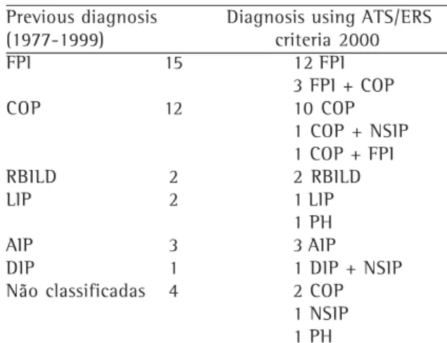
TABLE 2
Histopathological diagnosis before and after review
Previous diagnosis Diagnosis using ATS/ERS (1977-1999) criteria 2000 FPI 15 12 FPI
3 FPI + COP
COP 12 10 COP
1 COP + NSIP 1 COP + FPI RBILD 2 2 RBILD
LIP 2 1 LIP
1 PH
AIP 3 3 AIP
DIP 1 1 DIP + NSIP Não classificadas 4 2 COP
1 NSIP 1 PH
IPF: idiopathic pulmonary fibrosis; COP: cryptogenic organizing pneumonia; RBILD: respiratory bronchiolitis interstitial lung disease; LIP: lymphocytic interstitial pneumonia; AIP: acute interstitial pneumonia; DIP: desquamative interstitial pneumonia; HP: hypersensitivity pneumonia;
NSIP: non-specified interstitial pneumonia.
A study carried out in 2004(13) analyzed the
pathological concordance between pathologists in the diagnosis of cases of interstitial lung diseases of known and unknown etiologies treated between 1996 and 1997, using the ATS/ERS current criteria. The authors demonstrated that there was greater concordance between the diagnoses in cases of 'usual' interstitial pneumonia diagnosis (κ = 0.42), COP (κ = 0.57) and sarcoidosis (κ = 0.76), and that there was lower concordance between the diagnoses in cases of NSIP (κ = 0.23).
The identification of different morphological patterns in the same specimen can pose difficulties for pathologists, preventing them from reaching a definitive conclusion, and might have an impact on the clinical evolution, the therapeutic approach and the prognosis.(14-15) The combination
of patterns in the same sample leads us to believe that one pathological pattern can be transformed into another, or, at least in some cases, that the spectrum of injury might be more complex than suggested by the simplicity of the existing classification systems.(16-22) This was suggested in
a study conducted in 2002, which verified molecular similarity between 'usual' interstitial pneumonia and NSIP in familial interstitial diseases.(23) The results of other studies have also
indicated evolutive and morphological similarities between fibrotic NSIP and 'usual' interstitial pneumonia, thus confirming the hypothesis of an injury continuum.(7-8, 24-26)
One study(27) demonstrated that the lesion of
epithelial cell DNA occurred was similar in NSIP and 'usual' interstitial pneumonia, although with more intensity in the latter, suggesting an initial process of pulmonary epithelial lesion.
The establishment of the ATS/ERS classification system was an attempt to organize idiopathic c h r o n i c i n t e r s t i t i a l p n e u m o n i a s b y u s i n g histopathologic patterns applicable to clinical and radiological profiles. This classification system re-introduced COP as an interstitial entity and consolidated NSIP but maintained previously used standards, standardizing the language and the descriptions of patterns for pathologists.
The morphological patterns, together with clinical and radiological information, can confer relative specificity on the diagnosis of idiopathic interstitial lung diseases. According to the ATS/ ERS classification, the diagnosis was confirmed in
28 (71.8%) of the 39 cases analyzed, and superimposition of patterns was seen in 6 cases. These combinations indicate a lack of specificity in the pulmonary response to aggression and suggest that the pathogenesis is based on a continuous injury spectrum. The ATS/ERS classification system is a useful instrument for pathologists that analyze lung biopsies. However, in our study, the use of this system did not substantially modify the previously established pathological diagnoses.
REFERENCES
1. American Thoracic Society. Idiopathic pulmonary f i b r o s i s : d i a g n o s i s a n d t r e a t m e n t . I n t e r n a t i o n a l consensus statement. American Thoracic Society (ATS), and the European Respiratory Society (ERS). Am J Respir Crit Care Med. 2000;161(2 Pt 1):646-64. 2. American Thoracic Society; European Respiratory Society.
American Thoracic Society/European Respiratory Society International Multidisciplinary Consensus Classification of the Idiopathic Interstitial Pneumonias. This joint statement of the American Thoracic Society (ATS), and the European Respiratory Society (ERS) was adopted by the ATS board of directors, June 2001 and by the ERS Executive Committee, June 2001. Am J Respir Crit Care Med. 2002;165(2):277-304.
3. Carrington CB, Gaensler EA, Coutu RE, FitzGerald MX, Gupta RG. Natural history and treated course of usual and desquamative interstitial pneumonia. N Engl J Med. 1978;298(15):801-9.
4. Gross P. Chronic interstitial pneumonitis: a histogenetic study. Arch Pathol. 1960;69:706-15.
5. Liebow AA, Carrington CB. Interstitial pneumonia. In: Simon M, Poetchen EJ, LeMay M, editors. Frontiers of pulmonary radiology. New York: Grunne & Stratton; 1969. p.102-41.
6. G ro s s P, B e n z E J . T h e c o n c e p t o f o rg a n i z i n g pneumonia. Arch Pathol. 1961;72: 607-19.
7. Travis WD, Matsui K, Moss J, Ferrans VJ. Idiopathic nonspecific interstitial pneumonia: prognostic significance of cellular and fibrosing patterns: survival comparison with usual interstitial pneumonia and desquamative interstitial pneumonia. Am J Surg Pathol. 2000;24(1):19-33. 8. Katzenstein ALA, Myers JL. Nonspecific interstitial
pneumonia and other idiopathic interstitial pneumonias: classification and diagnostic criteria. Am J Surg Pathol. 2000; 24(1):1-3.
9. Katzenstein AL. Fiorelli RF. Nonspecific interstitial pneumonia/fibrosis: histologic features and clinical significance. Am J Surg Pathol. 1994;18(2):136-47. 10. Katzenstein AL, Myers JL. Idiopathic pulmonary fibrosis:
clinical relevance of pathologic classification. Am J Respir Crit Care Med. 1998;157(4 Pt 1):1301-15. 11. Flaherty KR, Travis WD, Colby TV, Toews GB, Kazerooni
12. Colby TV, Leslie KO. Interstitial lung disease and bronchiolitis. In: 22 International Congress of the International Academy of Pathology 13th World Congress of Academic and Environment Pathology (short course-handout) France 1998 (october); p. 1-41. 1 3 . Nicholson AG, Addis BJ, Barucha H, Clelland CA, Corrin
B, Gibbs AR, et al. Inter-observer variation between pathologists in diffuse parenchymal lung disease. Thorax. 2004;59(6):500-5.
14. Flaherty KR, Thwaite EL, Kazerooni EA, Gross BH, Toews GB, Colby TV et al. Radiological versus histological diagnosis in UIP and NSIP: survival implications. Thorax. 2003;58(2):143-8.
15. Selman M, King TE, Pardo A; American Thoracic Society; European Respiratory Society; American College of Chest Physicians. Idiopathic pulmonary fibrosis: prevailing and evolving hypotheses about its pathogenesis and implications for therapy. Ann Intern Med. 2001;134(2):136-51. 16. Fukuda Y, Mochimaru H, Terasaki Y, Kawamoto M,
Kudoh S. Mechanism of structural remodeling in pulmonary fibrosis. Chest. 2001;120(1 Suppl):41S-3S. 17. Selman M, Pardo A. The epithelial/fibroblast pathway in the pathogenesis of idiopathic pulmonary fibrosis. Tying loose ends. Am J Resp Cell Mol Biol. 2003;29(3 Suppl): S93-7.
18. Kamp D. Idiopathic pulmonary fibrosis. The inflammatory hypotheses revisited. Chest. 2003;124(4):1187-9. 19. Nagai S, Kitaichi M, Itoh H, Nishimura K, Izumi T, Colby
TV. Idiopathic nonspecific interstitial pneumonia/fibrosis: comparison with idiopathic pulmonary fibrosis and BOOP. Eur Respir J. 1998;12(5):1010-9.
20. Coalson JJ. The ultrastructure of human fibrosing alveolitis. Virchows Arch A Pathol Anat Histol. 1982;395(2):181-99.
21. Thomas AQ, Lane K, Phillips J 3rd, Prince M, Markin C, Speer M, et al. Heterozygosity for a surfactant protein C gene mutation associated with usual interstitial pneumonitis and cellular nonspecific interstitial pneumonitis in one kindred. Am J Respir Crit Care Med. 2002;165(9):1322-8.
22. Madi K, Lima RJ, Rufino R. Diagnóstico histopatológico. In: Rufino R, Madi K. Fibrose pulmonar idiopática. Rio de Janeiro: Revinter; 2005. p. 83-105.
23. Meyers JL. Idiopathic interstitial pneumonias. In: Churg AM, Meyers Jl, Tazelaar HD, Wright JL. Thurlbeck's Pathology of the Lung. New York: Thieme Medical; 2005: p.563-601.
24. Travis WD, Colby TV, Müller NL, King TE. Infiltratitve lung diseases. In: Travis WD, Colby TV, Koss MN, Rosado-de-Christensen ML, Müller NL, King TE. Atlas of nontumor pathology, non-neoplastic disorders of the lower respiratory tract. Washington, DC: Armed Forces Institute of Pathology; 2002.p.857-990.
25. Coker RK, Laurent GJ. Pathogenesis of pulmonary fibrosis: implication for pharmacological interventions. In: Walters EH, duBois RM. Immunology and management of interstitial lung disease. London: Chapman & Hall Medical; 1995. p.19-36.
26. Katzenstein AL, Zisman DA, Litzky LA, Nguyen BT, Kotloff RM. Usual interstitial pneumonia: histologic study of biopsy and explant specimens. Am J Surg Pathol. 2002;26(12):1567-77.