Universidade de Lisboa
Faculdade de Medicina de Lisboa
COUPLING
OF
ATHEROSCLEROSIS
PROGRESSION
AND
BONE
DISTURBANCES
IN
INFLAMMATORY
RHEUMATIC
DISEASES
Diana de Almeida Carmona Fernandes
Orientador(es):
Prof. Doutor João Eurico Cortez Cabral da Fonseca
Prof. Doutora Helena Cristina de Matos Canhão
Tese especialmente elaborada para obtenção do grau de Doutor em Ciências
Biomédicas, Especialidade de Ciências Biopatológicas
Júri:
Presidente: Doutor José Augusto Gamito Melo Cristino, Professor Catedrático e Presidente do Conselho Científico da Faculdade de Medicina da Universidade de Lisboa
Vogais:
− Doutor José Miguel Andrade de Oliveira Bernardes, Professor Auxiliar Convidado da Faculdade de Medicina da Universidade do Porto;
− Doutora Ana Filipa de Sousa Pestana Mourão, Professora Auxiliar Convidada da Faculdade de Ciências Médicas da Universidade Nova de Lisboa;
− Doutor Jacinto Manuel de Melo Oliveira Monteiro, Professor Catedrático da Faculdade de Medicina da Universidade de Lisboa;
− Doutor João Eurico Cortez Cabral da Fonseca, Professor Catedrático da Faculdade de Medicina da Universidade de Lisboa (Orientador);
− Doutor Luís Alberto da Cunha Mendes Pedro, Professor Associado com Agregação da Faculdade de Medicina da Universidade de Lisboa;
− Doutora Maria José Parreira dos Santos, Professora Auxiliar Convidada da Faculdade de Medicina da Universidade de Lisboa.
A impressão desta tese foi aprovada
pelo Conselho Científico da
Faculdade de Medicina de Lisboa
em reunião de 16 de Outubro de 2018.
As opiniões expressas nesta
publicação são da exclusiva
responsabilidade do seu autor.
Dissertação apresentada à Faculdade
de Medicina da Universidade de
Lisboa, para obtenção do grau de
Doutor em Ciências Biomédicas.
ix
T
ABLE OF CONTENTS
Table of contents ... ix
Figures Index ... xi
Acknowledgements ... xiii
List of abbreviations ... xvii
Resumo ... xxi
Abstract ... xxv
Introduction ... 1
Vessel inflammation and atherosclerosis ... 3
Bone inflammation and osteoporosis ... 9
Atherosclerosis and Osteoporosis interplay ... 19
Vessels, bones and systemic immune mediated diseases ... 20
Rheumatoid arthritis bones and vessels ... 21
Systemic lupus erythematosus bones and vessels ... 24
Animal models of atherosclerosis ... 28
Aims ... 31
Material and Methods ... 35
Results ... 39
I. Early vascular alterations in SLE and RA patients - a step towards understanding the associated cardiovascular risk ... 43
II. Incidence and predictors of cardiovascular events in a cohort of patients with rheumatoid arthritis ... 51
III. Soluble receptor activator of nuclear factor κB ligand/osteoprotegerin ratio is increased in systemic lupus erythematosus patients ... 61
IV. Bone disturbances and progression of atherosclerosis in ApoE knockout mice ... 69
V. Atherosclerosis and Bone Disease in Humans – results from cadaver donors and endarterectomized patients ... 105
x Discussion ... 145 Conclusions ... 152 References ... 156
xi
F
IGURES
I
NDEX
Figure 1 – Representation of atherogenesis and an unstable atherosclerotic plaque ... 5 Figure 2 – Representation of bone remodeling ... 12
xiii
A
CKNOWLEDGEMENTS
Uma tese de doutoramento é um projecto para a vida. Não é “só” um PhD, é um percurso que se inicia com a preparação do projecto mas que nos dá ferramentas e valores para sempre. E se hoje assim o sinto, em muito o devo a todas as pessoas que me acompanharam durante este percurso, e a quem agradeço do fundo do coração.
Aos meus orientadores, Professor Doutor João Eurico Fonseca e Professora Doutora Helena Canhão, por aceitarem orientar-me como estudante de doutoramento, por acreditarem que eu era capaz e por não me deixarem nunca duvidar disso, pela liberdade de me deixarem fazer as coisas no meu tempo sem nunca me deixarem desamparada, por me ajudarem a manter o foco e a pensar criticamente no meu trabalho.
João, uma vez pediram-lhe que escrevesse uma frase que me definisse e guardo até hoje esse papel: “persistência perante a adversidade”. Obrigada por estas palavras que já reli várias vezes ao longo dos anos e me têm ajudado em várias ocasiões. Obrigada por me aceitar na UIR (agora já não se chama assim, mas será sempre a UIR!), por tudo o que me ensinou nestes 9 anos e por me ajudar a crescer como pessoa e como investigadora.
Lena, nem sei por onde começar... pela orientadora sempre disponível, mesmo que em alguns momentos fisicamente mais distante; pela mulher com uma força e capacidade de trabalho inesgotáveis e inspiradoras; pela amizade e carinho em todos os momentos. Obrigada por acreditares em mim, mas principalmente por me ensinares a acreditar em mim e nunca desistir, até ao fim!
A todos os membro do meu comité de tese, Professora Doutora Maria José Santos, Professor Doutor Luis Mendes Pedro e Professor Doutor Moises Mallo, muito obrigada por terem acompanhado este projecto, pelas reuniões, discussão e sugestões que em muito ajudaram a melhorar este trabalho e a levá-lo a “bom porto”.
Maria José, a “culpada” de tudo isto! Quando me escolheu para realizar os ensaios laboratoriais do seu doutoramento, semeou o bichinho desta relação entre o osso e o vaso. A partir daí sempre soube que tinha de estar ao meu lado nesta aventura, e nada melhor que tê-la como minha tutora. No diccionário diz que tutor é aquele que ampara, protege, defende... e eu posso dizer que é tudo isto e muito mais. A amizade, a preocupação, o carinho, o incentivo, a palavra de calma nos momentos de confusão. Muito obrigada por tudo!
xiv Ao Professor Doutor Luis Mendes Pedro e a todos os elementos do Departamento de Cirurgia Vascular do Hospital de Santa Maria agradeço a disponibilidade para colaborarem neste trabalho, permitindo a recolha das amostras dos doentes com aterosclerose e ajudando na obtenção das informações clínicas e realização de exames complementares aos seus doentes.
À Doutora Margarida Ivo e a todos os elementos do Departamento de Transplantação do Hospital de Santa Maria agradeço a disponibilidade para colaborarem neste trabalho, permitindo a recolha das amostras dos dadores cadáver, muitas vezes colhidas pela equipa noutros hospitais e garantindo o correcto acondicionamento das mesmas para que pudessem ser utilizadas nos ensaios laboratoriais.
To Mikko Finnilä and Antti Koskela, from Finland, thank you for having accepted our proposal for this collaboration regarding mice bones mechanical tests. Although we didn’t have the opportunity to meet personally, it was a great pleasure to have this collaboration with you.
To Ara Nazarian, thank you for having received me in Boston and opened the doors of your lab for me in a phase where we were still understanding the possibilities of our work. You gave me a highly valuable help in understanding the potential of our samples.
Muito obrigada à Sílvia Cardoso do IGC por me ter ensinado tudo sobre as aortas dos ratinhos.
Do IMM, não posso deixar de agradecer a todos os elementos das unidades de Histologia, Bioimagem, Citometria de Fluxo, Biobanco e Biotério pela disponibilidade e ensinamentos que permitiram a realização nas melhores condições de muitas das técnicas utilizadas neste trabalho. Agradeço também a todos aqueles que me ajudaram a encontrar o caminho certo, seja por auxílio com certos procedimentos, por momentos de brainstorming, ou simplesmente por me ajudarem a ganhar forças quando parecia que tudo estava a correr mal: Irina Alho, Ana Água-Doce, Ângelo Calado, Ângelo Chora, Marta Caridade, Lénia Rodrigues, Helena Cabaço, Diana Fontinha, Sofia Cerqueira, Elisabete Martins, Karine Serre, entre muitos outros.
Obrigada a todos quantos fazem parte da Unidade de Investigação em Reumatologia por me terem recebido, ajudado, apoiado, acompanhado ao longo destes anos. Joana Lopes, que saudades dos nossos cafés logo pela manhã, quando quase não havia mais ninguém no instituto, e as conversas que iam da programação cultural do momento aos mais intenso debate sobre os problemas da técnica que teimava em não resultar ou qual o melhor passo a
xv seguir. Inês Perpétuo, um dia escreveste que “amigos recentes podem ser os melhores amigos” e contigo aprendi que é realmente assim. Ana Maria Rodrigues, sempre acreditaste neste projecto e me incentivaste a levá-lo para a frente, ainda que por vezes por caminhos diferentes do que eu queria, já para não falar das partilhas de almoços, congressos, e muitos outros momentos de lazer (que são tão bons e tanta falta fazem para adoçar o dia-a-dia!). Obrigada também a vocês as três pela ajuda no delinear deste projecto. Rita Cascão, Rita Moura, Bruno Vidal, Inês Lopes, Vasco Romão, Joaquim Pereira, Elsa Sousa, Ana Filipa Mourão, Ana Lopes, obrigada por fazerem da UIR o que ela é e por todo o apoio.
Sofia Barreira, Pedro Santos, António Nicolau Fernandes, Filipe Cortes Figueiredo , Carolina Amaro Gonçalves, Rafael Cruz, Mariana Fernandes, os “meus meninos”! Foram várias as vossas incursões ao internamento da cirurgia vascular para recolher todas as informações e assegurar que os doentes cumpriam todos os critérios para os podermos incluir no estudo. Sem vocês não tinha sido possível! Obrigada também por me permitirem acompanhar-vos na realização dos vossos trabalhos de mestrado, aprendi muito com cada um de vocês. Obrigada também à Dra. Alice Castro pela ajuda na recolha de informação clínica e pelo apoio aos alunos nos procedimentos de recolha das mesmas.
Joana de Sá, a tua energia positiva é contagiante, e o teu entusiasmo com o trabalho é inspirador, ainda que um pouco fora daquilo que seria o normal para uma futura Farmacêutica.
Natacha e Renata, muito obrigada! Minhas companheiras de luta... O tempo que partilhámos no laboratório foi sem dúvida fantástico, cheio de bons momentos, companheirismo, risos, partilhas, mas também de grandes desafios no aperfeiçoamento e montagem de algumas técnicas. Os vossos “porquês” obrigaram-me a pesquisar mais e mais para vos poder dar a resposta certa, mesmo que por vezes demorasse um pouco a consegui-lo. Foi um enorme desafio ter de vos acompanhar às duas ao mesmo tempo e desculpem se nem sempre estive à altura, obrigada por acreditarem e confiarem em mim.
A todos os amigos que, ainda que nem sempre compreendessem, sempre me apoiaram neste percurso e acreditaram, e fizeram-me acreditar, que era possível chegar ao fim. Muito obrigada!
xvi Não posso concluir sem agradecer à minha família. Aos meus Pais, os meus pilares, obrigada por terem aceite com tanta naturalidade (ou não!) que a vossa filha queria ser cientista, por terem sempre os braços abertos para aquele abraço quando tudo parece desmoronar, sem vocês jamais seria possível ter chegado até aqui. Luis, entraste na minha vida já o meu doutoramento rolava, e ainda que tenhas apanhado a fase mais complicada, no meio dos teus silêncios, sempre acreditaste em mim e, ao teu jeito, soubeste demonstrá-lo com amor e carinho. E contigo vieram o Tiago, a Catarina e o Miguel, para agitarem a minha vida e desafiar-me a ser a melhor madrasta possível. Ao meu filhote Francisco, ainda não o sabes, mas reviraste a minha vida de uma forma muito boa e que faz de mim a mãe mais babada do Mundo (e com motivos para isso!) – esta tese é para ti!
xvii
L
IST OF ABBREVIATIONS
ACPA Anti-citrullinated protein antibodies
ACR American College of Rheumatology
ALP Alkaline phosphatase
Alx Augmentation index
AMI Acute myocardial infarction
Anti-dsDNA Antibodies against double-stranded DNA
Anti-Sm Antibodies against Smith antigen
ApoE Apolipoprotein E
BILAG British Isles Lupus Assessment Group
BMD Bone mineral density
BMI Body mass index
BMP Bone morphogenic protein
BMU Basic multicellular unit
BSP Bone sialoprotein
CALCR Calcitonin receptor
Cbfa1 Core-binding factor subunit alpha-1
cIMT Carotid intima-media thickness
CLCN Chloride channel
CRP C-reactive protein
CSF1R Colony stimulating factor 1 receptor
CTSK Cathepsin K
CTX Carboxy-terminal cross-linked telopeptide of type I collagen
CV Cardiovascular
DAS28 Disease activity score 28 joints
DC Dendritic cell
DKK1 Dickkopf-related protein 1
DXA Dual x-ray absorptiometry
EC Endothelial cell
ELISA Enzyme-linked immunosorbent assay
EPC Endothelial progenitor cell
xviii
EULAR European league against rheumatism
FRAX® Fracture risk assessment tool
FZ Frizzled
GSK-3β Glycogen synthase kinase-3β
HAQ-DI Health assessment questionnaire disability index
HDL High-density lipoprotein
HSC Hematopoietic stem cell
ICAM-1 Intracellular adhesion molecule-1
IFN-γ Interferon gamma
IGF Insulin-like growth factor
IgM Immunoglobulin M
IL Interleukin
LDL Low-density lipoprotein
LDLR Low-density lipoprotein receptor
LEF Lymphoid enhancer factor
LRP Low-density lipoprotein receptor-related protein
MCP-1 Monocyte chemotactic protein-1
M-CSF Macrophage colony stimulating factor
MGP Matrix gla protein
MSC Mesenchymal stem cell
NFATc1 Nuclear factor of activated T cell c1
NF-ƙB Nuclear transcription factor-ƙB
NTX Amino-terminal cross-linked telopeptide of type I collagen
OB Osteoblast OC Osteoclast OCL Osteocalcin OP Osteoporosis OPG Osteoprotegerin OPN Osteopontin Osx Osterix
P1CP Carboxy-terminal propeptide of type I collagen
P1NP Amino-terminal propeptide of type I collagen
PAT Peripheral artery tonometry
xix
RANK Receptor activator of nuclear factor-ƙB
RANKL RANK ligand
RF Rheumatoid factor
Runx2 Runt-related transcription factor 2
sFRP Secreted Frizzled related protein
SLE Systemic lupus erythematosus
SLEDAI SLE disease activity index
SLICC-DI Systemic Lupus International Collaborating Clinics / ACR Damage Index
SMC Smooth muscle cell
SOST Sclerostin
sRANKL Soluble RANKL
TACE TNF-α converting enzyme
TCF T cell factor
TF Tissue factor
TGF-β Transforming growth factor beta
Th T helper
TM Thrombomodulin
TNF Tumor necrosis factor
TNFR1 TNF receptor I
TNFR2 TNF receptor II
TRAF TNF receptor-associated factor
TRAP Tartrate-resistant acid phosphatase
VCAM-1 Vascular cell adhesion molecule-1
VE-cadherin Vascular endothelial cadherin
VLDL Very-low-density lipoprotein
VSMC Vascular smooth muscle cell
WHO World Health Organization
WIF Wnt inhibitory factor
xxi
R
ESUMO
As doenças cardiovasculares (CV) e a osteoporose (OP) estão entre os problemas de saúde mais comuns, contribuindo significativamente para o aumento da morbilidade e mortalidade da população. Segundo a Organização Mundial de Saúde, as doenças CV são a primeira causa de morte no mundo, e a aterosclerose é a principal causa de eventos CV. A OP é a doença óssea mais comum, caracterizada pela diminuição da densidade mineral óssea e aumento do risco de fraturas de fragilidade e da deterioração estrutural do osso. A incidência destas doenças aumenta com a idade e frequentemente coexistem no mesmo doente. Estudos epidemiológicos demonstraram claramente que, em indivíduos com patologia CV, o risco de fratura osteoporótica é 1,2 a 6,7 vezes maior do que na população geral. Essa relação também é observada em doentes com baixa massa óssea que apresentam um risco aumentado de eventos CV, calcificação vascular e progressão da aterosclerose.
A associação entre sistema vascular e esquelético ganhou destaque ao longo dos anos, especialmente devido ao paradoxo entre a calcificação vascular e a baixa densidade mineral óssea, sustentado por diversos estudos em ratinhos e humanos.
A calcificação vascular é um marcador do aumento do risco CV e tem sido apontada como um possível preditor de eventos CV. Acreditava-se que fosse um processo passivo, inevitável e natural do envelhecimento, mas essa deposição de minerais nos vasos mostrou-se mais complexa, ocorrendo de maneira organizada, não apenas como uma mera calcificação, mas como um processo semelhante à ossificação, com deposição de cristais de hidroxiapatite produzidos por células vasculares que têm a capacidade de mimetizar a diferenciação osteoblástica, formando uma estrutura semelhante ao osso no sistema vascular. Paralelamente, ocorre expressão de várias proteínas associadas ao desenvolvimento normal do osso, entre as quais OPG (osteoprotegerina), RANKL (receptor activador do factor nuclear ƙB), OPN (osteopontina), colagénio do tipo I e também citocinas pró-inflamatórias implicadas na modulação da imunidade adaptativa e consideradas potenciais reguladoras do desenvolvimento de lesões ateroscleróticas e calcificações. Apesar do sistema RANK/RANKL/OPG e da via Wingless (Wnt) serem característicos do metabolismo ósseo, estes foram identificados no desenvolvimento da aterosclerose e podem contribuir para a regulação da calcificação vascular. Além disso, o RANKL e a OPG são expressos não apenas por células relacionadas com o osso, mas também por muitos componentes do sistema CV, como as células endoteliais, as plaquetas e as células musculares lisas presentes nos vasos. Embora haja
xxii uma forte associação entre estas proteínas e lesões ateroscleróticas, o mecanismo pelo qual o processo de calcificação é regulado ainda é desconhecido.
A associação entre as doenças CV e a OP é particularmente relevante em doentes com doenças inflamatórias crónicas, imunomediadas, como Lúpus Eritematoso Sistémico (LES) e Artrite Reumatoide (AR). As doenças reumáticas imunomediadas são distúrbios inflamatórios que envolvem vários órgãos e que têm como principais comorbilidades a doença CV e a OP. Estes doentes apresentam taxas aumentadas de aterosclerose subclínica, com um risco 5-8 e 2-3 vezes maior de ocorrência de eventos CV no LES e na AR, respectivamente, e um aumento da prevalência de OP em torno de 5-25% no LES e 15-35% na AR.
Este trabalho tem por hipótese que os distúrbios ósseos e vasculares partilham mecanismos fisiopatológicos comuns e que existe uma relação entre a progressão de ambas as condições. Assim, esta tese teve como objetivo compreender a interação entre os vasos e os ossos no contexto de doenças reumáticas inflamatórias através do estudo do efeito da inflamação nos tecidos.
Na primeira parte deste estudo avaliámos como, nas doenças reumáticas inflamatórias crónicas, especificamente AR e LES, os vasos estão modificados, levando a alterações vasculares e à doença CV. Para tal avaliámos as alterações vasculares precoces observadas nesses doentes sem eventos CV prévios, através da determinação dos níveis séricos de biomarcadores vasculares e do estudo da função endotelial. Após 5 anos, avaliámos a incidência de eventos CV na coorte de doentes com AR, e verificámos o contributo de factores de risco de doença CV tradicionais assim como de parâmetros relacionados com a AR para a ocorrência de eventos CV. Mostrámos que a AR e o LES apresentam padrões distintos nas alterações vasculares precoces, mais pronunciadas nos doentes com LES, e que a atividade inflamatória foi muito relevante para o aumento dos biomarcadores da ativação endotelial e para a disfunção endotelial. Além disso, os doentes com AR têm uma incidência de eventos CV muito maior do que a incidência relatada para a população geral Portuguesa, para os quais os marcadores inflamatórios e de ativação endotelial contribuem significativamente.
Na coorte de doentes com LES, avaliámos como os biomarcadores do metabolismo ósseo, especificamente OPG e RANKL, são afetados pela doença. Constatámos que esses doentes têm uma relação sRANKL/OPG aumentada, devido à redução dos níveis de OPG. Além disso, os níveis de OPG e sRANKL estão relacionados com os anticorpos específicos do LES, mas não com
xxiii terapêutica com corticosteroides, sugerindo que o aumento dos estímulos osteoclásticos é impulsionado pelos mecanismos próprios da doença.
Finalmente pretendemos compreender o papel da inflamação como denominador comum para a interação entre os ossos e os vasos. Para isso, usámos primeiramente um modelo de aterosclerose em ratinhos (ApoE-/-) e avaliámos os distúrbios dos ossos e dos vasos e
verificámos a sua associação com marcadores inflamatórios. Os nossos resultados sugerem que a inflamação não é o principal fator para a progressão da aterosclerose e para os distúrbios ósseos neste modelo animal, mas que as alterações no transporte lipídico e a dieta rica em gordura, associada à idade dos animais, podem desempenhar um papel mais expressivo.
Em segundo lugar, utilizando amostras de osso e de aorta de dadores de órgãos e placas de ateroma de doentes com aterosclerose avançada submetidos a endarterectomia carotídea, avaliámos o papel da inflamação na relação entre alterações ósseas e a patologia vascular. Os nossos resultados sugerem que a relação entre as alterações observadas nos ossos e nos vasos em contexto da doença aterosclerótica e da osteoporose podem depender de uma relação intrínseca entre os tecidos envolvidos, independentemente da progressão de ambas as doenças.
Os resultados obtidos sugerem que a relação entre a aterosclerose e a osteoporose depende de uma conexão intrínseca entre os tecidos e que a contribuição dos fatores de risco para a progressão da doença, incluindo a inflamação, afeta mais acentuadamente os doentes com doenças inflamatórias crónicas imunomediadas.
Palavras-chave: Aterosclerose, Osteoporose, Inflamação, Doenças Sistémicas Imunomediadas, Doenças Cardiovasculares.
xxv
A
BSTRACT
Cardiovascular diseases (CV) and osteoporosis (OP) are among the most prevalent health problems, significantly contributing to increased morbidity and mortality of the population. The incidence of these conditions increases with age and they frequently coexist in the same patient. Epidemiological studies have demonstrated that patients with CV diseases have an increased risk for osteoporotic fractures and that patients with low bone mass have an increased risk for CV events. The inflammatory process has been implicated in the pathogenesis of both diseases, particularly in patients with immune-mediated rheumatic diseases like Rheumatoid Arthritis (RA) and Systemic Lupus Erythematosus (SLE).
In this work we hypothesize that bone and vascular disturbances share common pathophysiological mechanisms and that a connection between the progression of both conditions exists. Therefore, the aim of this thesis was to understand the interaction between vessels and bones in the context of inflammatory rheumatic diseases through the study of the effect of inflammation on tissues.
In the first part of this study we evaluated how, in chronic inflammatory rheumatic diseases, namely RA and SLE, the vessels are disturbed, leading to vascular alterations and CV disease. For that we have assessed the early vascular alterations observed in these patients without previous CV events, through the determination of serum levels of vascular biomarkers and the study of endothelial function. We have then assessed the incidence of CV events in the RA cohort after 5 years and identified the contribution of traditional CV disease risk factors and RA-related parameters to CV events. We found that RA and SLE show distinct patterns of early vascular changes, more pronounced in SLE patients, and that the inflammatory activity was central to the increased endothelial activation biomarkers and to endothelial dysfunction. Additionally, patients with RA have a much higher incidence of CV events than the incidence reported for the general Portuguese population, for which the inflammatory and endothelial activation markers contribute significantly.
In the SLE cohort, we evaluated how bone metabolism biomarkers, specifically OPG (osteoprotegerin) and RANKL (receptor activator of nuclear factor-ƙB ligand), are affected by the disease. We have found that these patients have an increased sRANKL/OPG ratio, due to
xxvi reduced OPG levels. Also, both OPG and sRANKL levels were associated with SLE specific antibodies, but not with corticosteroids therapy, suggesting that the increased osteoclastic stimuli are driven by SLE disease mechanisms.
Finally, we intended to comprehend the role of inflammation as common contributor to the interplay between bones and vessels. For that we have first used a mouse model of atherosclerosis (ApoE-/-) and evaluated bones and vessels disturbances and verified their
association with inflammatory markers. Our results suggest that inflammation is not the principal driver for atherosclerosis progression and bone disturbances in this animal model, but that altered lipid transport and high-fat diet, associated with mice aging, may play a more significative role.
Secondly, using bone and vessel samples from organ donors and atheroma plaques from endarterectomized patients with advanced atherosclerosis, we have evaluated the role of inflammation in the relationship between bone changes and vascular pathology. Our results suggest that the relationship between the changes observed in bones and vessels in the context of atherosclerotic disease and osteoporosis, may rely on the intrinsic connection between the tissues involved, independently of the progression of both diseases.
Overall, our results suggest that the connection between atherosclerosis and osteoporosis rely on an intrinsic connection between the tissues and that the contribution of the risk factors for the diseases progression, including inflammation, affect more pronouncedly patients with immune-mediated rheumatic diseases.
Keywords: Atherosclerosis, Osteoporosis, Inflammation, Immune-mediated Systemic Diseases, Cardiovascular Diseases
1
I
NTRODUCTION
3 Cardiovascular diseases (CV) and osteoporosis (OP) are among the most prevalent health problems, significantly contributing to increased morbidity and mortality of the population. According to the World Health Organization (WHO), CV diseases are the first cause of death worldwide 1, and atherosclerosis is the major cause of CV events 2. OP is the most common
human bone disease, characterized by decreased bone mineral density (BMD) and increased fracture risk due to low bone mass and structural deterioration of bone 3,4. The incidence of
these conditions increases with age and they frequently coexist in the same patient. Epidemiological studies have clearly demonstrated that in patients with CV diseases, the risk for osteoporotic fracture is 1.2 to 6.7 times higher than in general population 5. This
relationship is also observed in patients with low bone mass who have an increased risk for CV events, vascular calcification and atherosclerosis progression 6,7. This association is particularly
relevant in patients with chronic inflammatory, immune-mediated diseases like Systemic Lupus Erythematosus (SLE) and Rheumatoid Arthritis (RA) that have increased rates of subclinical atherosclerosis 8 with respectively, a 5-8 and 2-3 fold higher risk of CV events 9, and an
increased prevalence of OP of about 5-25% in SLE 10,11 and 15-35% in RA 12.
Vessel inflammation and atherosclerosis
Atherosclerosis is the most common pathologic process that underlies CV diseases 2,13,14.
Atherosclerosis is a chronic inflammatory disease characterized by the accumulation of lipids, inflammatory cells, fibrous elements, cellular waste products and calcium 5,15 in
sub-endothelial regions, leading to narrowing of the arterial lumen (stenosis), which can evolve to atheroma plaque formation, plaque rupture and thrombosis. Atheroma plaques and progressive stenosis affect mainly large and medium-sized arteries 13,16,17. As a consequence,
the risk for CV events, like ischemia of the heart, brain or extremities increases 18, particularly
in the presence of instability of the atherosclerotic plaque 19. It begins with a disruption and
activation of endothelial cells (ECs) with increased surface adhesion molecules (including ICAM-1 (intercellular adhesion molecule 1) and VCAM-1 (vascular cell adhesion molecule 1)) leading to augmented vascular permeability that allows the attachment and infiltration of lipids and inflammatory cells into the sub-endothelial space. The increased connection of low-density lipoprotein (LDL) to endothelium and smooth muscle cells (SMCs) raises the transcription of the LDL-receptor gene, which is activated by proinflammatory cytokines like tumor necrosis factor (TNF) and interleukin (IL-) 1β 20. Oxidative modification of LDL is believed
4 to have an important role in atherogenesis. Even slightly changed LDL may induce the expression of adhesion molecules and cytokines by ECs. Increased formation of oxidized LDL in the vascular intima might lead to monocyte recruitment 21. The initial sign of atherosclerosis is
EC dysfunction/activation in response to oxidized lipids in the subendothelium, and expression of VCAM-1. This will start both the adhesion of leukocytes and the migration of activated platelets into the endothelium. Monocytes and T cells bind to ECs expressing VCAM-1 and migrate into the arterial tissue 22. Consequently, there is an increase in the production of
cytokines and growth factors, lipid accumulation, SMCs proliferation 15 and up-regulation of
adhesion molecules, with subsequent infiltration and diapedesis of monocytes through the arterial intima, leading to the formation of pro-inflammatory foam cells and atherosclerotic plaque 16. These inflammatory cells, specifically monocytes, will differentiate into
macrophages, which will then accumulate lipids forming the foam cells 22 and produce and
release pro-inflammatory cytokines (e.g. IL-1β and IL-6), creating an inflammatory environment and perpetuating this cycle 18. In early lesions, vascular smooth muscle cells
(VSMCs) proliferate/migrate and form a fibrous cap that encloses the growing lipid core, forming a more stabilized plaque. Recruitment, activation and differentiation of monocytes into macrophages and dendritic cells (DCs) are induced by the initial vascular activation, which is quickly followed by an adaptive immune response and recruitment of T cells 23. Intima
infiltration by activated T lymphocytes (CD4+, HLA-DR+ e IL-2R+), although in lower number than macrophages, has regulatory key functions on atherosclerotic plaque 24. B lymphocytes
and mast cells are scarce in the intima, but may be seen in the adventitia at sites of atherogenesis 25. However, the gradual loss of VSMCs by apoptosis and increase activity of
matrix-degrading enzymes in the cap, can result in plaque rupture, with subsequent platelet aggregation and thrombosis (Figure 1) 26. So, the process is more complex than just a passive
5
Figure 1 – Representation of atherogenesis and an unstable atherosclerotic plaque
Plaque formation involves a series of events: (1) EC dysfunction and activation; (2) attachment and infiltration of lipid and inflammatory cells into the subendothelial space; (3) Monocytes differentiate into macrophages, foam cells formation and release of proinflammatory cytokines; (4) VSMCs proliferation and fibrous cap formation, enclosing the growing lipid core; (5) VSMCs apoptosis and gradual degradation of the fibrous cap; (6) plaque rupture, with subsequent platelet attachment and thrombosis. (Adapted from Wang and Bennett, 2012) 26. EC – Endothelial cell; VSMC – vascular smooth muscle cell; ICAM-1 - intracellular adhesion molecule 1; VCAM-1 - vascular cell adhesion molecule 1; MCP-1 - monocyte chemotactic protein 1; IL – Interleukin; TNF – Tumor necrosis factor.
The highest impact of atherosclerotic disease occurs in adults over 45 years old 15 and
constitutes one of the top death causes in industrialized nations 27. Atherosclerosis and its
complications are the leading cause of death and productive loss of life in the Western world. This disease progresses silently over decades. There is even evidence of early vascular changes during the fetal period 21. Atherosclerosis is a multifactorial disease dependent on
environment, lifestyle, nutrition and other specific individual characteristics, more than just genetics and age.
Several risk factors for atherosclerosis have been identified. In addition to high plasma concentrations of LDL, there are other causes, such as free radicals caused by cigarette smoking, hypertension, diabetes mellitus and genetic alterations that lead to endothelial dysfunction, which is the initial step of the atherosclerotic process 17,28. This causes the
development of pores in inter-endothelial junctions. Then, the injured or dysfunctional endothelium develops a surface that is conducive to inflammatory cells adhesion, rolling and migration to subendothelial region 20. Even though LDL appears to be a significant factor, some
6 Epidemiological studies unequivocally showed an increased vascular risk in individuals with high levels of proinflammatory cytokines 29,30 and acute phase reagents 31–34. The elevation of
inflammatory parameters confers a risk similar or superior to that presented by hypercholesterolemia.
In addition, there is evidence that several infectious agents aggravate CV risk, probably exerting their effect through inflammatory mechanisms 35,36. In support of the relevance of
systemic inflammation are also the results of clinical trials demonstrating a significant reduction of CV risk obtained with statins in individuals with elevated inflammatory parameters 37.
The relationship of atherosclerosis with conditions like type 2 diabetes and obesity is also partially explained by interactions with inflammatory and immune mechanisms occurring in these diseases 38. The adipose tissue is responsible for the secretion of several hormone-like
molecules called adipokines. These molecules regulate the lipids and carbohydrates metabolism and act in the inflammatory response, immune regulation and on angiogenesis, inducing low-grade inflammation and having pro-inflammatory and atherogenic effects (e.g. leptin, resistin and ghrelin). This regulation is complex and at least one adipokine, adiponectin, has been associated with a vascular protective effect, showing an inflammatory and anti-atherosclerotic action 14,39 in the context of chronic inflammatory/immune-mediated diseases
(SLE and RA, for example) 40. This possible anti-atherogenic effect was recently corroborated
by the negative association between serum adiponectin levels and intima-media thickness in a large study 41.
Leptin and adiponectin are the best-studied adipokines in their relationship to atherosclerosis. Although there is some divergent data 42, most studies show a pro-atherogenic effect of leptin,
while adiponectin has been reported to have an athero-protective effect 43,44.
The key mechanism in atherosclerotic disease is inflammation 45, comprising a complex
interaction with various cell types and cytokines that relate CV risk factors with immune-inflammatory activation of the vascular wall 23. Inflammation is present and affects the
progression of the disease during all phases 27, from fatty streaks and plaque formation to an
acute episode caused by plaque erosion/rupture and subsequent thrombosis 16,20. As an
inflammatory disease, endothelial dysfunction and inflammatory lesions are mediated by a number of pro-inflammatory cytokines. There is evidence of an increase of IL-1β, IL-6 and TNF
13,46,47 that contribute to lesion progression 25 and are associated with an aggravation of CV
risk, as demonstrated by epidemiological studies 48.
Innate and adaptive immunity operates in the development of atherosclerosis since immune cells and their mediators lead to chronic arterial inflammation. Macrophages, T lymphocytes
7 and mast cells are responsible for the latent inflammatory response in the vessel wall 22.
Pro-inflammatory cytokines present in atherosclerotic plaques are produced by monocytes and macrophages 49.
Increased serum levels of proinflammatory cytokines favor activation and dysfunction of ECs. TNF, IL-1 and INFγ promote the expression of adhesion molecules by ECs, the recruitment and activation of inflammatory cells and may be responsible for triggering the cascade of inflammation in the arterial wall 50. IL-17, activates several downstream pathways, such as
induction of adhesion molecules, like ICAM-1. However, IL-17 also downregulates the expression of endothelial VCAM-1. IL-17 may vary its effects on CV disease depending on the cell type that produces it and the environment where it acts, suggesting that IL-17 could have both pro- and anti-atherogenic roles 22,23. Fibrinogen, often elevated in inflammatory diseases,
by binding to vascular receptors such as ICAM-1, acts as a key link to increase adhesion and transendothelial migration of leukocytes 51.
The pro-inflammatory cytokine IL-1β, produced by monocytes and macrophages, is responsible for activation and up-regulation of ECs and leukocyte adhesion molecules, proliferation of VSMCs and secretion of other cytokines associated with the progression of atherosclerosis and, in some human studies, high expression of IL-1β was found in arteries with atherosclerotic lesions 16. IL-1β is implicated in several diseases, such as diabetes and leukemia
but also in diseases with a strong inflammatory component like inflammatory bowel disease, RA 52 and also, atherosclerosis 53. In fact, according to the CANTOS study 54, IL-1β inhibition
with canakinumab reduces hs-CRP and IL-6 levels, as well as the incidence of myocardial infarction, stroke and CV death, without any detectable impact on lipids plasma levels 55,56.
Il-1β often acts together with IL-6, another essential cytokine in the progression of atherosclerotic lesions and its rupture, triggering inflammation and expansion of inflammatory response in atherosclerosis and other chronic systemic inflammatory diseases. It is secreted into the lesions by macrophages, ECs, fibroblasts and adipose tissues. Its actions lead to endothelial dysfunction, VSMCs migration and transition of macrophages into foam cells 16.
The activation of the inflammatory process leads to interaction with other molecules that modulate various mechanisms in other tissues, e.g. adipose tissue and blood vessels, increasing the plasma levels of inflammatory mediators like TNF and C-reactive protein (CRP), among others 57,58. TNF is also involved in the pathogenesis of systemic inflammatory diseases
and there is evidence of involvement in atherogenesis and vascular inflammation. The main producers of this cytokine are macrophages, monocytes, ECs, VSMCs and fibroblasts. It binds to its two receptors, TNF receptor I (TNFR1) and TNF receptor II (TNFR2) and this process induces the activation of nuclear transcription factor (NF)-kB. Its role in atherosclerotic disease
8 is mediated by an increase in the expression of vascular endothelial adhesion molecules, recruitment of macrophages and leaking of microvasculature. Moreover, it regulates nitric oxide synthesis and reactive oxygen species formation that cause endothelial dysfunction. In fact, some studies reveal that TNF administration leads to an increased expression of ICAM-1, VCAM-1 and E-selectin and production of reactive oxygen species 16. Cytokines such as TNF can
be up-regulated by NF-kB, which is activated under oxidative stress, creating a vicious cycle and maintaining a chronic low-grade inflammatory activity in the vessel. 25,59,60.
Chronic inflammation makes an important contribution to the proliferation of SMCs and remodeling of the vascular wall. However, the mechanical stimuli to which the endothelium is directly exposed (e.g. bloodstream forces) is another vector of vascular remodeling. In the case of endothelial injury, mechanical stimuli are perceived directly by SMCs, inducing profound changes in their properties. Middle layer cells and adventitial fibroblasts also have the capacity to undergo rapid phenotypic transformations, proliferate and migrate. In response to aggressions, SMCs produce extra-cellular matrix, including collagen and elastin, thus contributing to neointima and fibrous plaque formation. The progression of lesions, potentiated by various injuries to the vessel wall, is characterized by focal necrosis and by a repairing fibroproliferative process that reduces the vascular lumen (atheroma plaque), alters blood flow and causes chronic tissues ischemia. If there is thrombosis or embolism of the plaque there is a sudden interruption of the blood flow that gives rise to an acute ischemic event. Thrombosis is triggered by plaque rupture and exposure of thrombogenic material leading to platelet aggregation and activation. The most vulnerable plaques predisposed to rupture and atherothrombosis also present an intense inflammatory infiltrate 61.
The endothelium is the main regulator of the vascular wall, responsible for maintaining the balance between vasoconstriction and vasodilation, between stimulation and inhibition of proliferation and migration of SMCs and between thrombosis and fibrinolysis. Endothelial function is compromised in individuals with established atherosclerotic disease 62, but
alterations in endothelial function are also an early marker of vascular disease, and may precede structural changes in the arterial wall 63. The presence of CV risk factors is a strong
predictor of endothelial dysfunction 64 and subclinical inflammation correlates with endothelial
changes 65. These changes are potentially reversible with pharmacological therapies (statins,
cholestyramine, angiotensin converting enzyme inhibitors or hypoglycemic agents) and non-pharmacological (diet or exercise) 66,67.
As the plaque develops, deposition of calcium will also occur. This can happen in two ways: some authors defend that the increase of inflammatory cytokines will provide a VSMCs reaction, which will migrate, proliferate and turn into osteoblasts-like cells and will cause
9 instability and calcification of the atherosclerotic plaque 17. Others believe that this mechanism
is more like an endochondral bone formation due to chondrocyte-like phenotype expressed by VSMCs. This endochondral bone formation consists in the differentiation of a cartilage model, which is posteriorly invaded by osteoblasts and replaced by mineralized bone 68.
Bone inflammation and osteoporosis
Bone is one of the most dynamic human tissues, as it remains metabolically active throughout life and has pleiotropic functions in the organism. It not only protects the internal organs, supports the body and assists in its movement, but also functions as a mineral (calcium and phosphorus) and fat reservoir and harbors bone marrow, where hematopoiesis occurs 69.
Bone is composed of three different cell types: osteoclasts (OCs), osteoblasts (OBs) and osteocytes. These cells are responsible for the daily bone renewal in a tightly balanced process
70–72. On the other hand, the organization of bone as a tissue is dependent on complex
interactions between three main players: type I collagen, hydroxyapatite crystals and cells 72.
From a structural perspective bone can be classified in hierarchical levels of complexity, all affecting its mechanical properties. It must be stiff, slightly flexible and light to make loading possible and facilitate movement 72–74.
Additionally, bone has a unique characteristic that distinguishes it from other tissues: it can respond according to the location and extent of the biomechanical loading and microdamage, remove the damaged tissue and replace it with new bone 72,75,76. Disequilibrium of this system
is observed in bone metabolic diseases, such as primary OP, or as consequence of systemic inflammatory diseases such as RA 72,77. This will induce bone fragility and eventually low energy
fractures 78.
The bone mechanical properties describe the relationship between applied loads or forces (designated as stress) and bone deformation. Deformation is referred to as strain and is defined as a relative change in size or shape. Normal strains represent elongation or shortening while shear strains represent distortion. Therefore, a load-deformation curve or a stress-strain curve are often used to define the biomechanical properties of bone 79.
The load-deformation curve reflects the structural behavior of bone and describes the amount of load needed to produce a unit of deformation. The analogous stress-strain curve reflects the material behavior of bone tissue and this curve is generated through a mechanically standardized (size and shape) test. Load and deformation have a linear relationship until the
10 yield point is reached; at this time the slope of the curve is reduced. Before the yield point the material obeys to the Hooke’s law, which states that there is a proportional relationship between stress and strain. This linear relationship is translated by the Young’s modulus, or bone stiffness, that measures the resistance of the bone to deformation 80,81. The yield point is
the cross point of transition from an elastic behavior to a plastic one and corresponds to the occurrence of the first microfractures in bone tissue. The slope of the elastic region defines bone stiffness, while the strain energy, or toughness, is the energy required to initiate failure of the structure and is computed as the area under the curve 79,81.
Bone tissue is composed of two main patterns, cortical and trabecular bone. Cortical bone represents 80% of skeletal bone and is dense and compact with low turnover ratio; it constitutes the outer part of all bones, providing mechanical strength and protection 82. The
trabecular bone, also known as cancellous bone, only composes 20% of the whole skeleton and is a spongy-like tissue present in the interior of the bone, highly vascularized and is formed by a porous network called trabeculae 83. It is light, quickly adaptable to external biological
stimulus, flexible and capable of absorbing energy 82. Even though representing only 20% of
bone, its remodeling rate is much higher than cortical bone.
The bone matrix comprises an organic and an inorganic phase. The organic phase is composed mainly by type I collagen, glycoproteins, proteoglycans and bone cells. On the other hand, the inorganic part is formed by carbonated hydroxyapatite crystals (Ca10(PO4)6(OH)2) that are
distributed among the collagen fibers 80,84–86. The composition and organization of the bone
matrix gives this tissue unique mechanical properties such as stiffness, ductility, tensile strength and exceptional lightness 86,87. In vivo, bone is subjected to several different loading
modes producing tension, compression, shear, bending, torsion and combined loading. In bending, a combination of tensile and compressive loads is applied in such a way that causes the structure to bend. Bending tests can be produced by three (three-point bending) or four (four-point bending) forces and are mostly applied to long bones 88,89.
The organic component of bone is comprised by type I collagen (90%) and noncollagenous proteins. Type I collagen is synthesized by OBs. It self-assembles in fibrils and fibers that are deposited in parallel or concentric layers 90–92. There are several non-collagenous proteins on
bone, such as phosphoproteins, proteoglycans, glycosylated proteins and γ-carboxylated proteins. These include osteocalcin (OCL), matrix gla protein (MGP), osteopontin (OPN), bone sialoprotein (BSP), alkaline phosphatase (ALP) and osteonectin 79,93. These proteins are all
involved in the ordered deposition of hydroxyapatite by regulating the amount and size of the mineral crystals 91,93–96.
11 The type I collagen is the principal protein of bone, a heterotrimer comprising α1 and α2 protein chains encoded by the COL1A1 and COL1A2 genes. COL1A1 Sp1 polymorphism is associated with osteoporotic fractures and the density of bones 4.
In clinical practice, products of the collagen processing or breakdown, either for the formation or degradation, act as markers of collagen turnover and are used to assess bone remodeling 97.
Type I collagen propeptides, amino-terminal (P1NP) and carboxy-terminal (P1CP) result from post-translational cleavage of the pro-collagen molecule before its organization in fibrils 98. The
propeptides P1NP and P1CP are two small domains present in the procollagen molecule, which after secretion of the molecule into the extracellular space are enzymatically cleaved. Since both propeptides are stochiometrically produced from each new collagen molecule, P1NP and P1CP are considered quantitative measures of collagen formation 99. Moreover, P1NP serum
levels correlate with bone formation indices assessed by histomorphometry 100–102. The
strength of the collagen fibers depends on the formation of covalent cross-links between the telopeptides and the adjacent helical domains of collagen molecules. The cross-linked telopeptides of type I collagen, amino (NTX) and carboxy-terminal (CTX), are products of the collagen cleavage. Both NTX and CTX are products of cathepsin K (CTSK) action and represent collagen breakdown 101–104.
Osteocalcin is exclusively expressed in bone and dentin. It is a calcium-binding protein characterized by three gamma-carboxyglutamic acid residues. Due to its interaction with hydroxyapatite it is believed that OCL regulates the growth and maturation of bone crystals 105.
In fact, OCL measurements in serum have proven to be a valuable marker for bone turnover in metabolic bone diseases 97.
The endpoint for matrix mineralization is the deposition of crystals between collagen fibers, a process that comprises two phases, an initial formation of apatite crystals within matrix vesicles and a subsequent propagation phase within the matrix 106.
Both collagen and non-collagenous proteins regulate the expansion of crystals, while cells can affect their maturation status 106.
There are three main cell types present in bone: (i) OCs, giant multinucleated cells derived from macrophage-monocyte lineage that resorb bone by dissolving the mineral phase and by enzymatically degrading extracellular matrix proteins; (ii) OBs, cells of mesenchymal origin responsible for bone formation that are able to produce the organic bone matrix and aid in its mineralization; (iii) and osteocytes, that are mature OBs that become entrapped in the bone matrix and act as mechanosensors, a crucial function in the regulation of bone remodeling
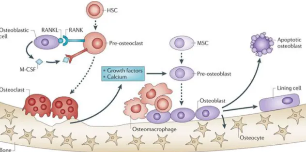
12 Bone resorption and formation not only should be quantitatively balanced but must also be coupled in time and space. This tight coupling is essential for the repair of microscopic damages that result from constant impact 109. Bone remodeling (Figure 2) is a dynamic and
continuous process that occurs throughout life, in which old bone is replaced by new one through the action of OBs, OCs and osteocytes 110,111. These cells have a central role in the
maintenance of the skeleton mechanical integrity and repair of fractures 111.
Figure 2 – Representation of bone remodeling
Osteoclasts (OCs) are derived from hematopoietic stem cells (HSCs) and become activated by RANK-RANKL binding and M-CSF stimuli, acquiring the ability to resorb bone and release growth factors and calcium, allowing mesenchymal stem cells (MSCs) differentiation into osteoblasts (OBs). OBs will replace the cavities with new bone, with the production of type I collagen and its subsequent mineralization to form the calcified matrix of bone. Osteocytes, terminally differentiated OBs embedded in bone, sense mechanical strain and signal to OCs and OBs, thus participate in the remodeling process. (Adapted from Weilbaecher K. et al., 2011) 112. M-CSF - macrophage colony stimulating factor; RANK - receptor
activator of NF-κB; RANKL - RANK ligand.
OCs, derived from hematopoietic lineage 109, are the unique bone resorbing cells, formed by
cytoplasmic, but not nuclear, fusion of mononuclear precursors of the monocyte/macrophage lineage of the bone marrow (hematopoietic stem cells - HSCs), peripheral circulation and tissue macrophage populations 113–116. Osteoclastogenesis is a complex process that includes many
stages, such as commitment, differentiation, multinucleation and activation of immature OCs
13 signaling pathways and depend on two major factors: macrophage colony stimulating factor (M-CSF) and receptor activator of nuclear factor-κB (RANK) ligand (RANKL) 115,119–121.
M-CSF is an hematopoietic growth factor expressed by OB lineage cells involved in the proliferation, differentiation and survival of monocytes, macrophages and bone marrow progenitor cell 122,123. M-CSF interacts with CSF1R expressed on OC precursors, which is
essential for their proliferation and survival and stimulates surface expression of RANK 124,125.
RANKL is a member of the TNF superfamily. It is expressed by bone marrow stromal cells, OBs, osteocytes, chondrocytes and immune system cells and interacts with its receptor RANK, expressed at the cell surface of OC precursors to induce OC differentiation 119,126–130. RANK is a
type I transmembrane receptor protein, member of the TNF receptor superfamily, expressed primarily on the cells of the monocyte/macrophage lineage including OC precursors and DCs but also on B and T cells and neutrophils 119,131–133.
RANKL exists both as soluble and membrane-bound forms 93. The soluble form (sRANKL)
corresponds to the c-terminal part of membranous RANKL that may be produced either directly by the stromal cells or OBs through alternative splicing followed by secretion to the extra-cellular medium, or by proteolytic cleavage of membranous RANKL by TNF- α converting enzyme (TACE) 110. Membrane-bound RANKL is expressed at the cell surface of OBs, bone
marrow stromal cells, fibroblasts, mammary epithelial cells and activated immune system cells
124. Deletion of the RANKL gene in mice resulted in severe osteopetrosis and a complete lack of
OCs, as a result of an inability of OBs to support osteoclastogenesis 134.
RANKL also binds to another TNF receptor family member, osteoprotegerin (OPG), which is produced by OBs, bone marrow stromal cells, immune system cells 124,135,136 and expressed in
normal VSMCs and downregulated in calcified VSMCs 137. OPG was first identified in 1997 as
being a protein that exhibits a protective effect on bone 138, although it is expressed
ubiquitously and abundantly in many tissues and cell types 139. When bound to RANKL, OPG
prevents its binding to RANK and thus inhibits the biological activity of RANKL, being the major negative regulator of bone resorption 119,140–143. It also exhibits a protective effect against
calcification in cells by reducing alkaline phosphatase 137. OPG-/- mouse exhibit a decrease in
total bone density with severe trabecular and cortical bone porosity, thinning of the skull, and a high incidence of fractures 144, contrary to OPG overexpression that causes severe
osteopetrosis in mice 138.
These studies demonstrated the importance of the RANKL/OPG ratio in OC differentiation and on its bone-resorptive function 125. The amount of RANKL and OPG expressed by OBs depend
on their stage of differentiation: pre-OB cells express high levels of RANKL and relatively low levels of OPG, thus stimulating OC differentiation and function. On the other hand, more
14 mature OBs express higher levels of OPG, in comparison to RANKL levels, inhibiting OC differentiation and function 145. Hence, the RANKL/OPG ratio in bone microenvironment is the
main molecular mechanism that determines osteoclastogenesis.
Together, M-CSF and RANKL are required to induce expression of OC lineage-specific genes, including those encoding TRAP, CTSK, calcitonin receptor (CALCR) and αvβ3 integrin, leading to the development of mature OCs 113.
OC precursors differentiate into TRAP+ mononuclear cells after RANKL stimulation. These cells
fuse with each other and differentiate into multinucleated OCs 146. Cell fusion is one of the
most distinctive characteristic properties of OCs. The OC also contains a unique and complex ruffled membrane, which is the resorptive organelle. Although the origin of this specialized membrane was enigmatic for some time, it is now considered to be the most specific marker of the OC, since it appears only when the cell is resorbing bone 147–149. Mature OCs express high
levels of tartrate-resistant acid phosphatase (TRAP), which has been widely used as a cytochemical marker of OCs and their precursors 150 and several other genes that regulate their
resorptive ability, including those encoding the chloride/H+ antiporter channel CLCN7 and CTSK 115,120. TRAP is a soluble acid resistant phosphatase secreted by the OC that plays a role in bone
resorption 151.
Bone resorption is a complex and specific process that occurs in three stages: (i) OC attachment to bone matrix and cell polarization, (ii) resorption, and (iii) cessation of resorption. The first study about the basic mechanism of bone degradation was performed in 1985 and established that the resorptive space is a highly acidified microenvironment [183]. Bone collagen degradation occurs through the action of a lysosomal enzyme, CTSK, which also polarizes to the ruffled membrane upon OC attachment to bone 152. After removal of the
inorganic bone phase and exposure of the matrix proteins, procathepsin K is activated. CTSK is secreted by OCs to degrade several bone matrix proteins, among which are included type I collagen and OPN. CTSK degrades type I collagen in its non-collagenous termini (N- and C-telopeptide regions) and releases crosslinked N- and C-C-telopeptides (NTX-I and CTX-I), which can be detected in urine and serum by immunoassays and are currently used to assess bone turnover 153,154.
OBs are mononuclear, non-terminally differentiated, specialized cells that derive from the mesenchymal stem cell (MSC) lineage 109,155 and have the ability to secrete bone matrix where
hydroxyapatite crystals deposit 145,156,157. OBs, chondrocytes, adipocytes, myoblasts, tendon
cells and fibroblasts are all derived from MSC. The lineages are determined by specific transcription factors 157. The differentiation of a MSC into a functional OB requires the
15 as runt-related transcription factor 2 - Runx2), osterix (Osx) and Wnt/β-catenin 158. More than
a decade ago, Cbfa1 was established as essential for bone formation since mice deficient in this transcription factor showed no ossification 159. Cbfa1 is a member of the runt family of
transcription factors that is expressed by MSCs at the onset of skeletal development and is present in OBs throughout their differentiation 160. Osx is another essential transcription factor
for OB differentiation. Osx transcription is positively controlled by Cbfa1 161. Osx acts by
forming a complex with NFATc1 (nuclear factor of activated T cell c1) resulting both in the activation of the type I collagen promoter 162 and in the induction of Wnt signaling pathway
and bone formation 163.
The progressive development of the OB phenotype from a proliferating immature cell to a mature OB that is able to synthesize bone proteins is characterized by a sequential expression of specific genes that identifies three periods of OB phenotype development: proliferation, maturation and extracellular matrix synthesis, and matrix mineralization 164. OB differentiation
and function, and therefore bone formation, is under the control of bone morphogenic proteins (BMPs) and Wnt signaling pathways 107,158. The Wnt signaling pathway regulates
several aspects of OB activities, including commitment of MSCs, OB progenitor amplification and cell death. The first evidence that Wnt/β-catenin signaling plays an important role in bone formation came from studies in humans where mutations that inactivate the Wnt co-receptor low density lipoprotein receptor-related protein (LRP)5 were shown to cause OP 165.
In the absence of Wnt proteins, glycogen synthase kinase-3β (GSK-3β) phosphorylates β-catenin, which is degraded, and the OB signaling cascade is blocked, so the MSCs become chondrocytes or adipocytes 166,167. The Wnt/β-catenin pathway is frequently referred to as the
canonical pathway and it promotes cell fate determination, proliferation and survival through the increase of β-catenin levels and alteration of gene expression by the transcription factor lymphoid enhancer factor/T cell factor (LEF/TCF) 166,168,169. Activation of this signaling pathway
occurs with binding of Wnt to the receptor Frizzled (FZ) and the co-receptors LRP5/6 170–172.
Some molecules specifically block the Wnt pathway such as secreted Frizzled related proteins (sFRPs), Wnt inhibitory factor (WIF), the dickkopf-related protein 1 (DKK1) and sclerostin (SOST) 172–175. DKK1 is a soluble inhibitor of Wnt pathway produced by osteocytes and OBs 169.
It interacts with LRP5/6 and transmembrane proteins kremen1 and 2, leading to the internalization of this complex and degradation by the proteasome 176. Its overexpression leads
to inhibition of OB proliferation and impaired mineralization 173 and its blockade leads to
increase in bone mass 177–179. Another inhibitor of the Wnt pathway is SOST, a secreted protein
mainly expressed by osteocytes 180. This protein is secreted in response to mechanical stimuli,
16 SOST inhibits Wnt/β-catenin pathway by binding to LRP5/6 co-receptor; however, it binds to a different region of LRP5/6 and it does not mediate receptor internalization 169,186. SOST
knockout mice have increased BMD, bone volume, bone formation and bone strength 182 while
overexpression of SOST leads to osteopenia 180.
During the post-proliferative phase, which is characterized by high levels of ALP, the extracellular matrix progresses to the mineralization phase, in which OBs synthesize several proteins associated with the mineralized matrix 187 including BSP 188, OPN and OCL 189. Collagen
type I, ALP and OCL are specific phenotypic markers of OBs expressed during its differentiation
119. OCL is expressed only in the post-proliferative phase, showing highest levels during
mineralization 119,190.
Besides synthesis and deposition of extracellular matrix proteins OBs are also responsible for the synthesis and secretion of molecules responsible for initiate and control OCs differentiation 119. OBs have the ability to regulate bone resorption through the expression of
RANKL, which binds to its receptor, RANK, on the surface of pre-OC cells, inducing their differentiation. On the contrary, the soluble decoy receptor OPG, also produced by the OB, is able to block RANK/RANKL interaction by binding to RANKL and thus prevent OC differentiation and activation 191.
At the end of the bone formation phase OBs have one of three possible fates: they can be embedded in the matrix and differentiate into osteocytes, they can become bone lining cells or they can undergo apoptosis 158,192–195. When OBs become entrapped in the bone matrix a
dramatic shift in both shape and function occurs and the cell becomes an osteocyte. Osteocytes are non-proliferative, terminally differentiated cells and constitute the main cellular component of mammalian bone, representing more than 95% of bone cells. They reside both in mineralized matrix and in newly formed osteoid, locked inside small lacunae. Osteocytes have dendritic morphology 126,196,197. Once embedded into the bone matrix, the
osteocyte ceases its matrix synthetic activity and initiates the function as strain and stress sensor 198. Another function of osteocytes within the bone cell network is the ability to deposit
and resorb bone around the lacuna in which they are housed, thus changing the shape of the lacuna. This process is called osteocytic osteolysis and is limited to specific situations 199.
Osteocyte differentiation is accompanied by progressive reduction of several bone markers such as ALP, BSP, OCL, type I collagen and Cbfa1 198,200. Although osteocytes by themselves do
not resorb or form bone, except in the lacunar area, they signal OBs and OCs to perform their functions 201. Osteocytes have been shown to produce RANKL and M-CSF and to support the
generation of functional resorbing OCs from their progenitors 202,203. These cells also express