Corresponding author: Angela Kinoshita, Universidade Sagrado Coração – USC, Rua Irmã Arminda, 10-50, CEP 17011-160, Bauru, SP, Brazil, e-mail: [email protected]; [email protected]
the Polyurethane Resin (
Ricinus communis
– Based
Biopolymer) Implantation Assessed by Light and Scanning
Electron Microscopy
Gustavo Campos Belmonte, Sérgio Augusto Catanzaro-Guimarães, Tatiana Peixoto Telles de Sousa,
Renato Savi Carvalho, Angela Kinoshita
Universidade Sagrado Coração, USC
Gilberto Orivaldo Chierici
Instituto de Química, USP
Abstract: The tissue reaction of bone tissue accessed by light microscopy and scanning electron microscopy (SEM) images after polyurethane resin implantation is presented in this study. Twenty four male rabbits were used, divided into two groups of 12 animals each (experimental group and control group) in which full-thickness cranial defect was surgically created. At 30 and 90 days post operation 6 animals of each group were euthanized and bone samples were removed for analysis. The microscopic results indicated no inflammatory foreign body reaction, a perfect union between the polymer and surgical bone bed surface, lack of bone resorption and presence of a thin layer of osteogenic material covering the polymer surface in contact with the surgical bone bed. The SEM images demonstrate the porosity of the resin, with diameters from 120 to 500 µm. This important feature of this polymer is associated with its osteoconductivity, allowing the bone growth inside it, improving the integration between the material and bone tissue. These results confirm that polyurethane resin derived from Ricinuscommunis is an excellent bone substitute for use in repair surgery for great bone losses.
Keywords: Bone repair, polyurethane resin, osteoconduction, biocompatibility.
Introduction
Extensive bone defects resulting from surgery, trauma and congenital anomalies continue to pose significant reconstructive problems. In defects reconstruction there is a need for creating conditions to the stimulus of bone neoformation in order to avoid the fibrous connective tissue growth into the bone defect, which would hinder osteogenesis. For this reason numerous biologically compatible materials have been applied experimentally for use in reparing bone defects[1-4].
The ideal bone substitute material must be biocompatible, biodegradable, (that is, able to be gradually substituted by the bone tissue in formation) osteoconductive and, if possible, must possess osteoinductive property[5].
Autogenous bone grafts have osteoinductive and osteoconductive properties. They are not immunogenic and are easily incorporated to the recipient bed. Thus, they are frequently used in maxillofacial reconstruction due to their excellent osteogenic properties[6]. However,
autogenous bone graft are associated to morbidity at the donor site characterized by infections, chronic pain, hemorrhages, neurological alterations and local fractures which demand a new surgical intervention[7].
The utilization of allogenous or xenogenous bone is not advisable due to the possibility of transmitting infections and the development of antigenic response[8-11].
Numerous synthetic bone promoting biomaterials such as: tricalcium phosphate[12,13], porous hydroxyapatite[14],
biopolymer[15-17] and bioactive glass[18,19] have been
developed. In this context, injectable scaffolds represent a very interesting option because they are easily adapted in the shape of defect, allowing the tissue repair after minimally invasive administration. These materials present new challenges because they must be converted from an injectable material to a solid or gel with appropriate kinetics and without damaging surrounding tissues. Examples of injectable scaffold currently available for bone regeneration include Acrylic Bone Cements (ABCs), Calcium Phosphate Cements (CPCs), Calcium Sulfate Cements (CSC). Each material has advantages and disadvantages and the choice depends upon the size, location of the defect, among others. The ABCs are based on methyl methacrylate and harden by an exothermic reaction. Their main disadvantage is the high polymerization temperature, of over 70 °C, which may cause local cellular destruction. The (CPCs) are formed by a combination of one or more calcium phosphates (CaPO4), which, upon mixing with the liquid phase, form a paste that sets and solidifies. Their main advantage is lack of macroporosity which results in slow bone neoformation. CSC (CaSO4) in its known form is named as plaster of Paris or gypsum. It is the
A
R
T
I
G
O
T
É
C
N
I
C
O
C
I
E
N
T
Í
F
I
C
material with the longest clinical history. It is a highly biocompatible material but the lack of bioactivity is one of its disadvantages[20].
Another injectable alloplastic material that has been used for tissue regeneration is Polyurethane (PU). This material allows the association with other substances such as fluor-hydroxyapatite,[21] and biphasic calcium
phosphate[22] forming a scaffold; with vancomicin, acting
as a sustained release system, preventing the infection in the defect site[23]; with rh-BMP-2, recombinant
human bone morphogenetic protein-2 to enhance bone formation[24].
In Brazil, a polyurethane with vegetable origin, made from fatty acids extracted from Riccinus communis
(castor oil) was developed and is produced as biomaterial by Poliquil – Araraquara., Polímeros Químicos LTDA, São Paulo state, Brazil. This resin is commercially available with the brand name “COR – Composto Óleo de Rícino”[25]. Barros et al.[26] evaluated biocompatibility
of polyurethane resin (pure), the polyurethane resin with calcium carbonate and with calcium phosphate. The materials were made in cylindrical form and were randomly placed in defects created in rabbit femur. Histological analysis revealed absence of persistent inflammation or foreign body reaction type in all periods analyzed (8, 12 and 16 weeks). After 8 weeks it was observed regeneration of cortical bone and a thin layer of fibroblasts and collagenous fibers parallel to the surface of the cylinders implanted. There were not observed differences in the interface bone/material among the three compositions studied. Pereira-Júnior et al.[15] analyzed
the behavior of this polyurethane resin in granulated form in segmental defects created in rabbits radius at 15, 30, 60 and 120 days postoperatively. Histomorphometric analysis showed progressive bone regeneration in both types of treatment, however, in periods of 120 days, defects treated with autologous bone showed to be fully remodeled by newly formed bone (100%), whereas treated defects by polyurethane exhibited 79% of the total volume of the defect filled by new bone. These results reinforced the osteogenic and osteoinductive properties of autologous bone and demonstrate the biocompatibility of polyurethane resin, its ability to prevent the proliferation of connective tissue into the interior of the defects.
These previous studies have attributed most of the positive results in relation to bone regeneration to the osteoconductivity of polyurethane. So, the main purpose of this work is the analysis of the tissue reaction to the implantation of the polyurethane resin by light microscopy and the interface polymer-bone tissue by scanning electron microscopy (SEM) aiming to determine its biocompatibility and osteoconductivity.
Material and Methods
This study was previously analized and approved by the Ethics Commitee of the “Universidade Sagrado Coração” Bauru – SP.
Twenty four New Zealand male rabbits weighing 2.5 kg and aged between 3 to 4 months were used. The animals were kept in good environmental, food, temperature, hygiene and light conditions.
The animals were divided randomly into two groups (experimental group and control group) of 12 animals each. Each animal of the experimental group received the polyurethane resin as a graft material (COR - Poliquil – Araraquara., Polímeros Químicos LTDA, São Paulo state, Brazil). The control group animals had their defects filled only with autogenous blood clot. The assessment periods were on the thirtieth and the ninetieth postoperative days, and a total of 6 control and 6 experimental animals were euthanized in each group.
Anesthesia was accomplished by intramuscular injection of Cetamin (10 mg/kg, Syntec do Brazil, LTDA) and Xilazin 2% (4 mg/kg, Syntec do Brazil, LTDA).
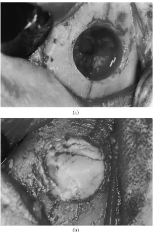
Local anesthesia (1 mL of Citocaine 3% and Felipressin 0.03 UI, Cristalia Pharmaceutical, Itapira, SP, Brazil) was administered to the soft tissues overlying the craniotomy site. The dorsal part of the craniun was shaved and asseptically prepared for surgery. A 30 mm long incision was made in the middle of the scalp parallel to the sagital suture. Subperiostal dissection was carried out and a bicortical bone defect with a diameter of 5 mm was prepared with a trephine in the parietal bones under saline irrigation (Figure 1A).
The polyurethane resin was prepared in a sterile plastic container using 5ml of prepolymer and 5ml of a poliol derived from the ricinoleic oil. The resin was implanted in the receptor bed and adapted to the bone
Figure 1. (a) Surgical bone defect created with a trephine 5 mm
surface (Figure 1B). Then, the periosteum was closed with resorbable suture and the skin with nonresorbable suture. Thirty minutes before surgery the rabbits were administered intramuscular injection of penicililin (2.500.000 IU/5 mL:0.1 mL/kg). The antibiotic treatment was continued postoperatively for 2 days. Following surgery rabbits received intramuscular injections of 0.5 mL of Dipirone for analgesia.
The control group animals were submitted to the same surgical procedure carried out with the experimental group animals, with the difference that the bone defect was filled only with autogenous blood clot (Figure 1A).
Twelve animals were euthanized in each period (30 and 90 postoperative days), six from the experimental group and six from the control group by lethal injection of sodium pentothal. The operated areas were exposed and carefully examined for eventual macroscopic signs of inflammation and/or infection. The specimens were removed by means of osteotomy with steel burs number 6 in low speed.
Twenty two of the 24 specimens obtained were submitted to light microscopy analysis. For this, pieces of skull containing polyurethane resin implants were then removed and fixed with 10% buffered formaline. After fixation, they were decalcified in solution of formic acid sodium citrate (equal parts of 50% formic acid and 20% sodium citrate). The decalcified bones were dehydrated and embedded in paraffin blocks. Thin sections – 6 µm thick - embedded in paraffin wax were cut and stained with haematoxylin and eosin and Masson’s Trichrome Stain. Two pieces – experimental cranium 30 and 90 days - and a piece of isolated fragment of polyurethane resin were prepared for the scanning electron microscopy analysis to verify the polymer/bone tissue interface and the physical structure of the polyurethane resin.
Results
Light microscopy
30-day period
In the control defects we observed residue of hemorrhage areas with mononuclear infiltration. Polimorphonuclear cells were eventually found. The surgical defect area presented entirely filled by mesenchymal immature granulation tissue, with unorganized collagen fibers in a fairly angiogenic activity. Moreover, immature bone callus extruding from the defects wall could be observed (Figure 2).
The implanted defects (Figure 3) were totally filled with the polyurethane resin. The polymer shows a porous aspect. The pores represent canals with variable diameters which interlace in the interior of the polyurethane resin in several directions. The most attractive aspect is the close link of the polyurethane resin to the surgical bone bed surface. It was also observed a thin layer of osteoid matrix covering the polymer surface in close contact with the wall of the bone bed surface. It is important to point out the lack of inflammatory cells, both on the defect bone wall and on the osteogenic connective tissue in contact with the polyurethane resin surface (Figure 3).
90-day period
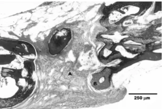
On the control defects we observed a bone callus in remodelling process. It is also noted connective tissue areas interlacing with the defect bone walls (Figure 4).
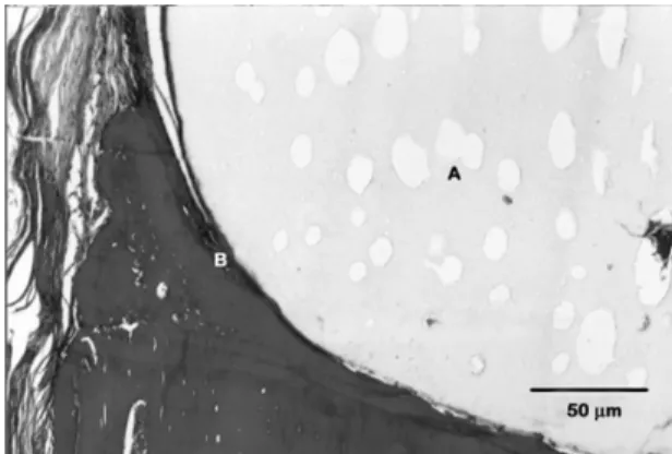
In the implanted defects it can be observed the porous polymer partially filled with non-lamelar newly formed immature bone. Non-lamelar immature bone is also found on the polyurethane resin surface. All polymer thickness is permeable by intercommunicating pores, allowing the penetration of newly formed bone inside the polymer’s interior. On this stage, the polyurethane resin shows greater fragmentation areas (Figure 5). The polymer fragmentation occurs without the action of the inflammatory cells.
Scanning Electron Microscopy (SEM)
For the SEM analysis in addition to the polyurethane resin isolated fragment, 2 specimens experimental cranium 30 and 90 were used.
Figure 6 shows in detail the polymer surface where it can be observed different pore sizes opening throughout
Figure 2. Photomicrograph of the control defect at 30 days
post-surgery. Mesenchymal granulation tissue (GT) with delicate collagen fibers interlacing anchoring discrete chronic inflammatory infiltrate (Masson Trichrome).
Figure 3. Photomicrograph of the experimental defect area at
can point out the guided tissue regeneration by using biological membranes, biological grafts and synthetics bone substitutes as implant materials.
An ideal material to be utilized as a bone substitute must present specific properties as biocompatibility, osteoconductivity and, if possible, osteoinductibility. Biocompatibility involves a perfect integration between bone and implant, that implies in lack of inflammatory reaction at the bone/implant interface[5,12,15,16,28]. Some
of these features were observed in this work such as: a perfect union between the polymer and surgical bone bed surface, lack of inflammatory reaction, lack of bone resorption and presence of a thin layer of osteogenic material covering the polymer surface in contact with the surgical bone bed, that is, at the bone/implant interface.
In none of the analyzed periods it was found inflammatory reaction such as foreign body chronic inflammatory. Osteoclastic bone resorption was totally absent either on the initial periods or at the end of experiment. Other reports have shown similar results[15,29,30]. As no necrotic tissue was noticed in contact
with the polymer, not even signs of cell degeneration,
Figure 4. Photomicrograph of the control defect area at 90 days
post-surgery. (A) Osteogenic mesenchymal connective tissue interposing between the bone walls. (B) Bone trabeculae exhibit a rich number of cells on the surface with osteoblasts. (Masson Trichrome).
Figure 5. Photomicrograph of the experimental defect area at 90
days post-surgery. Immature non-lamelar bone on the surface (A) and inside (B) the intercommunicating pores of the polyurethane resin. Absence of inflammatory reaction. (Masson Trichrome).
Figure 6. Scanning Electron Microscopy of the resin surface
showing the different pore sizes. Magnification 35×
the surface with diameters varying from 120 to 500 µm. It can be observed that the pore is actually a tunnel in which smaller intercommunicating tunnels are opening (Figure 7).
Figure 8 shows the bone defect (experimental 30-day group) in which details of the bone and implant boundaries are noticed. Polyurethane resin is on the top of figure. There is no sign of resorption at the bone/ polymer interface.
Figure 9 shows the bone defect (experimental 90 day group) filled by polyurethane resin. Some of the pores are partially filled by regenerated bone tissue.
Discussion
Bone regeneration failure is frequently observed on defects presenting great bone loss. When this process is not complete, the fibrous repair takes place which has no biologic value in the bone regeneration process. To solve this problem, several methods based on the different principles have been used with large surgical repercussion, when it comes to bone repair process in critical defects[27]. Among the various methods we
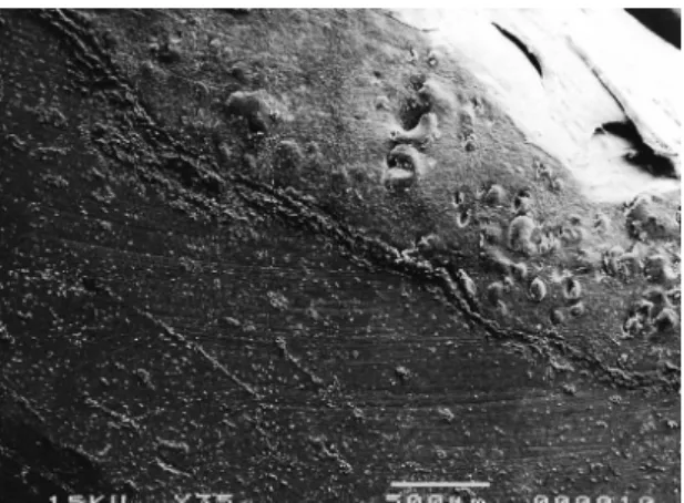
Figure 7. Scanning Electron Microscopy in higher magnification
it was concluded that polyurethane resin is devoid of citotoxicity. In other publications utilizing citotoxicity method in vitro by the release of radioactive chrome, a
certain citotoxicity in vitro in the polyurethane resin was observed[31]. This result tends to classify this resin
as tissue irritant. However, the same polyurethane resin utilized in this study showed to be totally non-toxic to the tissues, allowing us to conclude that the in vitro test must not be taken as totally reliable, when collecting data related to the biocompatibility of the material at the bone/ implant interface.
In the analysis of interface between polymer/bone tissue by SEM images, the osteoconductivity can by observed. Osteoconductivity is a feature of porous structures with diameters between 150 and 500 µm that allows the bone growth inside of them[32]. The SEM images
show that the polyurethane resin studied has porosity with diameters from 120 to 500 µm that characterizes the osteoconductivity of this resin (Figures 6 and 7). These micro porosities occurred at the resin, internally and at
the contact surface to the defect walls. In the periods of study, it was also noted osteogenic material covering the pore walls that were opening at the contact line with surgical bone bed. In the late periods it was possible to observe immature non-lamelar bone adhering to the wall or filling the polyurethane resin pores (Figure 5). The enlargement of the intercommunicating pore system diameter of polyurethane resin, with time, starts to permit the penetration of new formed blood vessels and undifferenciated cells, necessary to the immature bone deposition at the pore internal walls. The angiogenic activity and the infiltration of undifferentiated osteogenic cell population are the main determining factors of the osteoconduction in the alloplastics implants[33,34]. For
this reason this material is suitable for wide application in surgeries of large bone defects due to the perspective of this material to undergo gradual degradation when the new forming bone begins to replace it. This behavior is in agreement with the generalized notion that an ideal bone substitute is one that maintains its mechanical and volume stability in the initial phases of implantation and, subsequently, undergoes total resorption[35].
Conclusion
Our results indicate that polyurethane resin derived from Ricinus communis is an osteoconductive material
and behaves as a bone substitute. The polymer maintains the mechanical and volumetric stability in the initial periods of the repair process, undergoing subsequently a dismemberment and possibly substitution by newly formed bone.
References
1. Schmitz, J. P. & Hollinger, J. O. - Clin. Orthop. Rel. Res., 205, p.299 (1986). PMid:3084153.
2. Kawamura, M. & Urist, M. R. - Clin. Orthop. Rel. Res., 236, p.240 (1988). PMid:3180578.
3. Mardas, N.; Kostopoulos, L. & Karring, T. - J. Craniof. Surg., 13, p.453 (2002).
http://dx.doi.org/10.1097/00001665-200205000-00017
4. Haddad, A. J.; Peel, S. A. F.; Clokie, C. M. L. & Sándor, G. K. B. - J. Craniof. Surg., 17, p.926 (2006). PMid:17003622. http://dx.doi.org/10.1097/01.scs.0000230615.49270.d1 5. Jensen, S. S.; Aaboe, M.; Pinholt, E. M.; Hjørting-Hansen,
E.; Melsen, F. & Ruyter, I. E. -. Int. J. Oral Maxillofac. Implants, 11, p.55 (1996). PMid:8820123.
6. Jensen, O. T. - “Guided Bone Graft Augmentation”, in: Guided Bone Regeneration in Implant Dentistr, Buser D, Dahlin C, Schenck RK. Eds., Quintessence, Chicago (1994).
7. Arrington, E. D.; Smith, W. J.; Chanbers, H. G.; Bucknell, A. L. & Davino, N. A. – Clin. Orthop. Rel. Res., 329, p.300
(1996). PMid:8769465. http://dx.doi.org/10.1097/00003086-199608000-00037
8. Friedlaender, G. E. - J. Bone Joint Surg. Am., 69, p.786
(1987).
9. Goldberg, V. M. & Stevenson, S. - Clin. Orthop. Rel. Res., 225, p.7 (1987). PMid:3315383.
Figure 8. Scanning Electron Microscopy of the skull bone and
resin at 30 days post-surgery. Longitudinal section showing details of the bone and implant (resin) boundaries. Resin is on the top.
Figure 9. Scanning Electron Microscopy of the skull bone
10. Buck, B. E.; Malinin, T. I. & Brown, M. D.- Clin. Orthop. Rel. Res., 240, p.129 (1989). PMid:2645073.
11. Chapman, P. G. & Villar, R. N. - J. Bone Joint Surg. Br., 74,
p.398 (1992).
12. Calvo-Guirado, J. L.; Delgado-Ruiz, R. A.; Ramirez-Fernandez, M. P.; Mate-Sanchez, J.E.; Ortiz-Ruiz, A. & Marcus, A. - Clin. Oral Impl. Res., 23, p.667 (2012). PMid:21492238.
http://dx.doi.org/10.1111/j.1600-0501.2011.02193.x 13. Kim, H. J.; Park, I. K.; Kim, J. H.; Cho, C. S. & Kim, M.
S. - Tissue Eng. Regen. Med., 9, p.63 (2012).
14. Huang, X. & Miao, X. - J. Biomater. Appl., 21, p.351 (2007). PMid:16543281. http://dx.doi. org/10.1177/0885328206063905
15. Pereira-Júnior, O. C. M.; Rahal, S. C.; Iamaguti, P.; Felisbino, S. L.; Pavan, P. T. & Vulcano, L. C. - J. Biomater. Appl., 21, p.283 (2007). PMid:16543284.
16. Ereno, C.; Guimarães, S. A. C.; Pasetto, S.; Herculano, R. D.; Silva, C. P.; Graeff, C. F. O.; Tavano, O.; Baffa, O. & Kinoshita, A. - J. Biomed. Mater. Res., Part A, 95A, p.932 (2010). PMid:20845492. http://dx.doi.org/10.1002/ jbm.a.32919
17. Herculano, R. D.; Silva, C. P.; Ereno, C.; Guimaraes, S. A. C.; Kinoshita, A. & Graeff, C. F. O. - Mater. Res.,12, p.253 (2009).
http://dx.doi.org/10.1590/S1516-14392009000200023 18. Matsumoto, M. A.; Caviquioli, G.; Briguetti, C. C.; Holgado,
L. A.; Saraiva, P. P.; Muniz-Renno, A. C.; Kawakami, R. Y. - J. Mater. Sci. Mater. Med., 23, p.1447 (2012). PMid:22426745.
http://dx.doi.org/10.1007/s10856-012-4612-8
19. Wu, C.; Luo, Y.; Cuniberti, G.; Xiao, Y. & Gelinsky, M. - Acta Biomater., 7, p.2644 (2011). PMid:21402182.
http://dx.doi.org/10.1016/j.actbio.2011.03.009
20. Rahman, C. V.; Saeed, A.; White, L. J.; Gould, T. W. A.; Kirby, G. T. S.; Sawkins, M. J.; Alexander, C.; Rose, F. R. A. J. & Shakesheff, K. M. - Chem. Mater., 24, p.781 (2011). http://dx.doi.org/10.1021/cm202708n
21. Asefnejad, A.; Behnamghader, A. & Khorasani, M. T. - Int. J. Nanomed., 6, p.93 (2011). PMid:21289986 PMCid:PMC3025589. http://dx.doi.org/10.2147/IJN.S13385 22. Kim, H.; Park, I.; Kim, J.; Cho, C. & Kim, M. - Tissue Eng.
Regen. Med., 9, p.63 (2012).
23. Li, B.; Brown, K. V.; Wenke, J. C. & Guelcher, S. A. - J. Control. Release, 145, p.221 (2010). PMid:20382191. http://dx.doi.org/10.1016/j.jconrel.2010.04.002
24. Jerald, E. D.; Pamela, B. B.; Edna, M. P.; Teja, G.; Robert, G. H.; Joseph, C. W. & Scott, A. G. - Biomed. Mater., 7,
p.024112 (2012).
25. Poliquil Araraquara Polímeros Químicos. Disponível em: <http://www.poliquil.com.br/>. Acesso em: 18 dez. 2012. 26. Barros, V. M.; Rosa, A.L.; Beloti, M.M. & Chierice, G. - J.
Biomed. Mater. Res. A, 67, p.235 (2003).
27. Dahlin, C. - “Scientific background of guided bone
regeneration”, in: Guided bone regeneration in implant
dentistry Buser, D.; Dahlin, C.; Schenck, R.K. Eds, p.31-48, Quintessence, Chicago (1994). PMid:8150510. 28. Schlegel, K. A.; Sindet-Pedersen, S.; Hoepffner, H.
J. - J. Biomed. Mater. Res., 53, p.392 (2000). http://
dx.doi.org/10.1002/1097-4636(2000)53:4<392::AID-JBM13>3.0.CO;2-A
29. Carvalho, T. L.; Teofilo, J. M.; Araújo, C. A. & Brentegani, L. G. - J. Biomed. Mater. Res., 37, p.449. PMid:9407291. 30. Dumas, J. E.; BrownBaer, P. B.; Prieto, E. M.; Guda, T.;
Hale, R. G.; Wenke, J. C. & Guelcher, S. A. - Biomed. Mater., 7, p.S1 (2012).
31. Pascon, E. A.; Leonardo, M. R.; Safari, L. & Langeland, K. - Oral Surg. Oral Med. Oral Pathol., 72, p.222 (1991).
http://dx.doi.org/10.1016/0030-4220(91)90168-C 32. Sutherland, D. & Bostron, M. - “Grafts and bone graft
substitutes”, in Bone regeneration and repair. Biology
and Clinical applications, Liebernam, J.R.; Friedlaender, G.E. eds. Totowa, p. 133-156, Humana Press, New Jersey (2005).
33. Klawitter, J. J.; Bagwell, J. G.; Weinstein, A. M.; Sauer, B. W. & Pruitt, J. R. - J. Biomed. Mater. Res., 10, p.311 (1976).
PMid:1254618. http://dx.doi.org/10.1002/jbm.820100212 34. Daculsi, G. & Passuti, N. – Biomaterials, 11, p.86 (1990).
PMid:2397267.
35. Isaksson, S. - Swed. Dent. J. Suppl., 84, p.1 (1992). PMid:1334579.