UNIVERSIDADE DA BEIRA INTERIOR
Ciências da Saúde
Intestinal Polyposis Syndromes:
case reports of rare genetics variants and review
Ana Daniela de Oliveira e Silva
Dissertação para obtenção do Grau de Mestre em
Medicina
(ciclo de estudos integrado)
Orientador: Doutor Sérgio Bernardo de Sousa
Co-orientador: Doutora Sofia Maia
iii
Dedication
To my parents. To my brothers. To my friends.
v
Acknowledgments
This work symbolizes the end of an important period of my personal and professional development and a turning point to another one in which I wish to humbly implement everything that has been taught. Therefore, it is the moment to thank all the people who helped me to achieve this and I believe that will be the solid foundation that will allow me to approach the next new phase.
To Dr. Sérgio Bernardo de Sousa for the trust and commitment that has placed in this work, for the guidance and availability in its accomplishment and for the incessant sharing of knowledge.
To Dr. Sofia Maia for the collaboration and all the time made available in the accomplishment of this work.
To my parents and my brothers for the solid support they always give me. To my friends for the help and understanding they always have to me.
vii
Abstract
Introduction: Currently, colorectal carcinoma is the most prevalent gastrointestinal cancer in the world and the second cause of death from malignant disease. Hereditary polyposis syndromes account for about 1% of colorectal cancer. Familial Adenomatous Polyposis is the second most common inherited colorectal cancer syndrome and it is characterised by the early development of tens to thousands of adenomatous polyps and/or cancer in the colon and rectum. MUTYH-Associated Polyposis is associated with dozens or few hundreds of adenomatous polyps in the colon and an increased risk of colorectal cancer.
Objective: The present work will focus on adenomatous polyposis syndromes, in particular on the description of two families with rare presentations and review of the literature.
Clinical Cases: The first clinical case is a 17-year-old male who presented with a phenotype characterised by non-progressive bowing of the right leg detected at 18 months of age caused by a fibula malformation (pachydysostosis) and a large exophytic osteoma of the left radius, noticed at the age of 15 years, without gastrointestinal symptoms. Detailed clinical and radiological characterisation revealed multiple osteomas (of the left fibula, left ilium, metacarpals and mandible), skin lesions and dental abnormalities, raising the hypothesis of Gardner Syndrome. This diagnosis was confirmed by genetic testing (a de novo mutation in the
APC gene was identified) and endoscopic investigation, which identified the presence of
multiple adenomatous polyps throughout the colon, ileum and stomach. The second clinical case is the report of a 34-year-old male with adenomatous colonic polyps (45 polyps at the age of 33 years) and a family history of adenomatous polyposis and colon neoplasia. The patient's family history suggested an autosomal dominant inheritance pattern, which would be in favour of a mutation in APC gene (autosomal dominant inheritance) rather than mutations in MUTYH gene (autosomal recessive pattern), which ended up being the correct diagnosis.
Final Remarks: The clinical cases here described illustrate the diversity of presentations in patients with adenomatous polyposis syndromes and the challenges in their accurate recognition and diagnosis. The first case points out the difficulties in establishing an early diagnosis in a de novo APC mutation, which is essential for an appropriate management. It also emphasizes the importance of a detailed clinical characterization, including FAP extra-colonic manifestations, and the need of a multidisciplinary team in reference centres, articulated with international experts. The second clinical case highlights the importance of a detailed family history and of international guidelines, in particular for the appropriate genetic testing strategy.
viii
Key-words
Intestinal Polyposis Syndromes; Familial Adenomatous Polyposis; Gardner Syndrome; APC gene;
ix
Resumo Alargado
Introdução: Actualmente, o cancro colorrectal constitui a neoplasia gastrointestinal mais prevalente a nível mundial e a segunda causa de morte por doença maligna. As síndromes de polipose intestinal são responsáveis por 1% de todos os casos de cancro colorrectal. A Polipose Adenomatosa Familiar (FAP) é a segunda síndrome hereditária mais comum que predispõe para cancro colorrectal e é caracterizada pelo desenvolvimento precoce de dezenas a milhares de pólipos adenomatosos e/ou cancro a nível do cólon e recto. Esta entidade clínica está relacionada com mutações do gene APC. A Polipose Associada ao MUTYH está relacionada com o desenvolvimento de dezenas a centenas de pólipos adenomatosos cólicos e um risco aumentado para cancro colorrectal, mas numa idade mais tardia em comparação com a Polipose Adenomatosa Familiar. A Polipose Associada ao MUTYH deve-se a mutações no gene MUTYH. Objectivo: O presente trabalho foca-se nas síndromes de polipose adenomatosa cólica, em particular na descrição de duas famílias com apresentações raras destas síndromes e na revisão de literatura destas entidades clínicas.
Casos Clínicos: O primeiro caso clínico descrito é um jovem de 17 anos cujo fenótipo se manifestou inicialmente por arqueamento não progressivo da perna direita detectado aos 18 meses causado por uma malformação da fíbula (paquidisostose) e, posteriormente, um volumoso osteoma exofítico localizado no rádio esquerdo, detectado aos 15 anos de idade, que motivou a sua avaliação em vários hospitais e o seu posterior envio ao centro de referência de tumores ósseos do Hospital Pediátrico de Coimbra. Não tinha qualquer sintoma gastrointestinal. A caracterização clínica e radiológica detalhada neste centro revelou múltiplos osteomas de pequenas dimensões (mandíbula, fíbula esquerda, osso ilíaco esquerdo e metacarpos) e anomalias cutâneas e dentárias que, em reunião multidisciplinar, fizeram colocar a hipótese diagnóstica de Síndrome de Gardner (FAP com manifestações extra-cólicas proeminentes). Estudos endoscópicos subsequentes identificaram múltiplos pólipos adenomatosos a nível do cólon, íleo e estômago e o estudo do gene APC identificou uma mutação patogénica na região do gene conhecida como associada ao Síndrome de Gardner. O segundo caso clínico apresentado reporta um homem de 34 anos com diagnóstico de pólipos adenomatosos cólicos através de colonoscopia (45 pólipos cólicos aos 33 anos de idade) e com história familiar de polipose adenomatosa e neoplasia do cólon. A história familiar sugeria um padrão de hereditariedade autossómica dominante, o que seria a favor de uma mutação no gene APC (transmissão autossómica dominante). No entanto, o estudo genético do gene APC foi normal. Seguindo a estratégia diagnóstica recomendada, foi então proposto o estudo do gene MUTYH (transmissão autossómica recessiva), que identificou a presença de uma mutação patogénica já descrita em homozigotia.
x
Notas Finais: Os casos clínicos aqui descritos ilustram bem a diversidade de apresentações clínicas em pacientes com síndromes de polipose adenomatosa cólica e os desafios no seu reconhecimento e diagnóstico atempados. A descrição do primeiro caso clínico evidencia as dificuldades de um diagnóstico precoce num primeiro familiar afectado com uma mutação de
novo no gene APC. Este diagnóstico atempado é essencial para a implementação de um
seguimento adequado. É de referir ainda a importância de uma caracterização clínica e radiológica detalhada, da valorização das manifestações extra-cólicas que devem levar à suspeita de FAP e da necessidade de trabalhar em equipas multidisciplinares em centros de referência, articulados com peritos internacionais. No segundo caso clínico apresentado destacamos a presença de uma história familiar com um padrão de transmissão pseudo-dominante numa entidade clínica com hereditariedade autossómica recessiva. Perante o quadro clínico e história familiar, a primeira hipótese diagnóstica colocada foi FAP atenuada, mas o estudo genético do gene APC não confirmou esta hipótese. Como recomendado, prosseguiu-se com a realização do estudo molecular do gene MUTYH, que levou ao diagnóstico correcto, possibilitando a optimização no seguimento destes indivíduos, o aconselhamento genético preciso e o rastreio de outros familiares em risco.
xi
Index
Dedication iii Acknowledgments v Abstract vii Key-words viii Resumo Alargado ix Index xiList of Figures xiii
List of Tables xv
List of Acronyms xvii
1. Introduction 19
2. Methodology 21
3. Review of the Literature 23
3.1. Familial Adenomatous Polyposis 23
3.1.1. Colonic Manifestations 23
3.1.2. Extra-Colonic Gastrointestinal Manifestations 23
3.1.3. Extra-Intestinal Manifestations 24 3.1.3.1. Gardner Syndrome 25 3.1.3.2. Turcot Syndrome 26 3.1.4. Diagnosis 26 3.1.5. Surveillance 27 3.1.6. Prophylaxis 28
3.1.7. Treatment of Specific Lesions 29
3.1.8. Genetic Counselling 29
3.2. Attenuated Familial Adenomatous Polyposis 31
3.2.1. Colonic Manifestations 31
3.2.2. Extra-Colonic Gastrointestinal Manifestations 31
3.2.3. Extra-Intestinal Manifestations 31 3.2.4. Diagnosis 31 3.2.5. Surveillance 32 3.2.6. Prophylaxis 33 3.2.7. Genetic Counselling 33 3.3. APC Gene 34
xii
3.4. MUTYH-Associated Polyposis 36
3.4.1. Colonic Manifestations 36
3.4.2. Extra-Colonic Manifestations 36
3.4.3. Diagnosis 37
3.4.4. Surveillance, Prophylaxis and Treatment 38
3.4.5. Genetic Counselling 38
3.4.6. Prevention and Prognosis 39
3.5. MUTYH Gene 40 4. Case Reports 43 4.1. Clinical Case 1 43 4.2. Clinical Case 2 50 5. Discussion 53 5.1. Clinical Case 1 53 5.1.1. Skeletal Manifestations 54 5.1.2. Skin 56 5.1.3. Teeth 56 5.1.4. Gastrointestinal Tract 57 5.1.5. Genotype-Phenotype Correlation 57 5.1.6. Follow-up 59 5.1.7. Genetic Counselling 60 5.1.8. Prognosis 60
5.1.9. Summary of Phenotypic Characteristics, Comparison with Literature and Follow-up 62
5.2. Clinical Case 2 63
5.2.1. Family History and Heredogram 63
5.2.2. Diagnostic 63
5.2.3. Genotype-Phenotype Correlation 64
5.2.4. Follow-up and Prognosis 64
5.2.5. Genetic Counselling 65
6. Final Remarks 67
xiii
List of Figures
Figure 1 - Flowchart of the literature search, study selection and results. 21
Figure 2 - Family History of M.A.V.N. 43
Figure 3 – Lower legs photograph (A) and radiographs (B, C) of the patient (IV.4) at 16 years of age. The right fibula malformation, detected at 18 months of age, is likely to be congenital and not significantly progressive. This fibula is thick, elongated and bowed with posteromedial convexity, more evident in the lateral view (C). After discussion with international experts, it fits best the classification of pachydysostosis of the fibula. There is a slight length discrepancy between the two legs. The tibia is normally shaped, despite some cortical thickness and
waviness, a known feature of FAP. 44
Figure 4 - Photograph (A), CT (B) and radiographs (C, D) of the patient’s (IV.4) right forearm at 16 years of age. Please remark the large exophytic osteoma of the left radius and associated
increased volume of the forearm. 45
Figure 5 - Craniofacial photographs (A, B) and radiograph (C) of the patient at 16 years of age. Please note the bilateral prominence of the mandible and, at least, one small palpable osteoma
at the right mandible angle. 46
Figure 6 – Photographs of the patient, at the age of 16, showing a small cutaneous lesion located at the right dorsum/scapular area, most likely an epidermoid cyst (A), and the
chilblain/erythema pernio of the hands (B). 46
Figure 7 - Radiographs of the left leg (A) and left hand (B) of the patient at the age of 16. Please note the presence of small osteomas at the left fibula (arrows, A) and some waviness of the tibial and fibula cortical. At the hand, note the diaphyseal exostosis/osteomas of the second
and third metacarpals (arrows, B). 47
Figure 8 - Lower limbs radiograph of the patients at the age of 16. Please remark the multiple osteomas at the left ilium and left fibula (arrows) and some slight cortical thickness and
waviness of the long bones. 48
Figure 9 - Family History of F.A.M.M. 51
Figure 10 - The APC gene, APC protein domains and FAP phenotype usually associated with the respective germline mutation position. (Adapted from Nieuwenhuis et al, 2007 (34)) 58
xv
List of Tables
Table 1 - Prevalence of certain types of pathogenic variants depending on the geographic
location (Adapted from Nielsen et al, 2012 (27)) 41
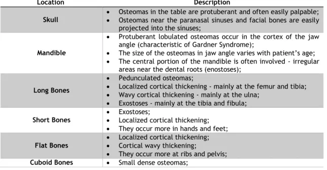
Table 2 - Bone lesions associated with Gardner Syndrome (Adapted from Chang et al, 1968 (33)) 56 Table 3 - Summary of Phenotypic Characteristics, Comparison with Literature and Follow-up
xvii
List of Acronyms
AFAP Attenuated Familial Adenomatous Polyposis APC Adenomatous Polyposis Coli
CHRPE Congenital Hypertrophy of the Retinal Pigment Epithelium CRC Colorectal Carcinoma
COX-2 Cyclooxygenase-2 CT Computed Tomography CK Creatine Kinase
CK-1 Casein Kinase-1
DCC Delected in Colon Cancer DNA Deoxyribonucleic Acid
FAP Familial Adenomatous Polyposis GSK3 Glycogen Synthase Kinase 3 HGMD Human Gene Mutation Data
HNPCC Hereditary Nonpolyposis Colorectal Cancer IPAA Ileal Pouch Anal Anastomosis
IRA Ileorectal Anastomosis
LEF Lymphoid Enhancer-binding Factor MAP MUTYH-Associated Polyposis MCR Mutation Cluster Region
MLPA Multiplex Ligation-dependent Probe Amplification MRI Magnetic Resonance Imaging
mRNA Messenger Ribonucleic Acid NGS Next Generation Sequencing
NSAIDs Nonsteroidal Anti-Inflammatory Drugs PCR Polymerase Chain Reaction
ROS Reactive Oxygen Species TCF T-Cell Factor
19
1. Introduction
Currently, colorectal carcinoma (CRC) is the most prevalent gastrointestinal cancer in the world and the second cause of death from malignant disease. (1) The incidence of CRC is higher in developed and western countries and a correlation has been made between the higher incidence and the typical dietary habits and lifestyle factors of these countries. (2)
Colorectal cancer can be, roughly, divided into sporadic, familial and hereditary. (3) About 20-30% of the cases show a positive family history. Nevertheless, only 5% are directly related to a known syndrome with a well-established Mendelian inheritance. (4-6) In familial CRC, the incidence is about two to three times higher than in the general population, suggesting a polygenic/multifactorial aetiology. (7)
Hereditary CRC predisposition syndromes can be, clinically, divided into those associated with colonic polyposis – namely adenomatous polyposis, which includes Familial Adenomatous Polyposis (FAP), Attenuated Familial Adenomatous Polyposis (AFAP) and MUTYH-Associated Polyposis (MAP) - and those not associated with polyposis – namely Lynch syndrome and Familial Colorectal Cancer Type X. (8-10) While syndromes that are not associated with polyposis contribute to about 5% of all CRCs, hereditary polyposis syndromes account for about 1% of CRC. (11, 12) Depending on the histological type of predominant polyps, they can be classified as adenomatous, hamartomatous and serrated/hyperplastic. (4)
The present work will focus on adenomatous polyposis syndromes, in particular on the description of two families with rare presentations and review of the literature.
21
2. Methodology
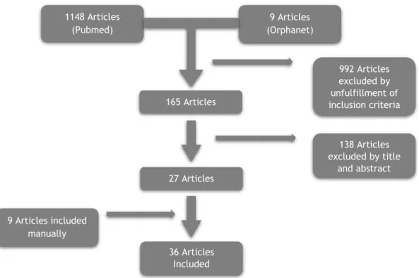
A comprehensive, computerized literature search of PubMed and Orphanet was carried between August and October 2016.
Potentially relevant studies were identified in the Orphanet site using “intestinal polyposis syndrome”, “familial adenomatous polyposis” and “MUTYH-related AFAP” groups of search terms. The search strategy in PubMed was based on Boolean operator combinations of MeSH terms and keywords including “hereditary colorectal cancer”, “genes, adenomatous polyposis coli” and “MYH-associated polyposis”.
Studies meeting all of the following inclusion criteria were deemed eligible and included in the analysis: (1) published in English, (2) published in the last five years, (3) case-report, review and journal articles and (4) human species studies. All studies that did not satisfy the inclusion criteria were excluded. After reading the title and/or abstract, articles were further excluded if they were about clinical manifestations not related to our two clinical cases or not relevant to this literature review. Finally, references from relevant papers were checked to identify studies overlooked in the original search.
A flowchart of the literature search, study selection and results of each step is presented in Figure 1. 1148 Articles (Pubmed) 9 Articles (Orphanet) 165 Articles 992 Articles excluded by unfulfillment of inclusion criteria 27 Articles 138 Articles excluded by title and abstract 36 Articles Included 9 Articles included manually
22
The clinical cases description was based on observation of clinics with these patients at the Hospital Pediátrico de Coimbra, analysis of the hospital medical records and discussion with the respective clinicians.
23
3. Review of the Literature
3.1. Familial Adenomatous Polyposis
Familial Adenomatous Polyposis (FAP) is the second most common inherited CRC syndrome with a prevalence of 2.29-3.2/10000 and equal gender distribution. (13, 14) It is a clinical entity characterised by the development of tens to thousands of adenomatous polyps in the colon and rectum during the second decade of life (mean age at diagnosis is 16 years). Affected individuals have a near 100% risk of developing CRC during their life time in the absence of treatment. (15) FAP is caused by germline heterozygous mutations in the APC gene, has an autosomal dominant transmission with complete penetrance and in 75-80% of the cases there is a positive family history of the disease. (16, 17)
3.1.1. Colonic Manifestations
Familial Adenomatous Polyposis is characterised by the development of adenomatous polyps throughout the colon and rectum during the adolescence (mean age at diagnosis is 16 years) with predominance in the distal colon (rectosigmoid). (17) By the age of 15, 50% of individuals already have polyps, and by the age of 35, 95% of the individuals will have polyps. The presence of colonic polyps rarely translates into symptoms until they become bulky or numerous. However, haematochezia, anaemia, constipation, diarrhoea, abdominal pain, palpable abdominal mass or weight loss might be observed. (18)
About a decade after the presence of the polyps, CRC develops. (18) Without prophylactic treatment, almost all patients develop colorectal carcinoma during their lifetime with a mean age at diagnosis of 39 years. The incidence rate of this cancer in individuals that are heterozygous for a germline APC gene mutation reaches 7% at the age of 21 years, 87% at the age of 45 and 93% at the age of 50. (17)
3.1.2. Extra-Colonic Gastrointestinal Manifestations
Approximately 50-90% of the individuals with FAP present stomach fundic gland polyps, which are hamartomatous tumours usually localized in the body and fundus of the stomach. (17, 18) Occurring at an average age of 38 years, fundic gland polyps are the most frequent polyps observed in the stomach of affected individuals, but they rarely progress to cancer. (1) The second most frequent gastric lesions are adenomatous polyps, usually localized in the antrum. (17) These lesions are estimated to be present in 40% of individuals. (18) Gastric adenocarcinomas develop in 0.5% of the patients, usually arising from adenomatous polyps and only rarely from fundic gland polyps. (17)
24
Fifty to ninety percent of FAP patients present adenomatous duodenal polyps, which are preferentially located in the 2nd or 3rd parts of the duodenum and in the periampullary area
and usually develop 10 to 20 years after the diagnosis of colorectal polyps. (17, 18) Spigelman’s classification, which is based on the number, size, histology and degree of dysplasia of the observed polyps, is used in the evaluation, surveillance, treatment and follow-up of duodenal polyps. (5)
It is estimated that, every 10 years, 5% of the duodenal polyps develop into cancer, namely duodenal and periampullary adenocarcinomas, and FAP patients have a 100-fold increased risk of these cancers. (18) The mean age at diagnosis is 45 and 52 years for duodenal and periampullary adenocarcinomas, respectively. The latter is the most common in colectomized individuals and the main cause of death in these patients. (7, 17, 18) Fifty percent of duodenal cancers occurs in the ampullary or periampullary regions. (14)
The incidence of adenomas and carcinomas of the small-bowel is estimated to be much lower than those of the duodenum. Individuals affected by FAP carry a lifetime risk of small bowel malignancy of 4-12% and there seems to be no correlation between duodenal polyposis and the number of colonic polyps. (17, 18)
3.1.3. Extra-Intestinal Manifestations
In comparison to the general population, individuals with FAP present an 800-fold increased risk of desmoid tumours and their lifetime risk is 10 to 30%. (11) Its occurrence is greater during the second and third decades of life (80% occur before the age of 40), its incidence is higher in women, and they may be located intra-abdominally (80%), in the abdominal wall (18%) or in an extra-abdominal region (2%). (8)
Desmoid tumours are considered to be benign lesions but may cause compression of the urinary and gastrointestinal tracts, nervous and vascular systems. Intra-abdominal desmoid tumours, in particular, can result in bowel obstruction and perforation, ureter obstruction, intestinal haemorrhage, enterocutaneous fistula, mesenteric blood vessels occlusion with intestinal ischemia and sepsis. (1, 8, 18) About 5% of the individuals experience morbimortality because of these tumours, particularly intra-abdominal desmoid tumours. (17)
The following factors are independent predictors of the development of desmoids tumours: an APC pathogenic variant located 3’ of codon 1399, a positive family history (7-fold increased risk), female gender and previous abdominal surgeries. (11, 17) Similarly, desmoid tumours can be induced by pregnancy and become evident within a period of 2 to 3 years. (8)
Some patients with FAP develop occult radio-opaque lesions, usually in the mandible, which translate themselves into osteoslerotic bone areas. These lesions are called osteomas and occur mainly in the mandible and skull. (7, 17) Its occurrence can precede the diagnosis of
25
or functional reasons only. (18)Another clinical manifestation of the FAP spectrum is the congenital hypertrophy of the retinal pigment epithelium (CHRPE), which is an area of discoloration in the ocular fundus that it is not specific to FAP. However, when multiple or bilateral, it becomes very suggestive of a familial adenomatous polyposis syndrome due to an APC mutation. (18) CHRPE is not age-related nor causes clinic problems. (7, 17)
Dental abnormalities are very common in individuals with FAP. These include unerupted teeth, congenital absence of teeth, supernumerary teeth, dentigerous cysts and odontomas, among others. These changes are reported in about 17% of the patients. (17, 18)
Fibromas, lipomas, sebaceous and epidermoids cysts are benign cutaneous lesions with no malignant potential that have already been described in FAP patients. (7, 17) In addition, other lesions have been reported, namely nasopharyngeal angiofibromas, benign thyroid disease (9.1-38%) with familial and female genre predominance and adrenal masses (usually asymptomatic) with a prevalence of 7.4% (2 to 4 higher than in the general population). (17, 18)
Compared to the general population, individuals with FAP present a higher risk of cancers, including extra-colonic cancers. The incidence of pancreatic adenocarcinoma (1-2%) and of bile ducts adenocarcinoma is low, but higher than in the general population. (17, 18) There is a 750 to 7500-fold higher risk of hepatoblastoma in children under the age of 5 with a germline mutation in the APC gene. The absolute risk of this cancer is around 1.6% and the majority develops before the age of 3. (17, 18) FAP patients, predominantly in women younger than 35 years, have been estimated to have 160-fold risk of thyroid tumours, particularly papillary carcinoma. (17, 18) Similarly, brain tumours have an increased prevalence in this population, although the absolute risk is low (1%). The most common brain cancer is medulloblastoma. (17)
3.1.3.1. Gardner Syndrome
Gardner Syndrome was historically regarded as a distinct clinical entity, but is now known to be a variant of FAP and it is caused by certain germline APC mutations. Currently, the term Gardner Syndrome is used for cases of FAP with prominent extra-colonic manifestations, namely osteomas, desmoid tumours, epidermoids cysts and dental abnormalities. (4, 17)
26
3.1.3.2. Turcot Syndrome
Turcot syndrome is the association of colonic polyposis/CRC and central nervous system tumours. It may be caused by APC mutations or by mismatch repair gene mutations and it can, therefore, represent either a FAP variant or a Lynch Syndrome variant. As in Gardner Syndrome, this entity was considered to be distinct from FAP and is currently incorporated into its spectrum of manifestations. (17) Medulloblastoma is the most frequent brain tumour in Turcot Syndrome associated with APC gene mutations, while glioblastoma is the most frequent one in the Lynch Syndrome variant. (4, 18)
3.1.4. Diagnosis
The diagnosis of FAP should be based primarily on family history and clinic findings. However, it should be confirmed, whenever possible, by genetic testing. (18) APC is the only gene in which pathogenic germline variants cause clinical manifestations of the FAP spectrum. (17)
The clinical diagnosis is based on the identification of signs and symptoms compatible with the colonic and extra-colonic manifestations that are associated with this pathology. Whenever FAP is suspected, sigmoidoscopy or total colonoscopy (depending on the patient’s age) should be performed to confirm or exclude the presence of colonic polyps. (18)
The diagnosis of FAP is clinically considered in an individual with one of the following: presence of at least 100 colonic adenomatous polyps before age 40 years or the existence of fewer than 100 colonic adenomatous polyps and a relative with FAP. Despite being the most accepted clinic criteria, they present some limitations, since the presence of more than 100 colonic adenomatous polyps is not specific to FAP. (17)
Genetic confirmation can be performed with the use of different tests and detection rates are highest when patients have a classic phenotype or a positive family history. The most commonly used test is sequencing of the entire APC coding region (all exons and intron-exon boundaries) (17, 18), either by Sanger sequencing or next generation sequencing (NGS). Gene sequencing is the most effective method to detect small intragenic deletions and insertions, missense, nonsense and splice site variants, and it establishes the diagnosis in up to 90% of the individuals with pathogenic variant. (17) However, it is unable to detect large rearrangements such as partial or whole gene deletions/duplications involving APC. (17, 18) In order to identify these variants, it is necessary to use techniques such as long-range quantitative PCR, chromosomal micro-array or, most commonly, MLPA (multiplex ligation-dependent probe amplification), which should include analysis of regulatory regions such as promoter 1B. If gene sequencing fails to detect a pathogenic variant in a FAP proband, a partial or whole APC deletion/duplication will be found in up to 8-12% of the cases.
27
that give rise truncated proteins. (17) In the near future, it is likely that NGS will be able to identify both point mutations, small indels and large rearrangements.The classic genetic strategy in intestinal polyposis was a sequential analysis of several genes associated with this phenotype. Genetic analysis was performed gene by gene depending on a negative result of the previous analysis. A more recent approach is the use of multi-gene panels, in which some or all genes known to be associated with intestinal polyposis syndrome are analysed. (17)
3.1.5. Surveillance
Individuals with genetically confirmed FAP are advised to initiate colorectal surveillance at the age of 10-12 with sigmoidoscopy or total colonoscopy every 1 to 2 years (according to the number and the degree of dysplasia of polyps). Once polyps are detected, polypectomy is recommended, and colonoscopy should be repeated every year until colectomy is performed. (17, 18)
It is recommended to perform routine esophagogastroduodenoscopy, beginning at the age of 25 or before the colon surgery. The recommended interval for screening is one to three years, according to the Spigelman stage for duodenal adenomas. (17) For surveillance of periampullary carcinoma, endoscopy should be performed with equipment that allows forward and side-viewing every 1 to 5 years, depending on the Spigelman stage. (18) Adenomatous tissue is commonly found in the papilla, even in the absence of polyps, and biopsy at this site may be warranted if it is enlarged. (17)
Small-bowel imaging is recommended as soon as duodenal adenomas are detected or before colectomy and should be performed every 1 to 3 years, according the findings and the presence of symptoms. (17)
Regarding hepatoblastoma screening, abdominal ultrasound and measurement of serum alpha-fetoprotein may be considered every 2-3 months from infancy to age 5. (17)
Annual physical examination, including thyroid gland palpation and, if clinically justified, ultrasound or fine-needle aspiration, is highly recommended. (17, 18) Routine ophthalmologic examination is considered optional. (17)
28
3.1.6. Prophylaxis
Prophylaxis of colorectal carcinoma should be offered to all patients with Familial Adenomatous Polyposis. There are several prophylactic surgeries available, including restorative proctocolectomy, proctocolectomy with ileal pouch anal anastomosis (IPAA), total colectomy with ileorectal anastomosis (IRA) and total proctocolectomy with permanent ileostomy, that should be considered and adapted case by case according to several factors, such as patient preference, polyps distribution and post-colectomy surveillance. (17) For example, proctocolectomy with IPPA is recommended for situations in which there is profuse colonic adenomatosis, particularly of the rectum, while colectomy with IRA is better suited for cases with a lower number of colonic polyps and scarce rectal polyps. (14)
Prophylactic colectomy should be performed between the end of the second decade of life and the beginning of the third. (18) The presence of multiple adenomas with >6mm, a significant increase in the number of adenomas (more than 20-30 adenomas), the presence of adenomas with high-grade dysplasia and the inability to perform polypectomy are relative indications for surgery. (14, 17) The presence of suspicious lesions, significant symptoms or the confirmation of cancer constitute absolute indications for immediate colorectal surgery. (14)
In the postoperative period, a complete physical examination and baseline abdominal ultrasound, CT or MRI are recommended to detect possible existing or future desmoid tumours. (18)
Regarding post-colectomy surveillance and depending on the type of surgery performed, the ileal pouch should be monitored every 2 years and the rectum should be monitored every 6 to 12 months. This surveillance is extremely important, as adenomatous polyps in the ileal pouch have been reported in up to 57% of FAP patients. (17) Although very rarely, cancer in the anastomosis has also been previously reported. (17)
In terms of pharmacological prophylaxis, nonsteroidal anti-inflammatory drugs (NSAIDs), particularly sulindac, celecoxib and aspirin, have been reported to cause regression of established adenomas and to decrease the number of polyps requiring excision in colectomized patients. Despite its effectiveness, none of these NSAIDs has currently FDA approval for FAP because of their considerable cardiovascular risk. (1, 17, 18)
After prophylactic colectomy, there is an increase in cases of male sexual dysfunction, namely erectile dysfunction (0-1.5%) and ejaculatory dysfunction (3-4%). These changes may occur after pelvic plexus lesion during surgery. (18) Dyspareunia may occur in 3 to 22% of women due to anatomical changes after surgery. After colectomy, surveillance of possible mineral and vitamin deficiencies is recommended. (17)
29
The diagnosis of duodenal/periampullary adenomas requires biopsy and histological study to determinate the degree of dysplasia and mucosal involvement. After this histological analysis, larger polyps should be resected endoscopically. Argon plasma coagulation is used to destroy small adenomas or post-mucosectomy polyps. There is also the possibility of performing endoscopic ampulectomy with maintenance of pancreatic duct in order to minimize the risk of pancreatitis. (18) Indications for surgical intervention include villous changes, severe dysplasia, rapid growth of an adenomatous polyp, polyps with >1cm diameter and the presence of symptoms. (17, 18) The surgical approach can be performed by different procedures, such as pancreaticoduodenectomy (Whipple procedure), duodenectomy with preservation of the pylorus or duodenectomy with preservation of the pancreas. (18)If a desmoid tumour is suspected, biopsy should not be performed due significant risk of haemorrhage. (18) The treatment of these lesions may include surgery, but there are common complications, such as incomplete resection of the tumour tissue and difficulties in the control of intraoperative haemorrhage. (17, 18) Furthermore, the surgical option is associated with a lesion recurrence rate of about 45%. (1) Desmoid tumours show a favourable response to pharmacological treatment with NSAIDs, anti-oestrogens, cytotoxic chemotherapy and radiation, and these may be a viable option in the control of these lesions. (17)
During pregnancy there is an increased incidence of desmoid tumours and adenomas. Due to the risk of foetal morbidity and mortality and whenever possible, treatment should be postponed after delivery. (18) Women with FAP have similar fertility, pregnancy and childbirth rates as the general population. However, the fertility rate decreases up to 54% after proctocolectomy with IPAA, probably due to adhesions resulting from surgery. (17, 18)
The presence of osteomas in an individual with FAP does not require an active attitude, since they are benign lesions. Nevertheless, these can be removed surgically for aesthetic or functional reasons. (17)
3.1.8. Genetic Counselling
Familial Adenomatous Polyposis is an autosomal dominant disease. When a specific APC mutation is identified in the proband, targeted genetic testing for this mutation should be offered to all first-degree relatives. Nevertheless, in some cases it is not possible to identify the responsible genetic mutation, so in these cases, it is not useful to provide genetic testing to family members who must follow a high-risk surveillance protocol while the individual with suspected FAP should be accompanied as if he was affected and discussed further genetic testing. (18)
30
Most germline APC mutations are inherited, with 75-80% of the individuals having at least one affected family member. Sometimes the family history is initially considered to be negative because of failure in recognizing the disease in affected relatives. The remaining 20-25% molecularly confirmed cases, are due to de novo mutations. The risk for siblings of affected individual depends on the genetic status of the parents. It should be noted that about 20% of individuals with a de novo mutation have somatic mosaicism. Moreover, APC sequencing in DNA extracted from peripheral blood may fail to detect somatic mosaicism and this might explain the lower variant detection rate in simplex cases than in probands with a positive family history. (17) Since germline mosaicism has already been reported in FAP families, if neither parent carries the mutation found in the proband, the risk to the sibs is low but greater than that of the general population. Therefore, genetic testing should be offered to the sibs of a proband carrying an apparent de novo mutation. (17, 18)
For those first-degree relatives who were unable or unwilling to perform a genetic test, high-risk surveillance is recommended starting at 10-12 years with annual flexible sigmoidoscopy or colonoscopy in adults. Surveillance intervals may be increased if adenomatous polyps are not identified until 50 years of age, in which, in the absence of polyps, they should follow the screening recommendations for colorectal cancer in the general population. (17, 18) Each descendant of an individual with FAP has a 50% chance of inheriting the mutated gene. The genetic test for classic FAP is, in fact, offered to children at the age of 10. However, these guidelines may change in the future as it may be appropriate to perform genetic testing at birth in order to implement an appropriate hepatoblastoma surveillance. There is a 750 to 7500-fold higher risk of hepatoblastoma in children under the age of 5 with a germline mutation in the APC gene. The absolute risk for this cancer is around 1.6% and the majority develops before the age of 3. (17, 18)
Reproductive options should be discussed with FAP patients. In families with a molecularly confirmed diagnosis, prenatal testing for the specific mutation may be performed after amniocentesis (at 16-20 weeks’ gestation) or chorionic villus sampling (at 10-12 weeks’ gestation). Another possibility is to perform preimplantation genetic diagnosis, through which mutation-free embryos are selected for implantation in the uterus, avoiding possible termination of pregnancy. (18) It should be noted that there is a significant variable expressivity in FAP and, therefore, the identification of a pathogenic APC variant on a foetus does not predict the severity of the disease. (17)
31
3.2. Attenuated Familial Adenomatous Polyposis
Attenuated Familial Adenomatous Polyposis (AFAP) is a clinical entity integrated in conditions associated with APC mutations such as FAP, but with a lower number of adenomatous colonic polyps (10 to 100), beginning at a later age (mean age 44 years), lower risk of colorectal cancer and diagnosis at a later age (mean age 56 years) compared to FAP, but higher than in general population. (18)
3.2.1. Colonic Manifestations
Attenuated Familial Adenomatous Polyposis is characterised by the development of adenomatous polyps in the colon and rectum with preferential involvement of the proximal colon (right). (8) The presence of polyps in the rectum is rare, which has practical implications in surveillance and prophylaxis. The number of polyps that develops is lower (average of 30 colonic adenomatous polyps). (13)
The risk of CRC is higher in relation to the general population, reaching 70% during lifetime. (13) The diagnosis of this neoplasm occurs, on average, 10 to 15 years later than in classic FAP, with a mean age of 56 years. However, it occurs earlier than sporadic cancer. (7)
3.2.2. Extra-Colonic Gastrointestinal Manifestations
As in FAP, polyps of the upper gastrointestinal tract are common, namely, gastric and duodenal adenomas. (7) With regard to gastric neoplasms, there is an increased risk of duodenal/ampullary adenocarcinoma (4-12%) and gastric adenocarcinoma (<1%). (7, 11)
3.2.3. Extra-Intestinal Manifestations
Patients with AFAP have an increased risk of breast adenocarcinoma, hepatoblastoma, and thyroid carcinoma (1-2%). (7, 11) Affected individuals have the same extra-intestinal manifestations of FAP, although CHRPE and desmoid tumours are rare. (17)
3.2.4. Diagnosis
Similar to FAP, the diagnosis of this entity is usually made based on family history and clinical findings but should, when possible, be confirmed by genetic testing. It should be noted that, in contrast to FAP, in most cases the clinical findings are sparse and the clinical suspicion of AFAP may not be obvious. (18) If AFAP is suspected, total colonoscopy, rather than sigmoidoscopy, is recommended, due to the proximal distribution of polyps in the colon. (18)
32
Several diagnostic criteria for AFAP have been proposed, namely those from Nielsen et al, 2007 (19) (no family member with more than 100 polyps before age 30 and at least two individuals with 10-99 adenomas diagnosed after age 30 or an individual with 10-99 diagnosed adenomas after age 30 and a first-degree relative with colorectal cancer with few polyps) and those from Knudsen et al, 2010 (20) (a dominant pattern of inheritance of colonic adenomatous polyposis and less than 100 colorectal adenomas at 25 years or older). Actually, AFAP should be considered in individuals with 10-99 colonic adenomatous polyps or more than 100 polyps in older individuals or a personal history of colorectal cancer before age 60 and family history of multiple adenomatous polyps. (17) It should be pointed out that all the proposed criteria had limitations and none of them obtained consensus as definitive diagnostic criteria for AFAP. (17) Genetic testing may facilitate diagnosis if an APC germline mutation is identified, as it is also the case for FAP. (18) However, a pathogenic APC variant is detected in less than 30% of individuals with attenuated phenotypes. (17)
Some genotype-phenotype correlations in AFAP have been reported. The attenuated phenotype is associated with the following pathogenic variants: 5' region (codons 1-177) of the gene (usually truncating variants), distal 3' region of the gene, exon 9, interstitial deletions at 5q22 that include APC, partial and whole-gene deletions and somatic mosaicism for APC pathogenic variants that are generally associated with classic FAP. (17)
Especially in phenotypes suggestive of AFAP, if APC sequencing and MLPA fail to identify a pathogenic mutation, MUTYH screening should be the next step (see below). (17)
3.2.5. Surveillance
Individuals with AFAP are advised to perform total colonoscopy every 2 to 3 years starting at the age of 18-20 in order to control colonic polyposis. In each colonoscopy, polypectomy of suspected polyps should be performed, and by doing this approximately 1/3 of the individuals are able to control the disease in the long term. (17) Esophagogastroduodenoscopy is recommended from the age of 25 or before colectomy at intervals of 1 to 3 years, depending on the severity of the duodenal adenomas (Spigelman criteria). Lateral observation is important for duodenal papilla control, where adenomatous tissue is frequent, and biopsy is recommended when it is enlarged. In certain cases, it is appropriate to perform endoscopic retrograde cholangiopancreatography to evaluate possible common bile duct adenomas. (17)
As for FAP, an annual general physical examination is recommended, including palpation of the thyroid gland and, when clinically justified, thyroid ultrasonography or fine-needle aspiration. (17)
33
Individuals with AFAP are advised to perform colectomy when there are more than 20-30 colonic adenomas or multiple adenomas with high-grade dysplasia. There are several types of colectomy available for these patients. Because of the more proximal distribution of polyps in the colon with a very small number of polyps in the rectal area, it is often possible to preserve the rectum without compromising the prophylaxis of colorectal cancer. (17)With regard to extra-colonic adenomas, these should be followed-up according to the recommendations for FAP, even though they have a lower risk of malignancy in AFAP. (18)
3.2.7. Genetic Counselling
Genetic counselling of probands and their families is similar in AFAP and FAP. It should be noted that in AFAP a germline APC mutation is found in only about 30% the probands. In asymptomatic individuals, genetic testing should only be offered starting at the age of 18, when it becomes clinically important for possible surveillance regimes. (17)
34
3.3. APC Gene
The APC gene is located on chromosome 5q.22.2 and its coding region is divided into 15 exons. It is a tumour suppressor gene, referred to as "gatekeeper" and involved in the Wnt pathway. (3) The frequency of pathogenic APC mutations in the general population is estimated to be 1/8000 with a penetrance of about 100% for FAP and 70% for AFAP at the age of 80. (17, 21)
The APC protein is normally located in the nucleus and/or membrane/cytoskeleton of human epithelial cells, where it binds to various intracellular proteins to participate in the processes of cell proliferation, differentiation, migration, adhesion, apoptosis, signal transduction, chromosomal stability and transcriptional activation. (17, 18, 22) It also plays a role in the organization of actin and microtubule network, formation of mitotic spindle and chromosomal segregation. (3)
Its participation in the Wnt pathway focuses on the control of beta-catenin degradation. In the absence of Wnt signal, the multi-protein complex consisting of beta-catenin, APC protein, GSK3, CK1 and two kinases induces phosphorylation of beta-catenin and subsequent ubiquitination and destruction by the proteasome. In the absence of the Wnt signal, the destruction of beta-catenin is inhibited and accumulates in the nucleus of the cell contributing to cell proliferation. (2, 23)
In the absence of a normal APC protein, the cytosolic destruction of beta-catenin is impaired, causing it to enter and accumulate in the cell nucleus. Intranuclearly it binds to Tcf and Lef family transcription factors that promote the transcription of genes such as c-myc and cyclin-D, which in turn induce the expression of proto-oncogenes. (18) Indirectly, the APC protein also regulates the transcription of genes involved in cell proliferation through its interaction with beta-catenin. Thus, loss of APC protein function increases the transcription of beta-catenin targets. (3, 18, 22)
Over 1,500 different APC germline mutations have been described so far. Most of them are nonsense or frameshift mutations that cause premature truncation of the APC protein. Some missense mutations have also been reported to predispose to FAP, and about 8-12% of the genetic mutations responsible for the FAP phenotype are larger deletions or insertions and complex rearrangements. (17) About 60% of all APC mutations are found in a mutation cluster region (MCR) located between codons 1284 and 1580 (5’ region of exon 15). Thirty percent of these are a deletion in codon 1309 or a deletion in codon 1061, representing the first and second most frequent mutations in the APC gene, respectively. (18) The most frequent pathogenic APC variant is c.3927_3931delAAAGA, which is located at codon 1309 and results in the following protein change: p.Glu1309AspfsTer4. (17) In addition to these "classic" mutations, certain rare alterations have been reported, such as promoter deletions, abnormal mRNA isoforms and profound intron base changes. (21)
35
cancers and represents an early stage in carcinogenesis. (18) In patients with a germline APC mutation, one somatic inactivation of the other allele (mutation or loss of heterozygosity) is sufficient to cause complete loss of protein function and to initiate the formation of adenomas. In sporadic cases, somatic mutations in both alleles are required to cause loss of function. In the case of a tumour suppressor gene, inactivation of both alleles is necessary, as there is still sufficient protein production with only one mutated allele. This theory is known as the Knudson hypothesis (or two-hit hypothesis) and is typical of tumour suppressor genes and most of the cancer predisposition hereditary syndromes. (24) This hypothesis was first described for retinoblastoma in which the two mutations produced a malignant lesion. In some cases, as in those associated with the APC gene, the two mutations lead only to a benign precursor lesion and another event (third hit) is required for a malignant lesion. Thus, the first hit (first APC gene mutation) is not phenotypically translated and the second hit (second APC mutation) leads to an increase in cell proliferation and adenoma formation. The increased rate of cell division predisposes to the occurrence of another mutation (third hit) that transforms this benign lesion into a malignant lesion. It seems that in FAP this third hit occurs predominantly in the TP53 andDCC genes. (25) However, following inactivation of the APC gene, the progression from
adenoma to carcinoma is similar in sporadic and hereditary cancers through the classic adenoma-carcinoma pathway. (14, 21) Neoplasms derived from the adenoma-carcinoma pathway traditionally exhibit chromosomal instability but microsatellite stability (unlike neoplasms associated with HNPCC). An APC mutation is almost invariably present in all tissues following the adenoma-carcinoma pathway. Mutations in the APC gene are found in about 80% of sporadic CRCs, and it is estimated that about 90% of CRCs develop from this classic pathway involving the transition from normal epithelial cells to adenomas and carcinomas due to genetic and epigenetic changes. (2, 26)
36
3.4. MUTYH-Associated Polyposis
MUTYH-Associated Polyposis is an autosomal recessive disease caused by biallelic
pathogenic variants of the MUTYH gene and it is associated with the development of dozens to few hundreds of adenomatous polyps in the colon and an increased risk CRC. The frequency of heterozygosity in the general population of Northern Europe reaches 1-2% and the incidence of the disease (homozygosity or compound heterozygosity) is 1/20,000-40,000. MAP contributes to 0.7% of all CRCs. (18, 27, 28)
3.4.1. Colonic Manifestations
MAP is associated with an attenuated form of adenomatous polyposis with the development of dozens to few hundred colonic polyps and a mean age at diagnosis of 45-50 years. (27) The number of polyps is similar to AFAP, with 60% of patients having 20-99 adenomas and 9% between 10 and 19. (16) Most colonic polyps are adenomatous but serrated/hyperplastic polyps may occur in 47% of individuals. (28)
The incidence of CRC is increased in patients with MAP (43 to 100% risk), and the penetrance increases with age: 19% at age 50, 43% at age 60 and 80% at age 70. There is some evidence of a low (2.5-fold) increased risk of CRC in patients with heterozygous germline mutation, with some authors proposing CRC screening in these individuals. (11, 16, 28) At diagnosis, CRC is present in around 50% of the individuals. (12) Colorectal cancer usually develops from polyps, but CRC has already been reported in individuals with few or no polyps. Colorectal cancer occurs in the right colon in 29-69% of the cases and synchronous or metachronous tumours are present in 23-27% of the cases. (27)
Individuals with the p.Y179C pathogenic variant have more profuse polyposis and an earlier (about 8 years earlier) age at onset of polyposis and CRC, when compared to carriers of the p.G396D mutation. (28, 29)
3.4.2. Extra-Colonic Manifestations
The spectrum of extra-colonic manifestations in MAP is more similar to those found in Lynch syndrome than those found in FAP. About 17-25% of the individuals have duodenal adenomas, but fundic gland polyps are rarer (11%). The risk of duodenal cancer is 4% (significantly increased compared to the general population) and the risk of gastric cancer is not significantly increased compared to the general population. Desmoid tumours are not reported. (1, 28)
Individuals with MAP have a global risk of extra-intestinal cancer of twice the one observed in the general population. There is a significantly increased risk for ovarian, bladder
37
cancer is also identified in men. Endometrial neoplasms, increased in these patients, have a mean age at diagnosis of 51 years. The risk for skin cancer is modestly increased for melanomas, squamous epithelial carcinomas and basal cell cancers. There is similarly an increase in the number of benign skin tumours, namely fibrous histiocytoma, capillary haemangioma, pilar cyst, dermatofibroma and follicle cyst. Tumours of sebaceous glands associated with MAP have also been described. (27)There are reports of thyroid abnormalities in these individuals, such as multinodular goitre, solitary nodules and papillary thyroid cancer, dental abnormalities, particularly temporomandibular joint bone cyst, and CHRPE in 5.5% of the cases. (27)
3.4.3. Diagnosis
The diagnosis of MUTYH-associated polyposis is based on family history, clinical findings and genetic testing. (27)
MAP should be suspected in the presence of the following findings: colonic adenoma and/or serrated/hyperplastic sessile polyp in the number of 1 to 10 (in an individual under 40 years); more than 10 (in an individual aged 40-60 years) or 20 (in an individual with more than 60 years); colonic adenoma and/or serrated/hyperplastic sessile polyp in a number between twenty and a few hundred; colonic polyposis (more than 100 polyps) in the absence of a pathogenic variant of the APC gene; CRC diagnosed in a patient under 40 years; or history of colon cancer (with or without polyps) consistent with an autosomal recessive pattern. The detection rate of MUTYH pathogenic variants increases with increasing number of polyps (greater when 10-99 polyps) and with decreasing age (before age 50) at onset of CRC. (12, 27) Regarding the histological and molecular criteria, the following findings support the suspicion of MAP: the identification of a KRAS pathogenic variant (64% of CRC in the context of MAP have the specific c.34G>T mutation at codon 12); low microsatellite instability (most of these CRCs have stability microsatellite, however 4% has microsatellite instability); preferential proximal localization; higher prevalence of synchronous tumours; higher rate of mucinous histotype; and higher frequency of tumour infiltrating-lymphocytes. (27)
Genetic testing for MUTYH is recommended for patients who have dozens or hundreds of adenomatous colonic polyps and a family history consistent with an autosomal recessive hereditary pattern and for those with a negative APC genetic testing. Some authors also suggest that individuals with attenuated phenotype and a family history compatible with AFAP are also tested for MAP, since there are reports of heterozygous MUTYH patients who developed polyposis (dominant component). (18) It is estimated that 25-30% of individuals with polyposis but without an identifiable APC mutation have MUTYH mutations. (1) Sequencing of the entire
38
deletions/insertions, missense mutations, nonsense and splice site. However, this genetic test fails to identify deletions/duplications of whole exons or the entire gene. (27)
3.4.4. Surveillance, Prophylaxis and Treatment
In Europe, individuals with a MUTYH pathogenic variant begin surveillance with total colonoscopy at the age of 18-20 and repeat it every one or two years. During colonoscopy, polypectomy of all suspected polyps should be performed. Upper endoscopy and duodenoscopy with lateral visualization beginning at age of 25-30 years and follow-up according to the Spigelman stage is also recommended. All polyps with apparent dysplasia or villous changes should be removed. A physical examination, such as palpation of thyroid gland and complementary examinations depending on the clinical case should be performed on a regular basis. (27)
Prophylactic colectomy should be performed when polyp excisions during screening colonoscopies are not effective in controlling colonic polyposis. Subtotal colectomy or proctocolectomy may be performed, depending on the distribution of the polyps and the preference of the individual. (1, 27) One half of the patients are diagnosed at a stage where CRC is already present, and colectomy is often necessary as a curative purpose. (28)
3.4.5. Genetic Counselling
As an entity with an autosomal recessive pattern, parents of those individuals with molecularly confirmed MAP are obligate carriers/heterozygotes but confirmation should be offered by targeted sequencing of the mutation(s) identified in the affected individual. Thus, the parents may present a risk of CRC 2 to 3 times higher than the general population and colonic surveillance based on family history should be decided. Similarly, the offspring of an affected individual with mutations in the MUTYH gene are/will be (at least) obligate carriers/heterozygotes. Since the frequency of carriers in the general population is 1-2%, each child has a probability of 0.5-1% of inheriting two mutated alleles and developing MAP. Genetic counselling is mandatory and, if this is their wish, specific genetic testing of the partner may be offered to assess carrier status. This test can include the targeted screening of the mutation(s) identified in the affected individual, screening for the more frequent mutations found in the respective population group or sequencing of MUTYH of the entire coding region. (27)
Each sibling of an affected individual has a 25% chance of being affected (biallelic mutation), a 50% chance of being a carrier (monoallelic mutation) and a 25% chance of being unaffected or being a non-carrier. Thus, all siblings should be offered genetic counselling and
39
before that age, the result would not have practical implications on follow-up. (27)Heterozygous individuals have a risk of CRC of 7.5% in men and 5.6% in women at 70 years regardless of family history. If there is a positive family history of CRC before age 50 in first-degree relatives, this risk increases to 12.5% and 10%, respectively. (27)
3.4.6. Prevention and Prognosis
There are currently no studies about the use of celecoxib or sulindac in individuals with MAP. However, there are preclinical studies that indicate a pattern of COX-2 expression in the mucosa of individuals with MAP similar to that observed in patients with classic FAP, which suggests a possible benefit in the use of NSAIDs. In any case, chemoprevention should not currently be proposed to patients with MAP. (28)
It should be noted that individuals with MAP have longer survival than their controls. The 5-year survival rate for patients with MAP is 78% while for controls is 68%. This is likely due to the detailed follow-up and monitoring provided to patients with confirmed MAP. (27)
40
3.5. MUTYH Gene
The MUTYH gene is located on chromosome 1p34.1, has 15 coding exons and it was first described as the causative gene of MAP in 2002. (18, 27, 28) This gene belongs to a DNA repair system called Base Excision Repair. The MUTYH protein (A/G-specific adenine DNA glycosylase) is located in the nucleus and cell mitochondria and is involved in the repair of DNA damaged by ROS generated by aerobic metabolism, radiation and chemical oxidants. (1, 18, 27) It also plays an important role in regulation of apoptosis by suppressing p53-related tumourigenesis. (27)
8-oxo-dG, the mutagenic species generated, tends to pair an adenine, rather than a normal cytosine leading to G:C>T:A transversions in DNA. A/G-specific adenine DNA glycosylase recognizes and excises these mismatched adenine bases preventing transversions and their subsequent somatic mutations in genes such as the APC. (1, 27)
MAP-derived CRCs have, in 64% of the cases, pathogenic variants of the KRAS gene involved in the regulation of cell division. Somatic pathogenic variants of the APC gene are found in 14-83% of CRC in the context of MUTYH-Associated Polyposis. The most frequently encountered are AGAA or TGAA demonstrating probably the susceptibility to guanine oxidation and the defect in MUTYH correction. (27) While G:C>T:A transversions in the APC gene are only found in adenomas, the G:C>T:A transversions of the KRAS gene are detected in 70% of the MAP serrated polyps. About 4% of CRCs developed in the MAP had shown microsatellite instability, probably associated with the serrated polyp pathway. (28, 29)
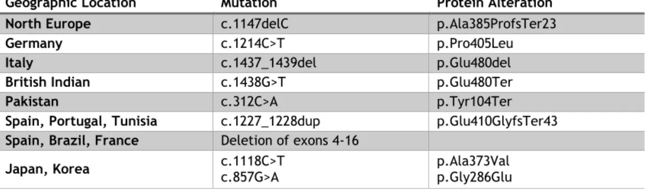
More than 300 different variants are described in the MUTYH gene, the majority being missense, but nucleotide substitutions, splice site, large genomic deletions and large intragenic deletions have been reported. (29) In the western population, the two major mutations are the missense c.536A>G (p.Y179C) or p.Tyr179Cys located in exon 7 and c.1187G>A (p.G396D) or p.Gly396Asp located in exon 13. (27, 28) These two specific mutations contribute to 90% of the pathogenic variants in Northern Europe. (27) For this reason, genetic testing should be directed primarily at these two specific mutations and, subsequently, in case of a negative result, extended to the whole coding region. If only one pathogenic variant is identified in one of the alleles, it is important to study the other in order to evaluate the presence of an inactivating mutation that constitutes a compound heterozygosity. (1)
41
depending on the geographic origin of the patients: (27)Table 1 - Prevalence of certain types of pathogenic variants depending on the geographic location (Adapted from Nielsen et al, 2012 (27))
Geographic Location Mutation Protein Alteration
North Europe c.1147delC p.Ala385ProfsTer23
Germany c.1214C>T p.Pro405Leu
Italy c.1437_1439del p.Glu480del
British Indian c.1438G>T p.Glu480Ter
Pakistan c.312C>A p.Tyr104Ter
Spain, Portugal, Tunisia c.1227_1228dup p.Glu410GlyfsTer43
Spain, Brazil, France Deletion of exons 4-16
43
4. Case Reports
4.1. Clinical Case 1
M.A.V.N. (IV.4), a 17-year-old male, was referred to the Paediatric Orthopaedic Unit of Hospital Pediátrico de Coimbra and later to the Medical Genetics Unit, with the clinical information of a large bone mass at the left forearm, detected at the age of 15.
Regarding the family history (Fig. 2), the father (48 years old) and the mother (47 years old) were non-consanguineous, healthy, had a normal stature and did not have bone deformities. M.A.V.N. was the youngest son of the couple, who had two previous pregnancies: the first resulted in a healthy female, now aged 22 years, and a hydatidiform mole that occurred at the second pregnancy. There was no family history of bone deformities, intestinal polyposis or other tumours, with the exception of a maternal grand-mother (II.5) who had pancreatic carcinoma.
Regarding the personal history, M.A.V.N.’s pregnancy and neonatal period were normal. Parents described as their first concerns problems with walking and an abnormal equine position of the right foot. The clinical and radiological evaluation led to the identification at the age of 18 months of a congenital malformation of the right fibula (see figure 3 and discussion), which did not require surgical correction. Growth and intellectual development were normal.
At the age of 15, the patient noticed that his left arm had an increased volume. He had no pain nor movements limitation, was observed in multiple hospitals and a bone mass was detected. In 2016 he was sent to the national reference centre for bone tumours at Hospital Pediátrico de Coimbra.
44
Figure 3 – Lower legs photograph (A) and radiographs (B, C) of the patient (IV.4) at 16 years of age. The right fibula malformation, detected at 18 months of age, is likely to be congenital and not significantly progressive. This fibula is thick, elongated and bowed with posteromedial convexity, more evident in the lateral view (C). After discussion with international experts, it fits best the classification of pachydysostosis of the fibula. There is a slight length discrepancy between the two legs. The tibia is normally shaped, despite some cortical thickness and waviness, a known feature of FAP.
A
B
45
bone consistency, normal painless pronation and supination and without skin or vascular local anomalies (Fig. 4). He had normal stature (1.73m) and normal head circumference (58cm). There was a bilateral prominence of the mandible, at the parotid region and mandible angle, with one palpable firm lesion at the right mandible angle (Fig. 5). The right leg was deformed, especially at its distal third, with bowing and posterior projection of the fibula, painless and without local skin or vascular abnormalities (Fig. 3). The right lower limb was 1 cm shorter than the left, with respective pelvis asymmetry. During walking, there was a slight rotation of the right foot. The patient had difficulty in walking in heels but referred no pain. The muscular development was normal with normal strength and capillary finger skin lesions considered very suggestive of erythema pernio were observed. Evaluating the skin in detail, we detected 5-10 small lesions (<0.5cm), tumefactions, at multiple locations, both in the limbs and trunk (Fig. 6). Dental enamel was abnormal.Figure 4 - Photograph (A), CT (B) and radiographs (C, D) of the patient’s (IV.4) right forearm at 16 years of age. Please remark the large exophytic osteoma of the left radius and associated increased volume of the forearm.
A
B
46
Figure 5 - Craniofacial photographs (A, B) and radiograph (C) of the patient at 16 years of age. Please note the bilateral prominence of the mandible and, at least, one small palpable osteoma at the right mandible angle.
Figure 6 – Photographs of the patient, at the age of 16, showing a small cutaneous lesion located at the right dorsum/scapular area, most likely an epidermoid cyst (A), and the chilblain/erythema pernio of the hands (B).
A
B
B
47
large exophytic osteoma of the radius, with similar features as previously identified in other hospitals (Fig. 4). There was also a significant number of other small osteomas with multiple locations: left fibula, left ilium, metacarpals and mandible. (Fig. 7). The right leg malformation fitted best the description of pachydysostosis of the fibula (see Fig. 3 and discussion). CT and MRI of the left forearm confirmed the mentioned osteoma without signs of malignity. Scintigraphy revealed a single uptaking lesion at the left forearm. Phosphocalcic metabolism and biochemical analysis, including CK, were normal.Figure 7 - Radiographs of the left leg (A) and left hand (B) of the patient at the age of 16. Please note the presence of small osteomas at the left fibula (arrows, A) and some waviness of the tibial and fibula cortical. At the hand, note the diaphyseal exostosis/osteomas of the second and third metacarpals (arrows, B).
48
Figure 8 - Lower limbs radiograph of the patients at the age of 16. Please remark the multiple osteomas at the left ilium and left fibula (arrows) and some slight cortical thickness and waviness of the long bones.