Marlene Sofia Belinho Nogueira
The impact of early life experiences on
oxytocinergic system modulation:
implications on social behavior development
Marlene Sofia Belinho Nogueira
The impact of earl
y life e
xperiences on o
xytocinergic sys
tem modulation:
implications on social beha
vior de
velopment
U n i v e r s i d a d e d o M i n h o Escola de Psicologia
Marlene Sofia Belinho Nogueira
The impact of early life experiences on
oxytocinergic system modulation:
implications on social behavior development
Dissertação de Mestrado
Mestrado em Genética Molecular
Trabalho efetuado sob a orientação da
Doutora Ana Raquel Mesquita
Doutora Ana Magalhães
DECLARAÇÃO
Nome
_______________________________________________________________________________________ Endereço electrónico: _______________________________ Telefone: _______________ / _______________ Número do Bilhete de Identidade: ______________________
Título dissertação □/tese □
_______________________________________________________________________________________ _______________________________________________________________________________________ _______________________________________________________________________________________ Orientador(es): _______________________________________________________________________________________ ____________________________________________________ Ano de conclusão: ___________
Designação do Mestrado ou do Ramo de Conhecimento do Doutoramento:
_______________________________________________________________________________________
Nos exemplares das teses de doutoramento ou de mestrado ou de outros trabalhos entregues para prestação de provas públicas nas universidades ou outros estabelecimentos de ensino, e dos quais é obrigatoriamente enviado um exemplar para depósito legal na Biblioteca Nacional e, pelo menos outro para a biblioteca da universidade respectiva, deve constar uma das seguintes declarações:
1. É AUTORIZADA A REPRODUÇÃO INTEGRAL DESTA TESE/TRABALHO APENAS PARA EFEITOS DE INVESTIGAÇÃO, MEDIANTE DECLARAÇÃO ESCRITA DO INTERESSADO, QUE A TAL SE COMPROMETE;
2. É AUTORIZADA A REPRODUÇÃO PARCIAL DESTA TESE/TRABALHO (indicar, caso tal seja necessário, nº máximo de páginas, ilustrações, gráficos, etc.), APENAS PARA EFEITOS DE INVESTIGAÇÃO, , MEDIANTE DECLARAÇÃO ESCRITA DO INTERESSADO, QUE A TAL SE COMPROMETE;
3. DE ACORDO COM A LEGISLAÇÃO EM VIGOR, NÃO É PERMITIDA A REPRODUÇÃO DE QUALQUER PARTE DESTA TESE/TRABALHO
Universidade do Minho, ___/___/______
Assinatura: ________________________________________________
Marlene Sofia Belinho Nogueira
[email protected] 934517930 13865577 4 ZZY
2016
Mestrado em Genética Molecular
05 02 2016
Doutora Ana Raquel Mesquita, Doutora Ana Magalhães e Doutora Paula Sampaio
912033495
The impact of early life experiences on oxytocinergic system modulation: implications on social behavior development
The work presented in this thesis was supported by FEDER through Programa Operacional Factores de Competitividade – COMPETE and by Portuguese founds through FCT – Fundação para a Ciência e a Tecnologia, in the framework of the projects FCOMP-01-0124-FEDER-029576 and PTDC/PSI-PCO/116612/2010. It was developed in the laboratory of Addition Biology at Institute of Celular and Molecular Biology (IBMC), Porto, and Neuropsychophysiology Laboratory, at Psychology School of University of Minho, Braga.
É profundamente reconfortante chegar ao final desta etapa e sentir um crescimento exponencial, tanto a nível científico como pessoal. Por isso mesmo não poderia deixar de agradecer a todos os que de alguma forma contribuíram para o sucesso deste projeto.
O meu profundo OBRIGADO às minhas “Anas” que são as orientadoras mais extraordinárias que poderia ter. À Ana Mesquita e à Ana Magalhães agradeço imenso por todo o apoio, atenção, disponibilidade, partilha de conhecimentos e competências. Pelo grande exemplo que são e porque nunca deixarão de ser modelos que inspiram qualquer pessoa! O meu grande agradecimento também á professora Paula Sampaio por ter estado sempre presente, pela disponibilidade e atenção. Muito obrigada por terem sido as melhores orientadoras que alguém pode pedir.
À Escola de Psicologia, em particular ao Laboratório de Neuropsicofisiologia, pelo acolhimento, pela simpatia, pelas palavras, por me terem incentivado a fazer sempre o melhor e por me terem mostrado o melhor ambiente que se pode ter dentro de um laboratório!
Ao Instituto de Biologia Molecular e Celular (I3S-IBMC) o meu grande obrigada por me ter deixado desenvolver parte da tese nas suas instalações! Um obrigada em especial á Doutora Teresa Summavielle por me ter acolhido da melhor forma no seu laboratório!
Ao Instituto de Investigação em Ciências da Vida e da Saúde (ICVS), em especial ao laboratório I1.03 e ás meninas dos Serviços Técnicos, o meu grande obrigada por me terem recebido e ajudado da melhor forma possível! Não posso deixar de agradecer, e muito, à Eduarda por toda a ajuda, ensinamentos, todo o acompanhamento, disponibilidade e toda a alegria! Sem ti, esta tese não seria a mesma coisa. MUITO OBRIGADA!
À Escola de Ciências da Universidade do Minho assim como ao meu mestrado, Genética Molecular, por me terem deixado “voar” e apoiado em todas as minhas escolhas. Obrigada por todos os ensinamentos que levo, pelo incentivo de procurar ser sempre melhor e nunca perder a vontade de investigar e aprender.
Um enorme obrigada à minha família por me ter acompanhado nesta viagem. OBRIGADA pais e obrigada manos! Isto também é vosso! Uma etapa concluída que sem vocês seria impossível.
Um GIGANTESCO OBRIGADA:
Aos fantásticos Nuno, Joana e Pedro por terem sido o melhor grupo de sempre! As coisas tornam-se sempre mais fáceis quando o ambiente que nos rodeia é absolutamente fantástico!
Ao Domingos, por ser um pilar importantíssimo, e “obrigar” a desacelerar quando necessário. Obrigada por teres acreditado sempre em mim!
Ao Campelo não posso deixar de dizer um grande “MERCI pour tout”! Pela força e pela compreensão em todos os momentos. A distância não nos separa, muito menos a nossa amizade.
Às minhas queridas Alice e Sofia, aos meus colegas de casa João e Miguel e á minha grande Ana Coelho. Obrigada por todas as palavras, pela força e pela ajuda. Por tudo.
A todos os meus colegas e amigos da Genética Molecular, ao Ivo, à Daniela, à Telma, ao Fernando, e ao Gustavo o meu enorme obrigada por me terem acompanhado e abrilhantado estes 2 anos!
Viver no mundo sem ter a consciência do significado desse mesmo mundo é como deambular por uma enorme
biblioteca sem tocar nos livros.
For all mammals, the relationship with the mother is the first social bond that it is crucial for the maintenance of the homeostasis. This bond along with the maintenance of the proximity between the dam and litter, and the stimulation of the maternal care has a particular role on the establishment of the infant’s stress coping style. It has been described that the mother-pup interaction has the intervention of many systems, namely the oxytocinergic. Both oxytocin (OXT) hormone and its receptor (OXTR) are widely distributed across the central nervous system and have clear impact on brain structures’ development and maturation. Maternal separation (MS) (for long and repeated periods) is a well-known and established model of early life stress induction that has been demonstrated as a severe modulator of the mother-pup interaction and capable to induce long-last impairments at behavior and neuroendocrine level that remain to adulthood. However, less is known about the consequences of short repeated periods of MS on adolescent social behavior and their implications on the oxytocinergic system maturation.
To explore this issue the present study aimed to: 1) Evaluate the impacts of short daily MS, during two different periods early in life, on the social behavior of adolescent rats. 2) Investigate the ability of environmental enrichment (EE) to protect from deleterious effects of maternal separation on the adolescent social behavior. The stimulation provided via EE applied early in life impacts both on brain and behavior and may be beneficial for the behavior development; 3) Determinate the expression profile of OXT and OXTR, and correlate it with the effects of MS.
Wistar rats’ litters were daily maternally separated for 2 hours at postnatal days (PND) 2-6 or 10-14, during which half of the litter was in EE and the other half in a standard environment. To assess the modulation of different components of social behavior during adolescence it was performed social recognition and interaction tests with familiar and unfamiliar subjects. It was also determined the expression profile of oxytocin and its receptor by RT-PCR in the same developmental period. Results showed that even subtle MS induced impairments in social behavior and oxytocinergic system. Rats exposed to MS from 2-6 PND did not expressed social affiliation/motivation neither preference for social novelty (impairment of social memory). The EE during this period of MS protected adolescent rat from this social memory impairment. Additionally, MS during 10-14 PND altered social interaction (mainly by increasing affiliative behavior) with the familiar peer but not with the unfamiliar one, whereas MS during 2-6 PND had more modest effects on social interaction in both cases. These behavioral data are positively and negatively correlated with an increased on the expression of oxytocin’s receptor expression on prefrontal cortex. Essentially, these data provide new evidence that early short periods of MS are able to shape adolescent social behaviors that are differentially sensitive to MS across ontogeny and modulate the oxytocinergic system, with some of those impairments being recovered by the EE.
Em toda a classe de mamíferos, a relação com a mãe é a primeira ligação social que é crucial para a manutenção da homeostasia. Este laço afectivo, para além de promover a proximidade física entre a portadora de cuidados e a sua ninhada, estimula o comportamento maternal, tendo um papel particular no estabelecimento de estratégias de adaptação da descendência. A interação entre mãe e cria tem a intervenção de diversos sistemas, nomeadamente o oxitocinérgico. Quer a oxitocina (OXT), quer o seu receptor (OXTR) estão plenamente distribuídos ao longo do sistema nervoso central e possuem um claro impacto no desenvolvimento e maturação de estruturas cerebrais. Os primeiros dias pós natais são um período de elevada plasticidade, especialmente para o sistema de stress, sendo isso, particularmente sensíveis a stressores. A separação maternal (SM) (por períodos longos e repetitivos) é um bem conhecido e estabelecido modelo de indução de stress precoce que tem demonstrado ser não só um modulador severo da interação mãe-cria, assim como capaz de induzir danos a nível comportamental e neuroendócrino, que permanecem até idade adulta. No entanto, pouco é conhecido sobre as consequências de separações maternais curtas e repetitivas no comportamento adolescente, assim como as suas implicações na maturação do sistema oxitocinérgico.
De forma a explorar este assunto, o presente estudo tem como objectivos: 1) Avaliar o impacto de curtas SM diárias durante dois períodos diferentes em idade precoce, no comportamento de ratos adolescentes. 2) Investigar a capacidade do ambiente enriquecido (EE) proteger dos efeitos tóxicos da SM no comportamento social adolescente. A estimulação providenciada via EE aplicada precocemente tem impacto tanto no cérebro como no comportamento, podendo ser benéfica para o desenvolvimento do comportamento. 3) Determinar o perfil de expressão da OXT e do OXTR correlacionando com os efeitos da SM.
Ninhadas de ratos Wistar foram separados diariamente por 2 horas entre os dias 2-6 e 10-14 pós natais, durante os quais metade da ninhada estava em EE e a outra metade em condições padrão. De forma a avaliar os diferentes componentes do comportamento social durante a adolescência foram executados testes de reconhecimento e interação social com sujeitos familiares e não familiares. Foi também determinado o perfil de expressão da oxitocina e do seu receptor por RT-PCR no mesmo período de desenvolvimento. Os resultados demonstram que até SM mais curtas induzem alterações no comportamento social e sistema oxitocinérgico. Os ratos expostos a SM entre o período 2-6 não expressaram nem motivação/afiliação social nem preferência pela novidade social (dano da memória social). O EE durante este período de SM protegeu o rato adolescente deste dano da memória social. Adicionalmente, a SM durante o período 10-14 alterou a interação social (principalmente o comportamento
modestos na interação social em ambos os casos. Estes dados comportamentais estão positiva e negativamente correlacionados com um aumento da expressão do OTR no córtex pré frontal.
Essencialmente, este estudo providencia novas evidências de que curtos perídos de SM podem modular o comportamento social adolescente, que são diferencialmente sensíveis pela descendência, modulando também o sistema oxitocinérgico, com alguns destes danos a serem revertidos pelo EE.
ACKNOWLEDGMENTS III
ABSTRACT VI
RESUMO VII
FIGURE INDEX XI
TABLE INDEX XIII
ABBREVIATION INDEX XIV
INTRODUCTION
1.1 FIRST SOCIAL INTERACTION IN MAMMALS: THE MOTHER-PUP BOND 3
1.2 OXYTOCINERGIC SYSTEM 5
1.2.1 OXYTOCIN 5
1.2.2 OXYTOCIN RECEPTOR 7
RECEPTOR STRUCTURE 7
GENE STRUCTURE 8
LOCALIZATION OF OXT AND OXTR WITHIN THE BRAIN AND GENE EXPRESSION THROUGH DEVELOPMENT 9
REGULATION OF OXTR 10
1.2.3 OXYTOCINERGIC SYSTEM AND SOCIAL BEHAVIOR 12
1.3 MATERNAL SEPARATION PARADIGM 13
1.3.1 MATERNAL SEPARATION PROTOCOLS 16
1.3.2 IMPACT OF MS ON SOCIAL BEHAVIORS 17
1.3.3 IMPACT OF MS ON OXYTOCINERGIC SYSTEM 18
1.4 REVERTING MS IMPACT: ENVIRONMENTAL ENRICHMENT 19
1.5 AIMS OF THE STUDY 21 MATERIALS AND METHODS 2.1 ANIMALS 25 2.2 MATERNAL SEPARATION PROCEDURE 25 2.3 BEHAVIORAL TESTS 26
2.3.1 SOCIAL RECOGNITION ABILITIES AND SOCIAL PREFERENCE 26
2.4 MOLECULAR GENETICS 29
3.1 SOCIAL RECOGNITION 35
3.1.1 SOCIAL AFFILIATION TEST (SESSION I) 35
3.1.2 SOCIAL PREFERENCE TEST (SESSION II) 35
3.2 SOCIAL INTERACTION 38 3.3 MOLECULAR BIOLOGY 46 DISCUSSION 51 REFERENCES 58
Figure 1 – Schematic model of the OXT’s aminoacid structure
Figure 2 – Oxytocin neurons in PVN and SON in rat brain revealed by green fluorescence-protein Figure 3 – Representation of the structural organization of the rat OXT gene (I)
Figure 4 – Structure and organization of the OXTR indicating the three potential sites for N-glycosylation in the extracellular N-terminal
Figure 5 – Structure of human OXTR: OXTR gene and OXTR cDNA
Figure 6 – The influence of stress in the HPA axis and how its metabolic effects ends regulating the stress response
Figure 7 – Illustration of the apparatus for Crawley's sociability and preference for social novelty test Figure 8 – Illustration of the Social Interaction tests
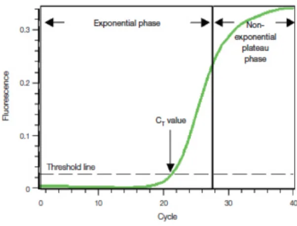
Figure 9 – Amplification plot of a typical real-time PCR output
Effects of MS and different environmental conditions on total time that the experimental rat Figure 10 –
spent in each compartment during the Social Affiliation test
Effects of MS and different environmental conditions on total time that the experimental rat Figure 11 –
spent in each compartment during the Social Preference test. Total contact duration between experimental rat and the stranger II or stranger I, during the social recognition test
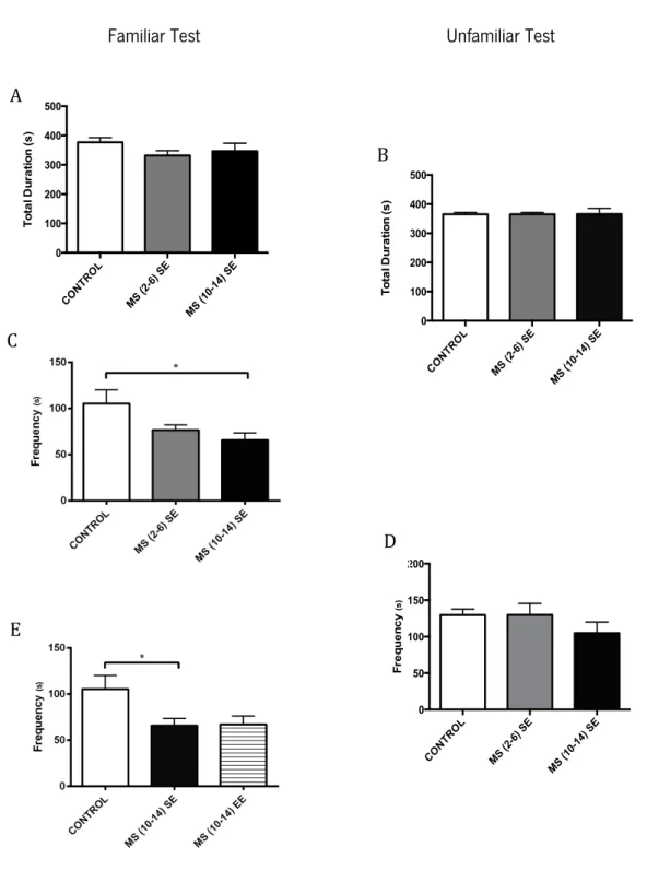
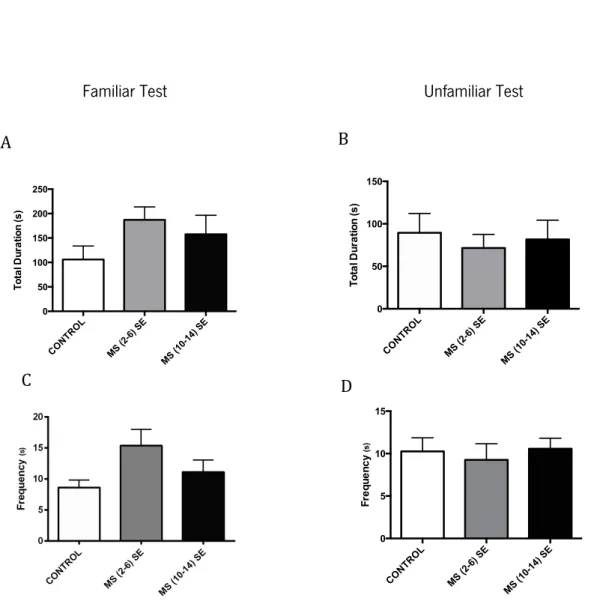
Figure 12 – Effects of MS and different environmental conditions during the social interaction tests: familiar and unfamiliar (B, D) for the investigatory behavior. Total duration of investigatory behavior and frequency
Figure 13 – Effects of MS and different environmental conditions during the social interaction tests: familiar and unfamiliar for the affiliative behavior. Total duration of affiliative behavior and and frequency
Figure 14 – Effects of MS and different environmental conditions during the social interaction tests: familiar and unfamiliar for the play behavior. Total duration of play behavior and frequency
Figure 15 – Effects of MS and different environmental conditions during the social interaction tests: familiar and unfamiliar for the non social activities behavior. Total duration of non social activities and frequency
familiar and unfamiliar for the self grooming behavior. Total duration of self grooming behavior and frequency
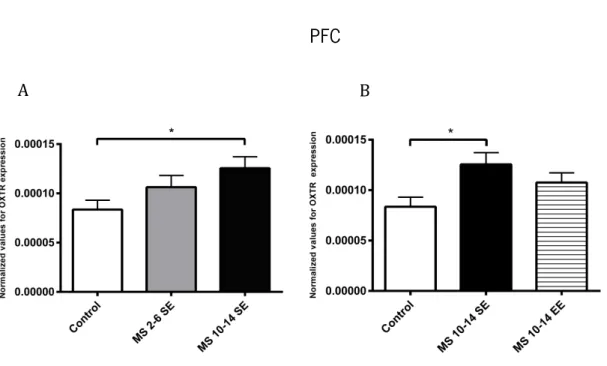
Figure 17 – Effects of MS and different environment conditions in OXTR expression in the PFC Figure 18 – Effects of MS in OXTR expression in the amygdala
Figure 19 – Effects of MS in OXT expression in the hypothalamus
Table Index
Table 1 - Distribution and Relative Densities of OXT-Binding Sites in the Adult (PND 90) and the Infant (PND 1O) in the rat CNS
Table 2 – Behavioral effects of OXT
Table 3 – Consequences of enriched environments in housed rats Table 4 - Rodent’s behavior assessment for social interaction tests
Table 5 – Correlational results between the familiar test and the molecular biology results from the OXT and OXTR expression
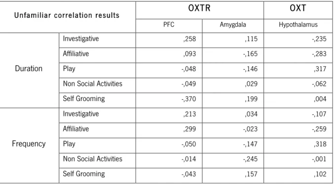
Table 6 – Correlational results between the unfamiliar test and the molecular biology results from the OXT and OXTR expression.
ACTH Adrenocorticotropic Hormone cDNA Complementary DNA
CNS Central Nervous System
CRF Corticotrophin – Releasing Factor CRH Corticotrophin – Releasing Hormone Ct Cycle Threshold
E Embryonic Day
EE Enriched Environments
FMRI Functional Magnetic Resonance Imaging HPA Hypothalamic – Pituitary – Adrenal
IL Interleukin
GAPDH Glyceraldehyde-3-phosphate dehydrogenase GPCR G Protein Coupled Receptors
mRNA Messenger RNA MPOA Medial Pre Optic Area MS Maternal Separation OXT Oxytocin
OXTR Oxytocin Receptor
OXT – IR Oxytocin Immunoreactive Neurons PCR Polymerase Chain Reaction PFC Pre Frontal Cortex
PND Postnatal Day
PVN Paraventricular Nucleus
RT – PCR Real – Time Polymerase Chain Reaction RNA Ribonucleic Acid
SON Supraoptic Nucleus
SHRP Stress Hypo Responsive Period VTA Ventral Tegmental Area
Introduction
1.1 First social interaction in mammals: the mother-pup bond
In mammals and rodents in particular, the mother is vital for homeostatic regulation and constitutes the first social bond in the life of offspring. Rats are a special model to study this specific social bond, because unlike other mammals that are promiscuously maternal, female rats only present interest in pups shortly before parturition (Insel, 2000). The first social interaction of a pup has the intervention of the dam and it is the result of specific interactions between them. This bond helps to maintain proximity, stimulates maternal care (Bowlby, 1969) and has a particular role on the establishment of the infant’s stress coping style (Lucassen et al., 2015). Indeed, the quality and quantity of maternal care was demonstrated to be very important for both humans and rodents considering it modulation of the expression of glucocorticoids and corticotrophin – releasing hormone (CRH) genes, which are involved in the appropriate stress response (Korosi & Baram, 2009).
During the weaning period, mammalian infants display motivation to seek out maternal contact and care producing affiliative behaviors and attachment bonds with their caregivers (Hofer et al., 1989). In turn, the dam adequately respond to litter needs, providing essential thermal, somatosensory, olfactory, visual and auditory stimulation, as well as maternal milk, for an extended period of postnatal development (Pryce, 1996). The maternal behavior reinforces infants’ innate bonding behavior that ensures their physical development and consequent adequate growth increasing the offspring’s probability of survival (Insel & Young, 2001).
Milk transfer is one of the primary functions of the mother-pup relationship. In fact, breastfeeding is frequently described as a unique form of social contact that promotes a strong affiliative bond (Febo et al., 2005) and has been shown to reduce activity and induce calmness on pups (Blass & Fitzgerald, 1988). Furthermore, the glucocorticoids present on caregiver’s milk have been connected with an enhance of the negative feedback on the hypothalamic – pituitary – adrenal (HPA) system (Ciruili et al., 1992). Interestingly, Kikusui and his colleagues proved that the dam has an important role on the regulation of the pups’ corticosteroid levels. Indeed, they observed high corticosteroid levels of pups, which where weaned at postnatal day 14 for 48 hours, return to baseline levels within 2 hours after pups return to the nest and reset interaction with the mother (Kikusui et al., 2009). The decrease of corticosteroid levels seems to be a side effect of the mother-pup bonding which has also been described as a social buffer that prevents infants from experiencing acute stress at least during weaning.
The mother-pup interaction depends on several elements to establish the bond depending on the olfactory learning, in which pups learn how to identify the mother through tactile stimulation from the dam and the dam’s odor, and it has the intervention of many hormones. The olfactory learning mechanism enables pups, that are born deaf and blind, to direct its behavior towards the mother but also for the mother to direct contact and licking (Moriceau et al., 2010; Moriceau et al., 2009; Reis et al., 2014). A crucial hormone for the mother-pup bond (Feldman et al., 2010; Nelson & Panksepp, 1998) as well as other social behaviors (Insel & Young, 2001; Insel, 2010; Lieberwirth & Wang, 2014) is oxytocin (OXT). This hormone has been proved to facilitate the onset of the maternal behavior (Nelson & Panksepp, 1998). In fact, studies of Pedersen and his colleagues reported a full profile of maternal behavior when administered OXT to virgin female rats (Pedersen et al., 1982). Besides, when female rats were administered with OXT antiserum (van Leengoed et al., 1987), injected with selective OXT antagonist into ventral tegmental area (VTA), medial pre optic area (MPOA) and the olfactory bulb (Pedersen et al., 1994; Yu et al., 1996) or presented lesions on paraventricular nucleus (PVN) ( Insel & Harbaugh, 1989) the maternal behavior was impaired. This suggest that the oxytocinergic system present in brain areas involved in the regulation of olfactory discriminations, emotions and reward, is extremely important to the expression of the maternal behavior and subsequently that it may strengthen mother-pup bond formation, which is also supported by studies with functional magnetic resonance imaging (FMRI) that shown that during breastfeeding, the release of OXT activated brain regions previously associated with the mother-pup bond (Febo et al., 2005). Literature suggests activation of hypothalamic OXT neurons immediately after birth, however its release has not been examined (Mogi et al., 2011). The expression the OXT receptors has been described as different in infants in different brain areas when compared to adults. In particular, the cingulate cortex is one of these altered brain sites and has been described in humans as an important area for emotional and emphatic responses (Compton, 2003; Singer et al., 2006)
.
Additionally, OXT and OXT receptor knockouts were described as emitting less ultrasonic vocalizations induced by isolation when compared to wild types. This might be result of a reduced sensitization of the knockout rats to social isolation, as maternal separation (MS) than wild type pups (Mogi et al., 2011; Takayanagi et al., 2005; Winslow et al., 2000), resulting in a lower exploration of the maternal contact and care. Notably, it has been also revealed that early-weaned rats display decreased maternal behavior in adulthood towards pups, which subsequently is transmitted to the next generation non-genetically (Mogi et al., 2011). On the contrary, oxytocin receptor (OXTR) overexpression has been associated with dysmenorrhea as demonstrated bystudies of Sun-Wei Guo (2013) (Guo et al., 2013), and increased expression has also being linked to enhanced alloparental responsiveness and accelerated partner preference formation as adults (Keebaugh & Young, 2011). Besides, studies from Lozic (2014) demonstrated for the first time that the overexpression of OXTR in the PVN was associated with the cardiovascular short-term variability and autonomic control of the circulation. In their research they found that rats overexpressing OXTR in this specific brain area had enhanced sensitivity of the baroreceptor reflex, which is meant to maintain blood pressure at constant levels and it buffered the stress induced baroreceptor variability response mediated by an increased outflow to blood vessels and stimulation of respiration (Lozić et al., 2014). This buffer effect might be mediated by the production of OXT by the magnocellular neurons and it supports other studies that suggest that OXT activates an anxiolytic response (Windle et al., 1997; Windle et al., 2004).
Together this data suggests that the OXT neural system might be also related to infant bonding and regulated by the presence of the mother (Mogi2011). Besides the mother infant bond might be an adaptive system that ensures infant’s sociality development.
1.2 Oxy tocinergic system
1.2.1 Oxytocin
Oxytocinergic system has served as a suitable example for discovering important molecular and cellular mechanisms of neuropeptide synthesis, precursor processing, and cellular trafficking. Sir Henry Dale first discovered it in 1906 during an experiment with extracts from human posterior pituitary that contracted the uterus of a pregnant cat (Viero et al., 2010). Since then, it has been explored in thousands of studies, being the first peptide hormone to be sequenced and synthesized by Vincent du Vigneaud in 1953, onwards having multiple interests and employments (du Vigneaud, et al., 1953).
OXT is a neurohypophyseal peptide synthesized in the hypothalamus and released into the blood stream via axon terminals in the posterior pituitary or neurohypophysis (Gainer, 1998). As all neurohypophyseal hormones it has a disulfide bridge between Cys residues 1 and 6, with hormonal effects and constituted of a 6 amino acid cyclic and a 3 amino acid C-terminal parts as seen in Figure 1. From an evolutionary point of view it is extraordinary that OXT homologs are still present in
invertebrates species (Knobloch & Grinevich, 2014) and that it is produced OXT or OXT-like hormones in all vertebrates (Hoyle, 1999). These are nonapeptides that differ from each other by the substitution of aminoacids in the polypeptide chain (Barberis et al., 1998).
Currently, several studies establish it’s important role in a wide spectrum of central and peripheral effects (Gimpl et al., 2001; Lee et al., 2009; Macdonald & Feifel, 2014; Ross & Young, 2009; Skopek et al., 2012; Uvnäs-Moberg, 1998) further it best known effects on parturition and lactation (Blanks & Thornton, 2003; Wakerley & Lincoln, 1973). Its effects come from the modulation of neuroendocrine responses (Stoop, 2012) to central mediation of social complex behaviors (Aoki et al., 2014) including attachment behaviors related to reproduction and care of the offspring (Caldwell & Iii, 2006; Neumann, 2008), but it also has an important role as a neurotransmitter in the brain (Insel & Young, 2001).
F i g u r e 1 - Schematic model of the OXT’s amino acid structure (Retrieved from Zingg et al., 1995)
Gene structure: anatomical synthesis and distribution
The OXT gene is expressed mostly in the PVN and supraoptic nucleus (SON) (see figure 2) of the hypothalamus and it is mainly released (by exocytosis) in response to multiple physiological stimuli (Gainer, 1998; Ivell & Richter, 1984; Viero et al., 2010). Actually, there were identified in the PVN two populations connected to the production of OXT: magnocellular neurons with projections that terminate in the neurohyphophysis and parvocelullar neurons with projections to specific areas of the central nervous system (CNS) (Ivell & Burbach, 1991).
F i g u r e 2 – Oxytocin neurons in PVN and SON in rat brain revealed by green fluorescence-protein (Retrieved from Knobloch & Grinevich, 2014).
In all species, OXT genes are located on the same chromosomal locus but are transcribed in opposite directions (Hoyle, 1999) as a result from the duplication of a common ancestral gene, which was followed by inversion of one of the genes. The OXT gene is found on chromosome 3 in rats (Caldwell & Iii, 2006; Ivell & Richter, 1984) and in chromosome 20p13 in humans (Dutil et al., 2001; Rao et al., 1992). Both genes consist in three exons: the first encodes a translocator signal peptide and the first nine amino acids of neurophysin, an important carrier peptide; the second exon encodes the central part of neurophysin (which primary function is the correct targeting, packaging, and storage of OXT) while the third encodes the COOH-terminal region of neurophysin (Marini et al., 1993; Sausville et al., 1985) (see figure 3). After transcription and synthesis as an inactive precursor, the OXT prepeptide is subject to cleavage and other post transcriptional modifications being conveyed axonally to the posterior pituitary where it can be released by appropriate stimulation (Gimpl et al., 2001; Ivell & Richter, 1984).
F i g u r e 3 - Representation of the structural organization of the OXT gene (Retrieved from Ivell & Richter, 1984)
1.2.2 Oxytocin receptor
Receptor Structure
The OXTR is a cell surface membrane protein of 43 kDa and it consists in 389 amino acids (Kimura et al., 1992). It is a classic member of the rhodopsin-type (class I) GPCR (G protein coupled receptors) family with seven transmembrane domains. This receptor display the structural hallmarks characteristic of most GPCRs: glycosylation on Asn residues present in the extracellular domains, a disulfide bridge between two highly conserved Cys residues in the second and third extracellular domains, and two relatively well-conserved Cys residues within the C-terminal receptor domain, which have been shown to be palmitoylated in other GPCRs (Barberis et al., 1998) (see Figure 4). The conserved residues among the GPCRs appear to be involved in a common mechanism for activation and signal transduction to the G protein. The switching from the inactive to the active conformation is
associated with a change in the relative orientation of transmembrane domains 3 and 6, (Bockaert & Pin, 1999). It has two (mouse, rat) or three (human, pig, sheep, rhesus monkey, bovine) potential N-glycosylation sites (N-X-S/T consensus motif) in its extracellular NH2-terminal domain, however it does not seem to be essential for proper expression and has no effect on the functional properties of the receptor (Kimura et al., 1997).
F i g u r e 4 – Structure and organization of the OXTR indicating the three potential sites for N-glycosylation in the extracellular N-terminus. Cysteines are shown as filled circles (Retrieved from Ivell et al., 2001).
Gene Structure
The OXTR gene is present in single copy in the human genome in the chromosome 3p25– 3p26.2, contains 3 introns and 4 exons (Inoue et al., 1994; Michelini et al., 1995; Simmons et al., 1995) and encodes approximately 389 amino acids. The rat OXTR gene is present in chromosome 4 (Caldwell & Iii, 2006), contains 3 exons and it is 93% identical to the human OXTR sequence (Kubota et al., 1996; Rozen et al., 1995).
Exons 1 and 2 correspond to the 5-prime noncoding region followed by exons 3 and 4 that encode the amino acid sequence of the OXT receptor. In fact, exon 4 contains the sequence encoding the seventh transmembrane domain, the COOH terminus, and the entire 3-noncoding region, including the polyadenylation signals (see Figure 6) (Kimura et al., 1992).
Localization of OXT and OXTR within the brain and gene expression through development
In the brain, OXT gene is essentially expressed in magnocellular neurons in the hypothalamic PVN, which terminate in neurohypophysis and SON that have terminations in other parts of the CNS. Despite this, OXT fibers and OXT neurons have been widely described in the CNS in rat (for more details see Buijs et al., 1985). The distribution and number of OXT binding sites changes in critical periods of development (Olazábal & Alsina-Llanes, 2015; Tribollet et al., 1992; Yoshimura et al., 1996). The OXTRs are abundantly distributed in several brain areas including the limbic system and the hypothalamus (Ostrowski, 1998; Yoshimura et al., 1993) (see table 1 for more details) although only a fraction of the OXTRs are constantly present through development. Independently of the sex, two critical periods were recognized, in which the expression differs from infant to older ages: the 3rd postnatal week, right after the weaning period, and puberty (Tribollet et al., 1992). In fact, during the early postnatal life, areas containing high densities of OXT-binding sites like the cingulate, retrosplenial cortex and substancia gelatinosa posteriorly drop, or are strongly reduced, between postnatal days 10 and 21. On the opposite, some OXT-binding sites only appear at the time of puberty, between postnatal days 40 and 45, at areas like the olfactory tubercle, hypothalamic ventromedial nucleus, bed nucleus of the stria terminalis and the central amygdaloid nucleus (for more details see table 1). This can be explained based on studies by Tribollet et al. (1992) and Yoshimura, (1996) in which they demonstrated that the developmental profile of OXTRs is classified as constantly or transiently expressed through development depending on the brain area and age of the animal. The transient expression corresponds to restricted intervals in which there is a detectable OXTR expression while the constant expression consists in a continuous expression until the adult age of the animal. Additionally, it is extremely important to have in concern the different circulation and release of OXT during the early development versus the adult, due to incomplete maturation of important structures. Actually, the OXTR mRNA appears for the first time at E (embryonic day) 13, representing the earliest expression in the CNS. Then, a stronger signal is detected at E 15, which is maintained throughout development being slightly reduced in adults (Yoshimura et al., 1996). Nevertheless, the ligand, OXT hormone, is only detected at time of birth (Buijs, 1992). The discrepancy in the timing of appearance between the OXTR and OXT might be due to the early expression of the receptor, which anticipates the release of OXT to express their function through development (Yoshimura et al., 1996).
Regulation of OXTR
As many members of the GPCR superfamily, OXTR expression undergoes through dramatic and cell-specific up and down regulation. The tissue-specific regulation of OXTR expression enables circulating OXT to switch its target organs. Besides, it appears to have both inhibitory and stimulatory influences acting upon a constitutive pattern of basal expression (Ivell et al., 2001).
There is plenty evidence suggesting that gonadal steroids can regulate various aspects of the OXT system (Insel et al., 1993; Patchev et al., 1993; Tribollet et al., 1990). In uterus as well as hypothalamus, the regulation seems to be indirectly correlated with gonadal steroids probably involving intermediate transcription factors or cofactors (Ivell & Walther, 1999). In rats, increased expression of OXT mRNA in certain areas of the hypothalamus are coincident with the onset of puberty and vary across the estrous cycle; when both of these events are associated with increased concentrations of circulating estrogens. Apart from regulating the transcription of OXT, gonadal steroids also affect serum and pituitary levels of OXT, axonal and dendritic release of OXT, as well as the electrical activity of OXT neurons (Dhakar et al., 2013; Insel et al., 1993).
It has also been proposed that human OXT receptor gene is associated with transcriptional gene suppression (Yoshimura et al., 1996) by a genomic element in the third intron of it’s sequence as it was hypermethylated in nonexpressing tissues and hypomethylated in myometrium when the OXT receptor gene was up regulated (Mizumoto et al., 1997).
Besides, though the 50-flanking regions of the rat and human OXTR contain several potential interleukin (IL). ILs were thought to represent potential regulators of OXT receptor expression and recent findings indicate that the human OXTR promoter is negatively regulated by IL-1b and IL-6, and that both cytokines decrease messenger RNA (mRNA) of OXTR accumulation in myometrial cells (Schmid et al., 2001).
Nevertheless and unlike others GPCRs, the OXTR undergoes desensitization by persistent agonist stimulation (Evans et al., 1997). This is, when these receptors are stimulated for a long period, they desensitize, being posteriorly phosphorylated and internalized. This process is very fast (within seconds or minutes) and can happen by numerous processes operating at transcriptional, translational, and protein levels (Evans et al., 1997; Gimpl et al., 2001).
T a b le 1 - Distribution and Relative Densities of OXT-Binding Sites in the Adult (PND 90) and the Infant (PND 1O) in the rat CNS (retrieved from Tribollet et al., 1992)
1.2.3 Oxytocinergic system and social behavior
Social behavior is the establishment of a pair bond between two animals and it is critical for social organization (DeVries et al., 1996). To perform any type of social interaction it is required the communication between conspecifics, in which it is used species-specific social and behavioral cues to to “understand” and “read” the intentions of the other (Veenema & Neumann, 2008). The development of social familiarity in rodents depends predominantly on olfactory or pheromonal cues, which define the capacity to recognize a conspecific, to determine the adequate response and formation of the “social memory” of individuals when within a social group (Neumann, 2008; Sanchez-Andrade & Kendrick, 2009; Winslow & Insel, 2004).
Adolescence is a gradual period (at about 40 days of age) in which the animals transit from childhood to adulthood characterized by certain characteristic behaviors, including increases in peer-directed social interactions and elevations in novelty-seeking and risk-taking behaviors (Spear, 2000). Social interactions and affiliation with peers take on particular importance during this time, and juveniles usually have more quality time of playful behaviors (Michael J. Meaney & Stewart, 1981; Panksepp et al., 2007; Spear, 2000). In fact, studies in animals that were deprived of play during the juvenile period indicate that social play is critical for normal social, motor, cognitive, and emotional (Argue & McCarthy, 2015; Graham, Burghardt, & Wiens, 2010; Pellis, Pellis, & Bell, 2010; Spinka, Newberry, & Bekoff, 2001; Van Den Berg et al., 1999).
Early social experience, stress or stress hormones have been described as important modifiers of the formation of the “social memory”, so they can modulate social behavior as well as the partner preference (DeVries et al., 1996; Young & Wang, 2004). OXT has been described has having a key role on regulating the formation of social memories and social recognition for a lot of species (Lee et al., 2009; Popik et al., 1992) as well as on mediating the benefits of positive social interaction and emotions (Uvnäs-Moberg, 1998). Indeed, the recognition of a familiar partner and subsequent pair bonding are essential to social organization. Therefore understanding the interaction between the OXT and social bonding has been and continues to be a source of stimulating studies in neuroendocrinology (DeVries et al., 1996). Several studies link OXT to pair bonding, which is mainly characterized by social preference (the subject spends more time in close proximity) of a familiar partner instead of a stranger one, being also referred in other studies as partner preference (Getz et al., 1981; Young & Wang, 2004). Interestingly, OXT-knock out male mice failed to recognize familiar conspecifics that have
previously encountered and social memory deficits were also found in mice lacking OXT or OXTR (DeVries et al., 1996; Winslow & Insel, 2002). Additionally, some pharmacological studies demonstrated that intracerebroventricular injections of small doses of OXT on medial amygdala could rescue the social recognition deficit (Ferguson et al., 2001). Nevertheless, some other brain areas as pre frontal cortex (PFC), nucleus accumbens and ventral pallidum have been also referred as essential to the formation of this pair bond using the reward brain system (Wise, 2002).
Furthermore, there are additional OXT effects related to rodent social behaviors, namely maternal care, aggression, affiliative behaviors, pair bonding, social recognition, cognition and sexual behaviors (Arakawa et al., 2015; Calcagnoli et al., 2015; Lukas et al., 2010; Miller & Caldwell, 2015; Ross et al., 2009; Uvnäs-Moberg, 1998; Young & Wang, 2004). Besides, there are some evidence towards similar behavioral effects on humans and indirect evidence supports also the OXT anxiolytic and anti stress effects (Carter et al., 2001; Carter & Altemus, 1997; Kosfeld et al., 2005), (for more details see table 2).
T a b l e 2 – Behavioral effects of OXT (Retrieved from (Lee et al., 2009)
1.3 Maternal Separation Paradigm
The MS is a well-established and powerful model of early life stress, applied in different research areas that can be used to characterize alterations on behavior and biological development.
Events occurring in early life are powerful modulators and can produce profound and long lasting changes in the development of different biological systems. Distinctive behavioral changes include impaired maternal care (Lovic et al., 2001), decreased maternal aggression in rats (Boccia & Pedersen, 2001), increased inter male aggression (Veenema et al., 2006), increased dominant juvenile play-fighting behaviors (Veenema & Neumann, 2009), impaired social recognition (Lukas et al., 2011) and play behavior (Arnold & Siviy, 2002; Zimmerberg & Sageser, 2011)) plus some evidences suggests impaired spatial learning and memory ability (Huang et al., 2002). It can also affect growth, metabolism, reproduction and inflammatory/immune responses (Chrousos & Gold, 1992; Darnaudéry & Maccari, 2008; Mesquita et al., 2007; O’Mahony et al., 2009; O’Malley, Dinan, & Cryan, 2011; Roque et al., 2014), increase susceptibility to addictions (Moffett et al., 2007; O’Mahony et al., 2009b; Ploj et al., 2003) and lead to psychiatric disorders (Charmandari et al., 2005; Chrousos & Gold, 1992; Chrousos, 2009). In particular, this stress induction model impacts the CNS that regulates the stress responsiveness (Hennessy et al., 2009; Lukaset al., 2010; Nishi et al., 2014; Zhang, & Zhao, 2013; Zhang et al., 2012) altering the trajectory and programming the response to stress (Darnaudéry & Maccari, 2008).
The optimal regulation of stress response is essential to adaptation, health and maintenance of homeostasis (De Kloet & Derijk, 2004; Ulrich-Lai & Herman, 2009). When the homeostasis is disturbed, the organism rapidly assemble a response in order to restore it (De Kloet et al., 1998). The physiological response to stress involves the activation of “classical” neuroendocrine mechanisms, from an efficient and highly conserved set of interlocking homeostatic systems, and aims to maintain physiologic integrity even in the most challenging situations (Ulrich-Lai & Herman, 2009). In order to restore the physiological and behavioral homeostasis, the autonomic nervous system and the HPA system are activated culminating with activation of stress centers in the brain (Cerqueira et al., 2007; De Kloet et al., 1998; Herman et al., 2003). There are two forms in which the stress response occurs. Initially, in an immediate response of a “proactive” mode, the corticotrophin – releasing factor (CRF) is liberated from the hypothalamic PVN in response to the stimuli. CRF is a very important molecule to organize the response to stressors and when it is released into hypophysial portal vessels it accesses the anterior pituitary gland. The binding of CRF to its receptor induces the release of adrenocorticotropic hormone (ACTH) into the systemic circulation (Charmandari et al., 2005; Smith & Vale, 2006). This immediate reaction is compensated by the parasympathetic nervous system activity
and in the endocrine dominion, ACTH targets it main focus, the adrenal glands, where it leads to an elevation of circulating levels of glucocorticoids (De Kloet et al., 2005; Herman et al., 2003; Kellendonk et al., 2002; Smith & Vale, 2006). Next, a slower approach takes place, the “reactive” mode, which facilitates the adaptation and helps re-establishing homeostasis (De Kloet et al., 2005). The corticosteroids feedback helps to terminate the stress-induced HPA activation by negative feedback regulation (Kellendonk et al., 2002) (Figure 6).
F i g u r e 6 – The influence of stress in the HPA axis and how its metabolic effects ends regulating the stress response (Adapted from ThePaleoMom, 2014).
In rodents, during the first two weeks of life there is a marked reduction of corticosterone secretion, which ensures low and stable levels of glucocorticoids (Levine, 2001; Sapolsky & Meaney, 1986). This is very important because it acts as a protective mechanism during this postnatal developmental window, designated as the stress hypo responsive period (SHRP) (De Kloet et al., 1998; Sapolsky & Meaney, 1986). Furthermore, it was demonstrate that the exposure to high levels of glucocorticoids, during the neonatal period leads to irreversible reduction of the brain weight (Huang et al., 1999), influences cerebellar development showing a total decrease in DNA content in the brain (Velazquez & Romano, 1987), impaired neuronal myelination (Dunlop et al., 1997), widespread
reduction of dendritic spines (Antonow-Schlorke et al., 2003) and inhibition neural precursor cell proliferation in brain areas that show postnatal neurogenesis (Scheepens et al., 2003).
1.3.1 Maternal Separation Protocols
The diversity of MS protocols is vast since they can vary in length and according to different developmental periods. MS protocol can occur in single or repeated periods of separation with sessions of few minutes to several hours but the majority of them occur during the SHRP (Schmidt et al., 2011). Additionally, for MS protocol, not only the periodicity of the separation is very important but also the amount of time that the pups remain separated from the dam. Literature suggests that a minimum of 2 hours is necessary to induce an immediate effect on HPA responsiveness (Kuhn & Schanberg, 1998; Pihoker, 1993) and more than 8 hours to produce an effect that is measurable numerous days after the manipulation (Rosenfeld et al., 1992). Many MS protocols are often used as a model for early life adversity mimicking maternal neglect where the pups are deprived from maternal care for several hours (Bulbul et al., 2012; Carlyle et al., 2012; Roque et al., 2014; Slotten et al., 2006). Nevertheless, there are MS paradigms that do not mimic maternal neglect. Instead, there is a physical separation between animals for lower time periods comparing to the MS protocols described above. In his studies, Levine manipulated the litter for approximately 15 min, where the dam was separated from pups during this period revealing several differences between non handled vs. handled animals (Levine, 1957). In one hand, early stress experience result in greater infant’s capability to adapt to psychological and physiological stress in adulthood and so presenting better stress coping. On the other, more recent studies have shown that this short periods of MS has a stimulating effect of maternal care, providing higher levels to pups (Pryce et al., 2001). However, this is not very consistent
with literature since there are studies that use repeated daily separations, even for shorter periods, that describe deleterious effects on pups development (Li et al., 2003; Rana et al., 2015; Roman et al., 2005). Additionally, longer separation periods are portrayed as capable to reduce the amount of maternal care for the pups and thereby modeling emotional as well as physical neglect (Schmidt et al., 2011) which may contribute to psychopathology in later life (Pryce & Feldon, 2003). Besides, pups that were maternally separated for long periods exhibit more anxious behaviors (Boccia & Pedersen, 2001), exaggerated behavioral and neuroendocrine responses to stress (Kalinichev et al., 2002) and display less maternal care in adulthood (Lovic et al., 2001). Indeed, a study from Mesquita and colleagues
(2007) suggest that MS acts on the serotoninergic system, and transiently impairs the acquisition of innate reflexes, which might contribute to the long-lasting effects of MS on emotionality and cognition (Mesquita et al., 2007).
1.3.2 Impact of MS on social behaviors
Early stressful events like the MS result in a withdrawal of regulatory processes that form the complex pattern of physiology and behavior responses (Hofer1994). The effects of the MS on social behaviors have been source of interest for more than 50 years for many mammallian species due to the alteration of the context of interactions between mother and offspring in the postnatal social environment (Hofer, 1973; Kaufman & Rosenblum, 1969). This early life stress protocol usually acts on critical time periods for the nervous system, like the SHRP during which the brain undergoes functional organization, neuronal proliferation, migration and differentiation, gliogenesis and myelination (Rice et al., 2000).
During the separation period the two-week-old rat pups show an immediate response consisting of repeated, high intensity vocalizations (in the ultrasonic range), accompanied by agitated searching behavior and high levels of self-grooming (Hofer & Shair, 1978). The body temperature declines, the HPA axis is activated and glucocorticoid secretion increases (Kuhn & Schanberg, 1998; Pihoker, 1993). Most of these manifestations are rapidly ameliorated when they return into contact with the dam or a familiar littermate (Hofer, 1994), or, even diminish as the pups gets older (Noirot, 1972).
Right after the MS period, rats demonstrated to respond to a novel environment with heightened levels of locomotion and rising behavior (Hofer, 1973), displayed more self-grooming, defecation, urination and a delayed sleep onset in comparison to their mothered littermates. Similar studies showed that rats were MS (Roman et al., 2005). Other studies have demonstrated to have neurobiological consequences in areas that significantly overlap with the neural circuitry involved in complex social behaviour, such as social recognition. There have been also studied the potential impact that MS may have on the long-term development of the neurobiology and behaviour involved in social interaction in rodents and higher primates (Lukas et al., 2011; Sabatini et al., 2007; Varlinskaya, 2008; Veenema, 2012; Zimmerberg & Sageser, 2011) further altered social preference patterns (Wang et al., 2015). It was also revealed an association between the duration of separation and attachment
disorder behaviors (O’Connor & Rutter, 2015).
The MS protocol disrupts the mother-pup bond by decreasing mother-pups contact and maternal care, changing temporal patterns. As described above, social proximity, olfactory learning, access to milk, and physical contact between animals are adversely affected (Reis et al., 2014). This disruption has also been proved to affect future pup’s HPA development and therefore future responses to stress (Liu et al., 1997). Other studies showed that maternally separated pups present maternal care deficits in adulthood (Lovic et al., 2001). Because OXT has been described to modulate the HPA axis, which has been proved to be altered on social behaviors, we can hypothesize that this is an extremely important hormone that has a particular role on behavior development.
1.3.3 Impact of MS on oxytocinergic system
As discussed above, MS can affect different biological systems. Indeed, MS induces alterations on the HPA axis, which is a major stress response system, and results in a hyperresponsiveness to stress from the animal in adulthood. Indeed, maternally separated rats tend to display more social avoidance and behavioral inhibition during social encounters (Cavigelli et al., 2007; Núñez et al., 1996; Roelofs et al., 2009). The oxytocinergic system has been described as one that it is disturbed by this stress-induction model (Kong et al., 2015; Lukas et al., 2010). In fact, OXT has been associated as a major HPA axis modulator (Neumann, 2002; Windle et al., 2004; Zheng et al., 2010), and also implicated in the expression of behavioral responses to stress (Ferguson et al., 2001; Ferguson et al., 2002; Lukas et al., 2010; Neumann, 2008). Indeed, brain OXT has a major role in the regulation of complex physiological stress responses including the stress-induced neuronal activation (Windle et al., 2004). Besides, the early exposure to relevant stimuli is known to permanently alter the responsiveness of the hormone receptor (Miller & Caldwell, 2015), so alterations by MS are probably noticeable at an OXTR distribution and expression levels.
Neurochemical data has been suggesting a key role of OXT in the development and maintenance of social cognition and social behavior. In fact, there are some studies in which OXT and OXTR knock out mice that were maternally separated that revealed decreased motivation to be in contact with the dam affecting their attraction towards the mother (Winslow & Insel, 2002). Nevertheless, at a molecular level it was found that presynaptic OXT was not essential to normal neuroanatomical distribution of OXTR. In accordance with this, there is studies in male rats from Lukas
and colleagues (2010) demonstrated that exposure to early life stress, using MS model, interferes with the normal development of OXTR binding in some specific forebrain regions (decreased levels in the agranular cortex, lateral septum and increased levels in the ventromedial hypothalamus) suggesting that environmental manipulations can, indeed, shape the developmental expression of OXTR through the brain. Also, the maternally separated rats displayed a decrease of OXTR binding throughout age, resulting in a lower OXTR binding density at adult age when compared with control rats (Lukas et al., 2010). Also, studies from Kong et al. (2015) in monogamous rodents revealed that repeated separation from pups affected the number of oxytocin immunoreactive neurons (OXT-IR) in PVN (Kong et al., 2015). Actually, long repeated periods of separation revealed an upregulation of the OXT-IR, while brief repeated periods of separation increased the central number of OXT-IR. The same was previous found in studies with rat dams after two weeks of MS from pups (Boccia & Pedersen, 2001).
Knowing that MS is a stress induction model that introduces several animal behavior and molecular impairments and that animal’s are particular sensitive to the surrounding environment, it becomes extremely important the understanding of how enriched environments (EE) provide extra comfort might be capable of attenuating some of those impairments.
1.4 Reverting MS impact: environmental enrichment
From all the different factors that influence social interaction, the environmental condition in which the animals are housed have been described to induce changes on it (Francis et al., 2002). The postnatal handling has been pointed out as a functional recover of the effects of prenatal stress through compensation on long-term impairment of the glucocorticoid feedback and induction of anxiety and even in reversing the effect of MS on the HPA function (Francis et al., 2002; Maccari et al., 1995; Vallée et al., 1997). In fact, there are studies that support that environmental handling is almost the opposite of MS when it comes to its effects (Caldji et al., 2010; Darlene D Francis et al., 2002).
Environment enrichment (EE) can be divided in two forms: physical and social. Social enrichment consists in housing animals in groups wherever possible, to promote social interaction. The physical includes bedding enhanced with natural materials (paper parchment), plastic tunnels, wooden objects to gnaw, ropes, swings, running wheels, balls, ramps, ladders and other appropriately sized animal toys (Simpson & Kelly, 2011).
Actually, it is believed that EE can improve animals’ well-being, brain weight, and it is used to demonstrate learning and brain plasticity in response to different environments (Baumans, 2005). There are also multiple studies in which we can observe that there is significant differences when it comes to EEs, specially in social interactions: increase on social play and social rest (Morley-Fletcher et al., 2003) and a decrease on social exploration (Magalhaes et al., 2007; Magalhães et al., 2006) (for more alterations see table 3).
T a b l e 3 – Consequences of EEs in housed rats (adapted from Simpson & Kelly, 2011) Attribute Effects of enriched housing vs
Standard Conditions
Body weight ↓ (males) – (females) (Moncek et al., 2004) (Harati et al., 2011) General behavior ↑ habituation, ↓ activity ↑ grooming, ↓ rearing (Brenes et al., 2008; 2009) Anxiety Elevated Plus Maze:
↑ open arm entries ↑ open arm movement
↑ percent open arm entries and time Social Interaction:
↑ social play, ↑ social rest and social exploration after prenatal stress Pre-weaning EE ↓ social exploration, social behaviour and play
(Pena et al., 2006) (Peña et al., 2009) (Galani et al., 2007)
(Morley-Fletcher et al., 2003) (Magalhaes et al., 2007; Magalhães et al., 2006)
Depressive- like Behaviours
Forced Swim Test:
↑ swimming, climbing, diving, ↓ immobility (Brenes et al., 2008; 2009) Learning and
Memory
Morris Water Maze: ↓ escape latencies
Novel Object Recognition: ↑ novel object preference Radial Arm Maze: ↓ errors and time in arms
(Leggio et al., 2005; Zhong et al., 2009)
(Bruel-Jungerman et al., 2005; Pamplona et al., 2009)
(Hoffmann et al., 2009; Leggio et al., 2005)
1.5 Aims of the study
At early ages all interactions and experiences are determinant for acquiring competences and adapt to environmental challenges. Early life stress events have been shown to play a critical role on social behaviors and act as severe modulators of the oxytocinergic system having long lasting implications throughout adulthood.
The MS model has been typically performed during the SHRP with establishment of “temporal windows” of vulnerability to the stress programming effects. Contrary to the most MS protocols, our MS paradigm involved short separations and was used as a non-negligent model in which physical MS is necessary. Instead of the typical MS group we decided to have two different periods (within the SHRP): from 2nd
to 6th
PND and 10th to 14th
PND, corresponding approximately in humans to the period, in which children initiate the nursery at age of five month and at age of 3 years, creating a bridge between rodents and humans.
The current knowledge in this field has yet many open questions, some of which will be addressed in this thesis.
The study developed in the scope of this thesis aims to:
- Evaluate the impact of short daily MS, during two time points periods of early life, on the modulation of different components of social behavior of adolescent rats;
- Investigate the effect of early interventions that can be designed to overcome or reduce the effects of these environmental insults and challenges.
- Determine the expression profile of OXT and OXTR during adolescence, and correlate it with the behavioral impacts of MS.
Materials and Methods
2.1 Animals
Wistar Han females (Rattus norvegicus) (Charles River, Barcelona, Spain) during estrous were placed overnight with males in same cage under controlled environmental conditions (ambient temperature 22ºC, 60% humidity, 12 h light/12 h dark cycle, lights on at 00:00 a.m.) with free access to food and water. Males were removed from the cage when a vaginal plug was confirmed; this day was designated as embryonic day 0 (E0). Pregnant Wistar females were left undisturbed during pregnancy. At the end of pregnancy (E22), all females were provided with nesting material and remained singly housed. Litters were delivered on gestation day 22. At birth (considered the postnatal day (PND) 0), pups were culled to 8 pups per litter, normalized in terms of gender and placed in a new cage. The experiments carried out were approved by local ethics committee and by the Portuguese Agency for Animal Welfare, general board of Veterinary Medicine, in compliance with the European Community Council Directive of September 22, 2010 (2010/63/UE). All procedures involving animals were conducted by FELASA C graded researchers, and all efforts were made to minimize the number of animals used and their suffering.
2.2 Maternal separation procedure
MS procedure consisted in a physical separation between the dam and the pups for 2h per day, during two different developmental periods, specifically between the 2nd
and the 6th
PND or between the 10th and the 14th PND. The mothers were removed from the home cage and put into another single cage (the same cage every day for each mother). Then the pups were removed from the home cage and put in another cage (the same cage every day) on heated sawdust at 30ºC, in another room, so as to prevent vocalization and auditory communication with the mother.
To further understand the effects of environmental manipulations, we compared the behavior of rats submitted to MS period under enriched or standard conditions. In this context, during MS period rats were housed in different environmental groups: standard or enriched conditions. The EE consisted in nest material such as paper towels, cardboard tubes and cotton.
According to this, the litters were randomly divided in 5 different experimental groups:
- MS 2-6 SE, Maternal separation from PND 2 to 6 in standard environment - MS 2-6 EE, Maternal separation from PND 2 to 6 in enriched environment - MS 10-14 SE, Maternal separation from PND 10 to 14 in standard environment - MS 10-14 EE, Maternal separation from PND day 10 to 14 in enriched environment
- Control pups from control litters were left undisturbed with their dams until weaning (PND 21)
After the two hours of MS, pups were returned to the home cage. At PND 22 all pups were weighed and housed in groups of two littermates of the same sex per cage until they achieve adolescence and the behavioral tests start.
2.3 Behavioral Tests
All testing procedures were conducted in the dark phase of the light/dark cycle between 13:00 and 20:00h under dim light (15–20 lx). All behavior tests were performed during the rat adolescent period when rats show a particularly strong tendency to be social (around 40 PND) (Terranova, Cirulli, & Laviola, 1999). They were designed to access the social recognition and the social interaction.
2.3.1 Social Recognition Abilities And Social Preference
Sociability is defined as the time that the animal spends with another rat, comparing to time spent alone in an identical but empty chamber (Kaidanovich-Beilin et al., 2011). Social memory is commonly examined in rodents through various social recognition tasks that utilize the innate preference of adult rodents to spend more time with novel over familiar conspecifics (Macbeth, Edds, & Young, 2009). One test that is used is the three-paradigm test or Crawley's sociability and preference for social novelty test. This test can be used to study social affiliation and social memory with novel conspecifics. Rats tend to spend more time interacting with a novel rat versus one they have previously encountered. The basic measure is the amount of time the subject spends with either a completely novel animal or an animal to which they have been introduced to in a previous session, or the animal can also choose to be in the middle section, that is, alone. The apparatus for Crawley's sociability and preference for social novelty test consists in a rectangular,
three-chamber box. Each chamber is 40 x 60 cm and the dividing walls are made from clear Plexiglas, with an open middle section, which allows free access to each chamber (Figure 7). Both tests were conducted as described by Kaidanovich-Beilin (2011).
Social Affiliation test (session I)
At 45 PND, each experimental rat started the test in the central compartment and was left there for 5 min for habituation. After this period the experimental rat was removed and another animal (Stranger I) was placed in one of the containers within the compartment of the apparatus while the other compartment remained empty. Then, the experimental animal was again placed in the center compartment and videotaped for 10 minutes. The widely spaced wire bars of the container permit olfactory, visual, auditory and some tactile contact while preventing aggressive and sexual interactions, thus ensuring a pure measure of simple interest in approaching and remaining in physical proximity to another.
Social Preference test (session II)
After social affiliation test, an additional rat (Stranger II) was placed in the empty container and the behavior of the experimental rat was again videotaped for additional ten minutes to record its behavior.
Videotapes were analyzed using the Noldus Observer® software (Observer 5; Noldus Information Technology, Wageningen, Netherlands). The following parameters were recorded: duration and number of direct (active) contacts between the experimental rat and Stranger I and empty containment cup or Stranger II. The following behaviors were analyzed 1) direct contact is define as stretching of the body of the experimental rat in an area 3-5 cm around the container; 2) number of other behaviors by the experimental rat in each compartment, including walking, self-grooming, lack of any body movements for more than 5 seconds (freezing); 3) duration and number of entries to each compartment (the animal is considered to be in the chamber when its head and four paws have entered into the chamber).