Maria Fernanda Dias Miranda Bernardo
Biological Evaluation of New Compounds as Potential
Aromatase Inhibitors for Estrogen-Dependent Breast
Cancer
Dissertação de 2º Ciclo de Estudos Conducente ao Grau de Mestre em Toxicologia Analítica Clínica e Forense
Trabalho realizado sobre a orientação do(a) professor(a)
Doutora Georgina Correia da Silva, Faculdade de Farmácia da Universidade do Porto
Doutora Natércia Aurora Almeida Teixeira, Faculdade de Farmácia da Universidade do Porto
É AUTORIZADA A REPRODUÇÃO INTEGRAL DESTA DISSERTAÇÃO/TESE APENAS PARA EFEITOS DE INVESTIGAÇÃO, MEDIANTE DECLARAÇÃO ESCRITA DO INTERESSADO, QUE A TAL SE
Author´s Publications
Abstracts published in congress books
F. Bernardo, C. Amaral, C. L. Varela, E. Tavares da Silva, F. M.F. Roleira, S. Costa, N. Teixeira, G. Correia-da-Silva, Antitumor efficacy of new steroidal AIs: AR and ER dependency on MCF-7aro cell. 2nd ASPIC International Congress, 28-29 April, 2016, Porto, Portugal
F. Bernardo, C. Amaral, C. L. Varela, E. Tavares da Silva, F. M.F. Roleira, S. Costa, N. Teixeira, G. Correia-da-Silva, Breast cancer growth-inhibitory efficacy of new steroidal compounds as aromatase inhibitors. IJUP’16 - 9º Encontro de Investigação Jovem da Universidade do Porto, 17-19 February, 2016, Porto, Portugal
Aknowledgments/Agradecimentos
E assim termina mais uma etapa da minha vida. Foi uma jornada com altos e baixos mas tive sempre ao meu lado as pessoas certas para me apoiar e mostrar que por mais difícil que a vida seja o importante é não desistir. Porque no jogo da vida o importante é não ter medo de ir á luta, porque a derrota é garantida mas a vitória só se conquista quando se joga. Por isso eu escolhi jogar sempre, porque no dia que deixar de jogar deixo de viver. Mas não joguei sozinha tive sempre ao meu lado pessoas que acreditaram em mim, me apoiaram e deram força para continuar e por isso não posso deixar de lhes agradecer.
À Dra Georgina Correia-da-Silva agradeço todo o apoio e disponibilidade para me ajudar, não só na tese como no seminário. Obrigada pela dedicação e incentivo nesta etapa da minha vida. Agradeço todos os ensinamentos e conselhos que foram úteis para o meu crescimento científico e pessoal.
À Dra Natércia Teixeira agradeço todo o carinho, atenção, apoio que demonstrou sempre ao longo deste meu percurso. Acima de tudo agradeço a disponibilidade que sempre teve, pois no meio de todo o trabalho e responsabilidade que tem, encontrou sempre um tempinho para me receber e acompanhar.
À minha equipa de trabalho, Cristina Amaral e Tiago Augusto foi um prazer partilhar este ano convosco. Cristina, obrigada por tudo o que me ensinaste, pela dedicação e tempo despendido comigo, pela ajuda, pelo apoio, pela paciência, pelos conselhos. Obrigada por teres sempre uma palavra de conforto para me dar nos dias mais dificeis. Mas principalmente, obrigada pela confiança que sempre depositaste em mim. Mais que uma “mãe” és uma amiga que eu vou guardar com carinho. Tiago, agradeço-te toda a paciência, inter-ajuda, companheirismo, boa disposição. Sobretudo obrigada por seres quem és pois isso deu-me motivação para querer me tornar melhor e ajudou me a crescer.
Agradeço ainda a todos os outros membros do laboratório, Marta Almada, Renata Fernandes, Maria Miguel, João Maia, Lia Costa, Susana Rocha, Ana Paula, Bruno Fonseca e Sandra Ribeiro que de alguma forma me ajudaram ao longo deste ano, desde a confiança, à ajuda, à amizade, ao apoio, aos bolinhos, às brincadeiras e bons momentos passados dentro e fora do laboratório.
Não posso deixar de agradecer a uma das pessoas mais importantes deste laboratório. Aquela que põe ordem e respeito e permite o trabalho dos investigadores com a sua organização, rigor e cuidado com todo o material. D. Casemira, obrigada por todos os “não há” que depois de confirmados haviam sempre. Pela preocupação e carinho. Por
todas as risadas e brincadeiras por eu “estar nua"que tornaram cada dia diferente e mais alegre.
Aos meus amigos, os de longa data e os que fiz ao longo desta jornada académica, um sincero obrigado por todos os bons momentos partilhados, pelo apoio, pela motivação, pela preocupação, pelo carinho, pela amizade e pela força, pois foi ela que muitas vezes me ajudou e deu forças para seguir em frente.
Por ultimo, agradeço à pessoa mais importante de todas, a minha mãe. Obrigada por todos os sacrifícios. Todas as lutas que ultrapassamos. Toda a paciência nos dias mais difíceis. Obrigada por me acompanhares em todas as vitórias, todos os sorrisos e lágrimas, que não foram poucas. Obrigada pelo orgulho que sentes por mim. Contigo aprendi que por mais difícil e dura que a vida seja o importante é lutar sempre. O caminho tem obstáculos que por vezes nos deitam ao chão mas não importa, isso só nos torna mais fortes. Obrigada por teres criado uma Menina que é uma guerreira e sem medo de lutar pelos seus sonhos. Obrigada por teres feito com que o que ontem era um sonho hoje seja uma realidade. Como te digo sempre, uma casa só se segura de pé porque tem uma base forte. Tu és a base da minha vida e agradeço-te imenso por isso.
Resumo
O cancro da mama é a principal causa de morte por cancro em mulheres em todo o mundo, sendo os tumores recetor de estrogénio positivo [ER+] os de maior prevalência. Os
estrogénios são responsáveis pelo desenvolvimento destes tumores. Deste modo, várias estratégias terapêuticas foram desenvolvidas para bloquear a ação estrogénica, tais como o uso dos modeladores seletivos dos recetores de estrogénio (SERMs), os inativadores seletivos dos recetores de estrogénio (SERDs) e os inibidores da aromatase (AIs). Os IAs têm provado ser uma das melhores opções para o tratamento e prevenção destes tumores em mulheres na pós-menopausa, pela sua capacidade de bloquear a enzima aromatase, fundamental na biossíntese de estrogénios. Embora muito eficazes, os AIs utilizados na clínica induzem alguns efeitos colaterais graves, incluindo a perda de massa óssea e o desenvolvimento de resistência à terapia. Deste modo, é importante o desenvolvimento de novos compostos potentes, com menos efeitos secundários.
Este trabalho focou-se no estudo de novos compostos como potenciais AIs, sintetizados a partir de modificações estruturais na molécula da androstenediona, um dos substratos da aromatase. Para isso foi avaliada a atividade anti-aromatásica, os efeitos biológicos e os mecanismos anti-tumorais de quatro novos compostos (57, 58, 59 e 60). Os estudos in vitro foram realizados numa linha celular não-tumoral de fibroblastos (HFF-1), numa linha celular humana de cancro da mama recetor de estrogénio positivo com sobre-expressão da aromatase (MCF-7aro) e numa linha celular humana de cancro da mama recetor de estrogénio negativo (SK-BR-3).
Os resultados demonstraram que todos os novos esteróides são potentes IAs sendo capazes de reduzir a viabilidade das células de cancro da mama hormono-dependente, sem afetar as células não tumorais. Os novos IAs induziram ainda paragem do ciclo celular e morte celular por apoptose pela via mitocondrial.
Em conclusão, os efeitos anti-proliferativos dos novos esteróides são devidos a uma paragem na progressão do ciclo celular e a mecanismos de morte celular. Este trabalho poderá contribuir para o desenho e síntese de compostos mais eficazes e para a compreensão dos mecanismos de supressão tumoral associados ao tratamento com IAs.
Palavras-chave: inibidores da aromatase, cancro da mama
Abstract
Breast cancer is the most common cause of cancer death in women worldwide, being the most prevalent the estrogen receptor-positive [ER+] breast tumors. Estrogens
are responsible for the development of ER+ breast tumors. Thus, several therapies have
been developed to block estrogen actions, such as the selective estrogen receptor modulators (SERMs), the selective estrogen receptor downregulators (SERDs) and the aromatase inhibitors (AIs). The AIs have proved to be one of the best options for the treatment and prevention of these tumors in postmenopausal women, since the enzyme aromatase is fundamental for estrogens biosynthesis. Though the AIs used in clinic are effective, they induce serious side effects including bone loss and the development of therapy resistance. For this, the search for novel potent compounds, with fewer side effects, is currently needed.
The present work focused on the study of new steroidal compounds as potential AIs, synthesized from structural modifications on androstenedione molecule, an aromatase substrate. The anti-aromatase activity, the biological effects and the underlying anti-tumor mechanisms of four new compounds (57, 58, 59 and 60) were evaluated. The in vitro studies were performed in a non-tumor fibroblastic cell line (HFF-1), an estrogen receptor-positive (ER+) human breast cancer cell line that overexpresses aromatase
(MCF-7aro) and an estrogen receptor-negative (ER-) human breast cancer cell line (SK-BR-3).
The results revealed that all the steroids are potent AIs, capable of decreasing the viability of the hormone-dependent breast cancer cells without affecting the non-tumor fibroblastic cells. In addition, these new AIs induced cell cycle arrest and cell death by apoptosis via the mitochondrial pathway.
In conclusion, the anti-proliferative effects of these new steroids are mainly due to cell cycle arrest and cell death mechanisms. This work might contribute to the design and synthesis of more effective compounds and to the elucidation of the tumor suppressor mechanisms associated with AIs treatment.
Keywords: aromatase inhibitors, estrogen-dependent breast cancer, endocrine
Table of contents Author´s Publications ... ii Aknowledgments/Agradecimentos ... iii Resumo ... vi Abstract ... viiiiii Table of contents ... x
Index of figures ... xii
Index of tables ... xii
Abbreviations List ... xiv
CHAPTER I – Introduction……….1
1. Hormone-dependent Breast Cancer ... 3
1.1 Breast cancer:incidence and risk factors ... 3
2. Estrogens ... 4
2.1 Estrogen Biosynthesis ... 5
2.2 Aromatase.………...……….6
2.3 Estrogen Receptors (ER) ... 8
2.4 Pathways of Estrogen Signaling ... 11
3. Hormonal therapy in breast cancer treatment ... 13
3.1 Seletive ER modulators (SERMs) and seletive ER downregulators (SERDs) .... 13
3.2 Aromatase Inhibitors (AIs) ... 15
3.2.1 Non-steroidal AIs ... 17
3.2.2. Steroidal AIs ...19
AIM of study ... 23
CHAPTER II - Materials and methods ... 25
1. Materials ... 27
2. Compounds ... 27
3. Preparation of the AIs, Testosterone and Estradiol ... 28
4. Cell Cultures ... 28
5. Preparation of charcoal heat-inactivated fetal bovine serum (CFBS) ... 29
6. Preparation of charcoal pellets ... 29
7. In cell aromatase assay ... 29
8. Cell viability assay ... 30
9. Western Blot analysis ... 31
11. Cell cycle analysis ... 32
12. Cell death analysis ... 33
12.1 Caspase activity ... 33
12.2 Intracellularr reactive oxygen species (ROS) ... 34
13. Statistical analysis ... 34
CHAPTER III - Results ... 35
1. Evaluation of aromatase inhibition ... 37
2. Cell viability assay in HFF-1 cell line ... 38
3. Effects on MCF-7aro cell viability ... 39
4. Caracterization of the mechanism of AIs action: dependence on aromatase and androgen-receptor ...41
5. Western blot analysis ... 44
6. Cell viability in E2-treated MCF-7aro cells versus SK-BR-3 cells ... 45
7. Morphological studies ... 47
8. Cell cycle analysis... 49
9. Cell death analysis ... 50
CHAPTER IV - Discussion ... 53
Index of figures
Figure 1: Worldwide incidence of breast cancer. The incidence of new diagnoses per
100,000 women per year. ... 3
Figure 2: Biosynthesis of estrogens ... 5
Figure 3: Aromatase Gene – CYP19 ... 6
Figure 4: Tertiary structure of aromatase isolated from human placenta ... 7
Figure 5: The active site of aromatase ... 8
Figure 6: Distribution of ERα and ERβ by the different organs ... 9
Figure 7: Schematic representation of the human ERα and ERβ structures ... 10
Figure 8: Schematic representation of ER signaling pathways ...12
Figure 9: Selective ER modulators (SERMs) ...14
Figure 10: Chemical structure of the non-steroidal AIs aminoglutethimide, fadrozol, anastrozole and letrozole ...19
Figure 11: Chemical structure of the steroidal AIs testolactone, formestane and exemestane ...21
Figure 12: Aromatase activity in MCF-7aro cells ... 37
Figure 13: Effects of steroidal compounds on HFF-1 cell viability analysed by MTT .... 38
Figure 14: Effects of steroidal compounds on MCF-7aro cell viability analysed by MTT39 Figure 15: Effects of steroidal compounds on MCF-7aro cell membrane integrity analysed by LDH assays ... 40
Figure 16: Effects of steroidal compounds on viability of E2-treated MCF-7aro cells, analysed by MTT ...41
Figure 17: Comparison of the effects of compounds 57, 58, 59 and 60 (1 -25 µM) in viability of MCF-7aro cells stimulated with testosterone (T) or estradiol (E2), during 6 days ... 42
Figure 18: Effects of steroidal compounds on CDX-treated MCF-7aro cells viability analysed by MTT ... 43
Figure 19: Comparison of the effects of different concentrations of compounds 57, 58, 59 and 60 (1 -25 µM) in viability of MCF-7aro cells stimulated with testosterone (T) and with or whit out Casodex (CDX), during 6 days ... 44
Figure 20: Western Blot analysis of Aromatase ... 45
Figure 21: Effects of steroidal compounds on SK-BR-3 cell viability analysed by MTT 46 Figure 22: Comparison of the effects of compounds 57, 58, 59 and 60 (1 - 25 µM) in viability of MCF-7aro cells stimulated with Estradiol (E2) and in SK-BR-3 cells, during 6 days ... 47
Figure 23: Effects of compounds (10 µM) on MCF-7aro cells morphology after 6 days of treatment cells ... 48
Figure 24: Effects of compounds on MCF-7aro cell cycle progression ... 49 Figure 25: Activation of caspase-7 and -9 in MCF-7aro cells treated with AIs (10 μM) after 3 days of incubation ... 50 Figure 26: Formation of ROS in MCF-7aro cells treated with compounds after 3 days . 51
Index of tables
Table 1 - Effects of the different treatments on cell cycle in MCF-7aro cells for 3 days of incubation……….4
Abbreviations List
AF-1 activation function 1 AF-2 activation function 2 AG aminoglutethimide AI(s) aromatase inhibitor(s) AKT protein kinase B
AP-1 activator protein 1 (transcription
factor)
Bcl-2 B cell lymphoma 2 BCS breast cancer survivors
BRAC1 breast cancer 1, early onset (gene)
BRAC2 breast cancer 2, early onset (gene)
cAMP cyclic adenosine monophosphate CCCP carbonyl cyanide
m-chlorophenylhydrazone
CDX Casodex
CFBS charcoal-treated bovine serum CoA co-activator CYP cytochrome P450 CYP19 aromatase DBD DNA-binding domain DCF 2’,7’-dichlorofluorescein DCFH2 2’,7’-dichlorodihydrofluorescein DCFH2-DA 2’,7’-dichlorodihydrofluorescein diacetate
DMEM Dulbecco’s Modified Eagle
Medium DMSO dimethylsulfoxide DR Dimerization Domain E1 estrone E2 17β-estradiol or estradiol E3 estriol
EDTA ethylenediaminetetraacetic acid
EGF epidermal growth factor
EGFR epidermal growth factor receptor ER(s) estrogen receptor(s)
ER+ estrogen receptor positive
(hormone-dependent or estrogen-dependent)
ER- estrogen receptor negative
ERE(s) estrogen response element(s) Exe Exemestane
FBS fetal bovine serum
FDA U.S Food and Drug Administration FSH follicle-stimulating hormone GF growth factor
GFR(s) growth factor receptor(s) GPCR(s) G protein-coupled receptor(s) GPR30 G protein-coupled estrogen
receptor 30
HER2 human epidermal growth factor
receptor 2
HFF-1 human foreskin fibroblasts-1 cell
line
HIF inducible hypoxia factors IGF1 insulin-like growth factor 1 IGFR1 insulin-like growth factor 1
receptor
LBD Ligand-binding Domain LDH lactate dehydrogenase
MAPK mitogen-activated protein kinase MCF-7aro estrogen receptor-positive
breast cancer cell line overexpressing aromatase
MEM Minimum essential medium MFI mean fluorescence intensity
MISS membrane-initiated steroid
signaling
mTOR mammalian target of rapamycin MTT tetrazolium salt
3-(4,5-dimethylthiazol-2-yl)-2,5- difenyltetrazolium
NADPH nicotinamide adenine
dinucleotide phosphate
NISS nuclear-initiated steroid signaling NLS nuclear localization signaling PGE2 prostaglandin E2
p53 tumor protein 53 (gene)
PI propidium iodide
PI3K phosphatidylinositide 3-kinase PMA phorbol 12-myristate 13-acetate PR progesterone receptor
PTEN tumor suppressor phosphatase
and tensin homolog
ROS reactive oxygen species
SERD(s) selective estrogen receptor
downregulator(s)
SERM(s) selective estrogen receptor
modulator(s)
SK-BR-3 estrogen receptor negative
breast cancer cell line
SP-1 specificity protein-1 Src tyrosine protein kinase STA staurosporine
T testosterone
TF(s) transcription factor(s) TNF-α tumor necrosis factor alpha 3β-HSD 3β-hydroxysteroid dehydrogenase 4-OHA 4-hydroxyandrostenedione or formestane 17β-HSD 17β-hydroxysteroid dehydrogenase ΔΨm mitochondrial transmembrane potential
Chapter I
Introduction
1. Hormone-dependent Breast Cancer
1.1. Breast cancer: incidence and risk factors
Breast cancer is the main cause of death in women worldwide (figure 1). Although it can also occur in men, it is 100 times more common in women (1). There is a wide geographic variation in incidence of breast cancer, and in 2012 the highest occurred in Europe. In Portugal, approximately 86% of new cases were diagnosed in that year, of which 18% women’s die victims of that disease (2). The majority of breast cancers are hormone-dependent and express the estrogen-receptor. Arround 65% of these tumors occur in premenopausal women and 75% in postmenopausal women (3). However, taking into account the advances in recent years to improve the screening and treatment of this disease, it has become possible to treat women with breast cancer at early stages, thus promoting the survival rate of women free of disease.
Figure 1: Worldwide incidence of breast cancer. The incidence of new diagnoses per 100,000 women per year. Adapted from (4)
Breast cancer is a heterogeneous group of diseases. According to the World Health Organization (WHO), breast cancer is subdivided into 21 different histological types based on cell morphology, growth and architecture standards (5). The majority of breast cancers develop from the epithelial cells lining the ducts or the lobules, being classified as ductal or lobular carcinomas (1). In the last decade, the research focused on the understanding of the heterogeneity of breast cancer and of the prediction of tumor behavior to improve therapeutic strategies. Using molecular parameters breast cancers were divided into Luminal A, which includes the estrogen or progesterone receptor (ER/PR) positive and
receptor negative human growth factor 2 negative (HER2-) while the ER+/PR+ and HER2+
belong to Luminal B. The triple negative breast cancer (ER-/EP-/HER2-) belongs to the
basal like subtype (1).
There are a number of known risk factors for the development of breast cancers. One of the most important is the occurrence of mutations in genes like: BRCA1, BRCA2, CHEK2, p53 and ATM (6),(7). Other important risk factors are the age, early menses, late menopause, null parity, lactation failure, hormone replacement therapy, oral contraception and treatment with the synthetic estrogen diethylstilbestrol. The obesity is also considered a risk factor. The reasons why women with higher body fat content are more likely to develop breast cancer are not yet clear, but it may be related to the higher expression of aromatase in adipocytes (4).
The treatment of breast tumors has advanced significantly in recent decades, improving the quality of life of women diagnosed with this cancer. Endocrine therapy is the standard cancer treatment for hormone-dependent breast cancer in post-menopausal women. The estrogen is responsible for the development of ER+ tumors, thus by
decreasing the amount of estrogens produced or by inhibiting their action it may be reduced the risk of developing such tumors. Among them, we highlight the selective estrogen receptor modulators (SERMs), selective estrogen receptor down-regulators (SERDs) and the aromatase inhibitors (AIs). However, for the triple negative breast cancer, the treatment options are more limited and there is still no specific treatment (8).
2. Estrogens
Estrogens are steroidal sex hormones that are synthesized from cholesterol and primarily secreted by the ovaries. These steroids circulate in the bloodstream bound to proteins, being the breast, uterus, ovaries, brain, heart and liver their major target tissues (9). Estrogens are divided into three types: estrone (E1), 17-β- estradiol (E2) and estriol (E3), being E2 the most potent (4, 9). Estrogens, in particular E2, participate in the regulation of the growth, differentiation and homeostasis of serial tissues (10). However, one of the most important effects of estrogens is associated with stimulation of breast tissue growth (11). Several studies, established a relationship between the development of breast cancer and estrogens (12). One explanation for this correlation was based on the fact that there is increased cellular proliferation (13), which can induce an increased risk of DNA lesions. This damage can result from the production of oxidative metabolites, DNA breaks and accumulation of genomic mutations (11). However, estrogens may have
beneficial effects as protective against cardiovascular disease, by regulating cholesterol level, maintaining bone density and preventing osteoporosis (14, 15). The effects of estrogen in target tissues can be mediated by the estrogen receptors (ER), ERα or ERβ (16).
2.1. Estrogen Biosynthesis
In premenopausal women the ovary is the principal source of estrogen, however during pregnancy it is also synthesized in the placenta (17). After menopause, the estradiol is synthesized in peripheral tissues, especially in adipose tissue, and from circulating steroid precursors from adrenal cortex (15).
The synthesis of estrogens are catalyzed by the action of selective and complex enzymes that belong to the cytochrome P450 enzyme family (CYP450) (18). The synthesis of estrogens starts with the production of pregnenolone from cholesterol (figure 2), which give rise to progesterone. Dehydroepiandrosterone (DHEA) and androstenedione are obtained from pregnenolone and progesterone, respectively. DHEA is then converted into androstenediol by 17β-hydroxysteroid dehydrogenase (HSDs; 17β-HSD-1, -7 and -12) and in androstenedione by 3β-hydroxysteroid dehydrogenase (3β-HSD-1). Androstenediol is converted into testosterone by 3β-HSD-1, whereas androstenedione is converted into testosterone by 17β-HSD-5. Finally, aromatase (CYP19) is responsible for the conversion of androstenedione to estrone and of testosterone to 17β-estradiol (E2) (19, 20).
Figure 2: Biosynthesis of estrogens.
2.2. Aromatase
Aromatase is a cytochrome P450 enzyme that is responsible by the biosynthesis of the estrogens, estrone and estradiol, being, therefore an important therapeutic target for breast cancer (21-23). This enzyme is found in the endoplasmic reticulum of cells producing estrogens, in particular, ovaries, placenta, testis, brain and skin (21, 24). The ovary is the organ where the aromatase concentrations are higher, in premenopausal women. While in postmenopausal women this enzyme has a higher expression in adipose tissue (21, 25, 26). The expression and activity of aromatase in adipose tissue increases with age, leading to the production of estrone (E1) and estradiol (E2) from androstenedione. The estrogen biosynthesis is regulated by paracrine interactions betweenmalignant breast epithelial cells, fibroblasts and endothelial cells, promoting the expression of aromatase (21, 27). Women with breast cancer sometimes show desmoplasias resulting from the accumulation of fibrous tissue around the epithelial cells (21).
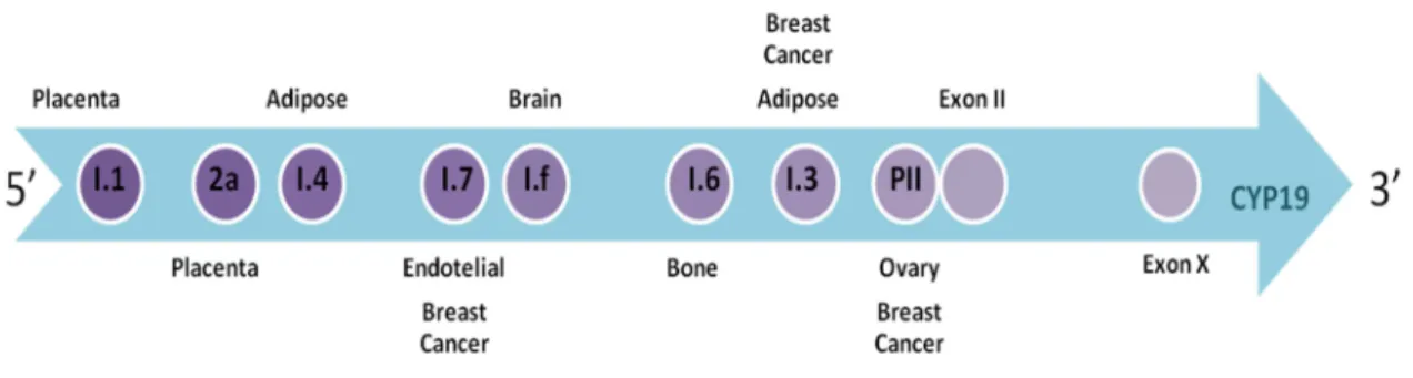
As mentioned, the aromatase is an enzymatic complex composed by a cytochrome P450 and a NADPH-cytochrome P450 reductase (28) and is encoded by the CYP19A1 gene (29) present in chromosome 15q21.1 (30). The coding region of the aromatase gene is composed by nine exons and its expression is regulated by the activation of specific promoters (21, 22, 24), (figure 3).
Figure 3: Aromatase Gene – CYP19
The promoter I.1 is expressed in the placenta, the promoter II in ovaries and the promoters I.3 and I.4 in adipose tissue. Besides I.3, the tumor cells of breast cancer also express promoters I.7 and II (31). Promoters I.f and I.6 are expressed in the brain and
bones respectively (32). There are also other factors that stimulate the expression of aromatase. The regulation of aromatase expression in the ovaries, is mediated by follicle-stimulating hormone (FSH) through a cyclic AMP-dependent pathway, while in adipose tissue the promoter I.4 is regulated by cytokines, such as tumor necrosis factor (TNF-α) (33, 34). Activation of abnormal promoters in breast and fat tissues by the action of malignant epithelial cells will induce the expression of aromatase, what makes that its concentration is higher in women with breast cancer (21, 35-41). Promoters I.3 and II can be activated by prostaglandin E2 (PGE2) (42). The mRNA levels that encode the enzyme aromatase are significantly higher compared to normal tissue where only intervenes promoter I.4 (43).
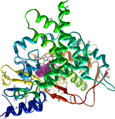
In 1987, Poulos et al presented a proposal for the structure of aromatase based on the similarity with the enzyme found in Pseudomonas putida (44), but, only in 2009, Ghosh et al were able to crystallize this enzyme from human placental microssomes (30) (figure 4).
Figure 4: Tertiary structure of aromatase isolated from human placenta.Colored in dark blue is the N terminus, starting at residue 45, and colored in red is the C terminus ending at residue 496. The α-helices are labelled from A to L and β-strands are numbered from 1 to 10. The heme group and the androstenedione molecule bound at the active site are also shown. Adapted from (30)
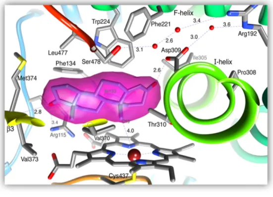
The tertiary structure of aromatase is constituted by 12 α helices and 10 β sheets (30). The androstenedione substrate binds to the heme iron localized in the aromatase active center by action of the carbon 19 resulting in two hydrogen bonds with Asp309 and Met374 residues (45) (figure 5). The supplementary cavity is formed by hydrophobic residues and porphyrin rings that contribute to the binding of enzyme-substrate. The Arg115 residues, Ile133, Phe134, Phe221, Trp224, Ala306, Thr310, Val370, Val373, Met374 and Leu477 residues establish van der Waals bonds with androstenedione (30, 45).
Figure 5: The active site of aromatase. Magenta shows the connection of androstenedione to the active center. Adapted from (45)
2.3. Estrogen Receptors
The discovery of a specific protein, in 1960, by Jensen and Jacobson, responsible for the response of estrogen in target tissues was denominated estrogen receptor (ER) (46). The estrogen receptor exists in two isoforms: ERα and ERβ (47-49) with a 56% homology between them (50). The ER belongs at a large family of nuclear receptors (16) and is synthesized by distinct genes (51), ESR1 and ESR2. The receptor alpha (ERα),
uncovered in 1962 (52), is considered one important receptor for the diagnostic of estrogen dependent breast cancer. The receptor beta (ERβ) was identified in 1996 (53) on prostate and ovaries of rats and on human testicle (54). However, its role in human breast cancer remains unclear (55).
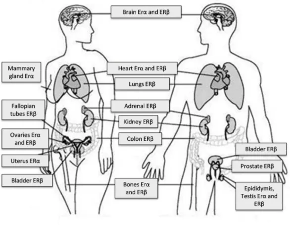
Estrogen receptors (ERs) play a decisive role in the development and function of the reproductive system and mammary gland (51) (figure 6) as well as in cancer development. ERα is expressed mainly in the liver, uterus and mammary gland, while in the lungs, kidneys, colon, gastrointestinal tract, there is a greater abundance of ERβ. In the ovaries, although both ERs are expressed, their cellular distribution is markedly different, since ERα is expressed mostly in the theca and interstitial cells, whereas ERβ is especially expressed in the granulosa cells (51).
Figure 6: Distribution of ERα and ERβ by the different organs
The clinical and in vitro studies allowed the understanding of the importance of these receptors in breast cancer disease (56). The α receptors are expressed in about 70% of breast tumors. Given the function of ER in regulating proliferation and differentiation of normal tissue, the study of cell signaling mechanisms is very important to control these processes in cancer situations. For this reason the ERα is used as a biomarker in the
progression of breast cancer and as an effective therapeutic target (51). Although the ERβ is present in fibroblasts of normal breast adipose tissue in ER positive breast cancer situations its expression decreases with tumor development, which may be related to the potential of these receptors for anti-proliferative action (55). ERβ has a decisive function on the gene expression of several matrix mediators, like the proteoglycans syndecans-2/-4 and serglycin, several matrix metalloproteinases, plasminogen activation system components and receptor tyrosine kinases (57).
The human ERα is a 66 kDa protein that contains 596 amino acids (58) and is located on chromosome 6 (6q25.1) (59). The ERβ has 59 kDa, contains 530 amino acids (60) and is situated on chromosome 14 (14q23.2) (61) (figure 7).
Figure 7: Schematic representation of the human ERα and ERβ structures. Both ERs are characterized by the A/B domain at the N-terminus, which contains the ligand-independent transcriptional activation domain (AF-1), the C domain that represents the DNA-binding domain (DBD) and contains a nuclear localization signal (NLS), the D domain, the E domain that harbors the ligand-binding domain (LBD) and the ligand-dependent transcriptional activation domain (AF-2) and lastly the F domain at the C-terminus. The percentage of homology for both ERs in relation to each domain is represented.
As with other nuclear receptors, ERs have multiple domains consisting in six functional regions. A A/B domain has the ligand-independent activation function (AF-1) (51), a DNA-binding domain (DBD) the C domain, a dimerization region (DR), the D domain, a ligand binding domain (LBD), the E domain (58). The C domain has a structure that allows the dimerization of the receptor and subsequent binding to DNA target sequences (62). The D domain is a region, with only 30% of homology between the ERs. The E region contains the ligand binding domain (LDB), the second nuclear localization signal and a ligand-dependent transcription activation function (AF-2). Lastly, the F
ER α
ER β
Domain/Homology
DR
AF-1
DBD
LBD/AF-2
AF-1
DBD
DR
LBD/AF-2
A/B
18%
C
97%
D
30%
E
47%
F
18%
domain contains the C-terminus wich may be associated with dimerization processes or protein interaction (16, 51).
2.4. Pathways of Estrogen Signaling
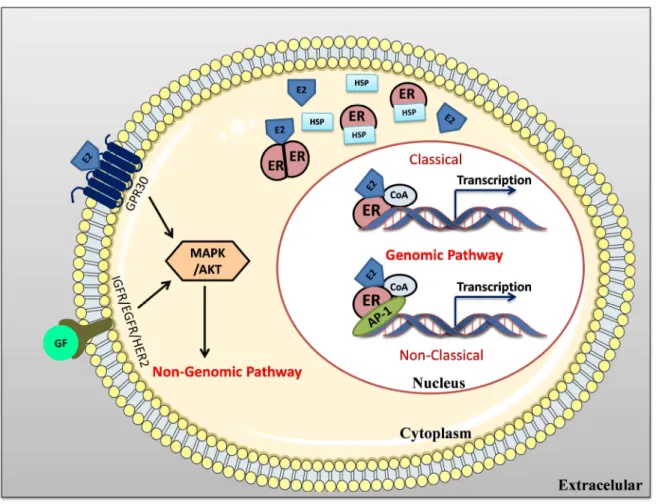
Various studies conducted in the last years have elucidated the molecular gene expression mechanisms regulated by ERs (63). The signaling pathways activated by ERs depend on their intracellularlocalization, since ERs can be localized in nucleus, cytoplasm or in cytoplasmicmembrane (64). There are two different mechanisms involved in the ER activation, the genomic and the non-genomic pathway (figure 8).
The genomic pathway or NISS (nuclear-initiated steroid signaling) may occur in two ways, the classical and non-classical pathways. In the classic or direct mechanism, the estrogens that have crossed the cytoplasmic membrane by diffusion bind to the ER receptor, resulting in its dimerization and activation (65) and allowing the translocation of ER to the nucleus. In the nucleus, in the presence of co-activators (CoA), it binds directly to specific regions of DNA, the estrogen response elements (ERE), located in the promoters of target genes (64). In the non-classic or indirect pathway, the ER binds indirectly to the DNA through other transcription factors, such as activation protein-1 (AP-1) and specificity protein-1 (SP-1). In both pathways, depending on the cell stimulus, the binding of ER promotes the recruitment of co-regulators or co-repressors (63).
The activation of the non-genomic pathway or MISS (membrane-initiated steroid signaling) induces a faster response than the genomic pathway (64). In the non-genomic pathway the ERs can also interact or activate other cell membrane receptors, such as the insulin-like growth factor receptor 1 (IGFR1), the epidermal growth factor receptor 1 (EGFR1) and human epidermal growth factor receptor 2 (HER2). This interaction leads to activation of intra-cytoplasmic kinases, such as phosphatidylinositide 3-kinases (PI3K), mitogen-activated protein kinase (MAPK) and protein kinase B (AKT) (65).
However, it is known that the genomic and non-genomic pathways are not completely independent, but, complementary and even synergistic (65). Several studies have demonstrated the occurrence of “cross-talk” mechanisms between the ER and the signaling pathways of EGFR / HER2 and of the IGFR1. Others studies showed that the G protein-coupled estrogen receptor (GPR30) can also induce the activation of EGFR (66). In addition, kinases can phosphorylate ER, co-activators and other transcription factors causing an increase in gene expression. These mechanisms of “cross-talk” between ER and
growth factors receptors (GFRs) promote cellular proliferation and progression of breast cancer, as well as the development of resistance to endocrine therapies (66, 67).
Figure 8: Schematic representation of ER signaling pathways. In genomic signaling of ER, considering the classical pathway, the ER binds directly to the DNA, to the ERE region, recruiting co-regulators. In the case of non-classical pathway, regulation and gene transcription occurs through receptor interactions with other classes of transcription factors such as AP-1. In the non-genomic signaling pathway the binding of E2 to the ER of citoplasmatic membrane associated to G protein or the binding of GF receptors will activate MAPK / AKT proteins, essential for cell proliferation.
E2-estrogen; ER, estrogen receptor; CoA co-activators; GF-growth factor; EGFR- epidermal growth factor receptor; IGFR- insulin like growth factor receptor; ERE- estrogen-responsive elements; MAPK- kinase activated by mitogenic agents; AKT- protein kinase B; AP-1 Activator protein-1; GPR-30-G-protein coupled receptors.
3. Hormonal therapy in breast cancer treatment
Treatment of breast cancer has advanced significantly in the recent decades (68). The chosen treatment plan depends mainly on the stage of disease, the type of tumor and the general health of the patient. In view of the needs of each patient it may be necessary to choose for one or even a combination of two or more treatments. Surgery and radiotherapy are considered local therapy, while the chemotherapy and hormonal therapy are examples of systemic therapy (69).
Endocrine therapy is considered the standard treatment for the hormone dependent breast cancer (70). Estrogens have important functions in breast cancer, so the development of a treatment able to block the estrogen signalling is a mark in history of breast cancer. Actually, there are two therapeutic strategies used to reduce the action of estrogens on the target organs. The first is the use of ER modulators (SERMs), as tamoxifen, that modulate the binding of estrogens to ER and of selective ER downregulators (SERDs), as fulvestrant that down-regulate ER protein levels. The second strategy involves the aromatase inhibitors (AIs) that bock the conversion of androgens to estrogens by inhibition of the enzyme aromatase (3, 71, 72).
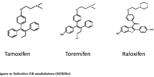
3.1. Selective ER modulators (SERMs) and selective ER downregulators (SERDs)
The SERMs are ER modulators that are partial agonists depending on the target tissue. In some cases have an agonistic effect while in others present an antagonistic activity. Tamoxifen was the first SERM approved through the U.S. Food and Drug Administration (FDA), in 1977, for treatment of breast cancer and in 1998 as quimio-preventive agent for women with high risk of developing this disease (73). More recently, other SERMs were approved, as toremifen and rolexifen (figure 9). FDA approved raloxifen for prevention and treatment of osteoporosis and tamoxifen, toremifen and raloxifen as preventive and treatment for breast cancer (74).
Figure 9: Selective ER modulators (SERMs)
Tamoxifen was the first endocrine therapy for breast cancer. It is prescribed for the treatment in premenopausal women for a period of at least five years, but it may be used in women after menopause supplemented with AIs. This drug is considered an antagonist in the breast tissues, since it blocks the action of estradiol, but also acts as an ER agonist in endometrium and bone, causing endometrial hyperplasia and increased risk of endometrial cancer, as well as the preservation of bone density in postmenopausal women (75). Tamoxifen was considered the treatment of choice during many years, because it can decrease by 50% the risk of recurrence. However, despite the demonstrated effectiveness, it can occur the development of resistance (74).
SERDs, like Fulvestrant, are considered pure antiestrogens (76). Fulvestrant binds to estrogen receptors, leading to rapid degradation of the receptors (77, 78). Several studies in vitro have shown that fulvestrant was more effective than tamoxifen (79). Studies have demonstrated that SERDs are effective in patients previously treated with tamoxifen or AIs (80). The fulvestrant administration route is parenteral, allowing better monitoring of treatment adherence and reduces pharmacokinetic interference from oral administration, by interaction with food or other drugs. Although being a good alternative for patients with advanced breast cancer when other endocrine therapies do not result, one cannot exclude the possible occurrence of resistance to fulvestrant in prolonged treatment situations (80).
3.2. Aromatase Inhibitors (AIs)
Aromatase inhibitors are very effective in inhibiting estrogen synthesis and are divided into two groups based on their chemical structure and mechanism of action, the non-steroidal and steroidal inhibitors (81). Steroidal AIs compete with androstenedione and testosterone to the active site of the enzyme and establish an irreversible binding, resulting in the inactivation and degradation of the enzyme, and for this reason, are also called suicide inhibitors (82). The non-steroids bind reversibly to the enzyme.
According the period of discovery and application, AIs can be divided as first-, second- and third-generation and each consecutive generation presented higher specificity to aromatase and lower adverse effects (83, 84). In 1980 in Europe, appeared the second generation aromatase inhibitors, formestane and fadrozol and a decade later, the use of the third-generation of aromatase inhibitors was approved. Currently, AIs used in clinic are those of the third generation, the non-steroidal, letrozole, anastrozole and the steroidal exemestane (83, 84). These have proven to be an alternative to tamoxifen for the treatment of ER+ breast cancer in postmenopausal women. It has been found further that
this therapy allows a better quality of life in these women (21). Aromatase inhibitors are also effective in the treatment of breast cancer resistant to tamoxifen (21, 85).
AIs showed to be advantageous for patients with breast cancer in postmenopause, because they are better tolerated and have a higher activity compared with tamoxifen (86). AIs inhibit cell proliferation causing retention in the cell cycle and apoptosis (87). This cell death is mediated by several mechanisms and regulated by the expression of different factors and proteins, especially of the Bcl-2 family (88).
Although AIs are considered a good alternative to tamoxifen for the treatment of hormone-dependent breast tumors, some are resistant before the endocrine treatment, due to intrinsic resistance or after an extended period acquired resistance may occur (89). Thus, it is crucial to understand the molecular mechanisms and characteristics of resistance to AIs and tamoxifen, to choose the most effective treatment (90).
There are several mechanisms proposed to explain the resistance to endocrine treatment, namely the dysregulation of various components of the ER signaling pathway, including the loss of expression of ERα; the activation of pathways that may promote cell proliferation and survival alternative stimulus, via EGFR, PI3K/AKT/mTOR pathway; changes in cell cycle and apoptosis and alterations in the epigenetic and microRNAs
modulation. The resistance to endocrine therapy may also be associated with a decrease or absence of the ER function.
Several other factors and pathway shave been referred to be involved in endocrine resistance, like HER2, cyclin E1, the inducible hypoxia factors (HIF) and MAPK pathway (91). The activation of other non-classical pathways may influence the resistance to hormone therapy as they can lead to changes in the expression of activators, co-repressors, and receptor tyrosine kinases (92). Relatively to AIs resistance there are differences between steroidal and nonsteroidal inhibitors. It is considered that the chemical structure of molecules and interaction with the active site of the enzyme may be one of the causes for the divergence in resistance behaviors (93).
Treatment with AIs, especially with exemestane, anastrozole and letrozole, leads to a reduction in estrogen levels, causing a decrease in bone mass resulting in increased bone fragility, which can ultimately induce osteoporosis (94). This disease is induced by treatments leading to premature menopause, chemotherapy, ovarian suppression and anti-estrogen therapies, causing bone loss and an increased fracture risk in breast cancer survivors (BCS) (95-97) Several studies demonstrated that in the first 5 years of natural menopause, it occurs an estimated 0,5-3% annual bone loss (98). However, this bone loss is accelerated with cancer treatments, as the endocrine therapy with AIs, because they effectively deplete residual oestrogen levels (99, 100). This bone loss ranges from 1,6-7,4% per year (98, 101-103) and 1,5-3 times of these women have bone fracture (104-106), which are associated with functional decline, fall risk and varying degrees of morbidity (107). In order to control and prevent the effects of this adverse action of AIs it is necessary the use of supplementary therapy with bisphosphonates administration, calcium and / or vitamin D (108).
Several studies show that treatment with these drugs leads to changes in cholesterol, triglycerides and glucose increasing the risk for cardiovascular diseases. In view of this data it is important to make a preliminary evaluation of other disorders such as heart failure, hypertension, arrhythmia, diabetes and obesity (109). Some studies indicate that therapy with AIs has fewer associated risks compared to tamoxifen, being better tolerated by patients (110).
3.2.1. Non-steroidal AIs
The non-steroidal AIs or Type II (figure 10) establish a non-covalent binding with the heme portion of the aromatase causing the saturation of the binding site (83). Inhibition per this type of AI is reversible and depends on a constant supply of drug (82). Some studies have shown that, given the reversible nature of these inhibitors, the enzyme aromatase activity is recovered after treatment (111).
Aminoglutethimide was the first AI used to endocrine treatment of breast cancer (82). Its introduction for therapeutic purposes occurred in 1960 as anticonvulsant, but, owing to the severe side effects, was withdrawn from the market by the FDA six years later. Its inhibitory capacity of aromatase, preventing the conversion of androgens to estrogens, was described in 1974 (112), however aminoglutethimide is unspecific and, therefore, can inhibit other CYP450 enzymes involved in the biosynthesis of other steroids, such as cortisol (113). In order to minimize the side effects of this inhibitor, its management was done in combination with corticosteroids (89).
Fadrozol a second generation AI (114), was more selective and potent, relatively to aminoglutethimide but also interferes with the biosynthesis of progesterone, aldosterone and corticosterone (115). Due to the lack of selectivity and to its adverse effects, this nonsteroidal inhibitor is not clinically used for the treatment of breast cancer, being replaced by the use of third generation non-steroidal AIs.
Anastrozole (Arimidex ®) a third generation AI is very selective. This compound
has a functional triazol group, which reversibly binds to the enzyme. This aromatase inhibitor has no progestogen, androgenic or estrogenic effect (116). It has an extensive hepatic metabolism by reactions of N-dealkylation, glucuronidation, and hydroxylation. After 72 hours of administration 10% of unchanged drug is excreted in the urine, while 60% is excreted as metabolites (117). Anastrozole has two principal metabolites, the hydroxy-anastrozole and anastrozole-glucuronide (118).
Anastrozole is indicated for the treatment of ER+ breast cancers in advanced or
early stages. The recommended daily dose is 1 mg. Anastrozole reaches peak plasma concentrations 2 hours after oral administration. It is estimated that anastrozol has a time half-life of 50 hours (119). The combination of anastrozole with other treatments requires special care, especially in the case of tamoxifen. Some studies demonstrated that the interaction with tamoxifen leads to a decrease of about 27% of plasma concentrations of anastrozole, thus decreasing its effectiveness (118).
The most common side effects of treatment with anastrozole are weakness, generalized pain, hot flashes, arthralgia, and elevated serum cholesterol levels. Like exemestane and letrozole, anastrozole causes a decrease of bone density as a consequence of the suppression of estrogen. The occurence of bone fractures during treatment with anastrozole is superior relatively to the observed with tamoxifen treatment. In treatment with anastrozole there is a decrease in the occurrence of relapses relative to tamoxifen (117). The resistance to anastrozole is associated with a upregulation of PI3K/AKT pathway and IGF-1 receptor (120)
Finally, letrozole (Femara ®) is the other highly selective third-generation AI,
which has a mechanism of action similar to anastrozole (121). This inhibitor is specific for aromatase, so it causes no changes in plasma concentrations of other hormones during treatment (122). This AI is used in early or advanced stages. In addition, letrozole is used for the treatment of patients with tumor progression after treatment with antiestrogens or in recurrence cases. Its administration is done orally at 2.5 mg daily (123) and it is rapidly and completely absorbed being widely distributed throughout the body. Letrozole has a half-life of 42 hours. The principal route of elimination is by via cytochrome P450, namely CYP3A4 and CYP2A6, with formation of inactive carbinol metabolites (124).
The adverse effects at bone level is very similar to those for anastrozole, being necessary the previous evaluation of bone density. The combination of letrozole with other drugs is another factor to consider, since drug interactions can occur and, consequently, reduce letrozole efficacy. Comparative in vivo studies of other AIs have shown that letrozole, relatively to anastrozole evidenced greater effectiveness. During treatment with letrozole it may occur cardiovascular problems such as thromboembolism (124).
Relatively to biological effects, Thiantanawat et al. demonstrated that both anastrozole and letrozole induced apoptosis in breast cancer MCF-7aro cells by down- regulation of Bcl-2, cyclin D1 and c-myc, up-regulation of Bax, p53 and p21 and activation of caspases-7, -9 and -6. These non-steroidal AIs caused a cell cycle arrest in G1-S phase transition (87, 125). Moreover, other studies have shown that anastrozole and letrozole are better than fulvestrant in suppressing tumor growth (122).
Figure 10: Chemical structure of the non-steroidal AIs aminoglutethimide, fadrozol, anastrozole and letrozole
3.2.2. Steroidal AIs
One of the earliest steroidal AIs used in the treatment of breast cancer was testolactone (figure 11). This AI has a structure similar to testosterone. Studies have shown that testolactone administration is associated with a decrease in serum levels of estrone. This may explain the anti-tumor activity of this steroid inhibitor (126).
The second-generation AI, the formestane or 4-hidroxiandrostenedione, is a selective, specific and effective AI. Formestane is administered intramuscularly, 200 to 500 mg every two weeks (127), but this form of administration causes several adverse reactions especially at administration site and, because of that, formestane is no more used in clinic (128).
Exemestane, discovered in the United States, is the only steroid third generation AI (129), binds irreversibly to the active site of aromatase leading to its modification and degradation (130). Therefore, its administration significantly reduces estrogen levels.
Exemestane (Aromasin ®) is prescribed for the treatment of postmenopausal women diagnosed with breast cancer ER+ in early or advanced stage and in situations
whose progression occurred after treatment with antiestrogens. Clinical studies have shown that suppression of estrogen levels is achieved by oral administration of 25 mg daily, resulting in 98% inactivation of aromatase (131). An oral daily dose of exemestane is rapidly absorbed with maximum plasma concentrations achieved after 2 hours. Its half-life is 27 hours, and then is eliminated by the liver and kidney (132). Its main metabolites are 17β-hydroexemestane and 6-hydroxymetylexemestane, which remain biologically active (133). The 17β-hydroexemestane metabolite has high affinity for the androgen receptor (AR) (84). In general, treatment with exemestane is successful, being possible to observe an improvement in clinical status and survival of patients diagnosed with breast cancer compared to tamoxifen treatment.
Like all AIs, exemestane has adverse effects but compared to tamoxifen the occurrence of thromboembolism (100) and gynecological symptoms are lower (134). It was also shown that this AI does not have major impact on cholesterol and lipoproteins levels (135). Although some studies have demonstrated that it induced the occurrence of bone fractures, others indicated that given its androgenic effect, this AI causes less bone loss than anastrozole or letrozole (84). The occurrence of resistance is another side effect verified during the treatment with exemestane. The mechanisms involved are an increase on MAPK pathway activity (136), a decrease on expression of ER (137) and a promotion of cell cycle progression (138).
In vitro studies demonstrated that exemestane induced antiproliferative effects in MCF-7aro cells, such as cell cycle arrest by blocking G0-G1 and G2-M phase transition and apoptosis. The latter was associated with loss of mitochondrial transmembrane potential, increase in caspases-9 and -7 activities and in reactive oxygen species (ROS) production. Moreover, this study showed that exemestane induced autophagy, though it was suggested that this process acted as a pro-survival mechanism (139).
Figure 11: Chemical structure of the steroidal AIs testolactone, formestane and exemestane
Aim of the study
One of the therapeutic strategies for estrogen receptor-positive (ER+) is the use of
aromatase inhibitors (AIs) that inhibit the enzyme aromatase, which catalyzes the final step of estrogens biosynthesis.
Although, the AIs used in clinic proved to be effective, they cause some serious side effects including bone loss and the development of resistance. For this, the search for novel potent compounds, with fewer side effects, is currently needed. In this sense, we aim to further study in breast cancer cell lines the effects of four new steroidal compounds (57,
58, 59 and 60) selected from a series of compounds, synthesized from structural
modifications on the aromatase substrate that previously demonstrated to be potent AIs in human placental microsomes. We intend to determine the anti-aromatase activity in breast cancer cells and study the biological effects of these compounds in an ER+
aromatase-overexpressing human breast cancer cell line (MCF-7aro cells), and in an ER-
human breast cancer cells (SK-BR-3)
With this study we pretend to contribute to the understanding of the most favorable structure modifications in androstenedione in order to design/synthesize new steroidal compounds as potential AIs. Furthermore, this study can clarify the mechanisms involved in the inhibition of growth and induction of cell death of breast cancer cells using AIs and contribute to the development of more potent compounds with lower side effects.
Chapter II
Materials and Methods
1. Materials
Eagles’s minimum essential medium (MEM), DMEM medium, fetal bovine serum (FBS), L-glutamine, antibiotic-antimycotic (10 000 units/mL penicillin G sodium, 10 000 mg/mL streptomycin sulphate and 25 mg/mL amphotericin B), Geneticin (G418), sodium pyruvate and trypsin were supplied by Gibco Invitrogen Co. (Paisley, Scotland, UK). Testosterone (T), estradiol (E2), ethylenediaminetetracetic acid (EDTA), dimethylsulfoxide (DMSO), tetrazolium salt 3-(4,5-dimethylthiazol-2-yl)-2,5- difenyltetrazolium (MTT), Höechst 33258, Casodex (CDX), propidium iodide (PI), Triton X-100, DNase-free RNase A, staurosporine (STA), charcoal, carbonyl cyanide m-chlorophenylhydrazone (CCCP), 2′,7′-dichlorodihydrofluorescein diacetate (DCDHF2-DA), phorbol 12-myristate 13-acetate (PMA), Trypan blue, protease inhibitor cocktail, Fluoroshield mounting medium, dextran and formestane were from Sigma–Aldrich Co. (Saint Louis, USA). Giemsa stain was purchased from Merck (Damnstadt, Germany). DPX mounting medium was from VWR (Radnor, PA, USA). Caspase-Glo® 3/7, Caspase-Glo® 9 luminometric assays, Cyto-Tox 96 nonradioactive cytotoxity assay kit and Reporter Lysis buffer were from Promega Corporation (Madison, USA). [1β-3H] androstenedione was
obtained from Perkin-Elmer (Boston, MA, USA) and liquid scintillation cocktail Universol from ICN Radiochemicals (Irvine, CA, USA). Bradford assay reagent was from Bio-Rad (Laboratories Melville, NY, USA). Exemestane was from Sequoia Research Products Ltd. (Pangbourne, UK). Z-VAD-FMK was from BD Biosciences Pharmingen (San Diego, CA, USA). Rabbit polyclonal β-tubulin, goat polyclonal CYP19, goat anti-rabbit IgG and mouse anti-goat IgG antibodies were from (Santa Cruz Biotechnology, CA, USA).
2. Compounds
In this work we focused on the biological evaluation of four new potential steroidal AIs (57, 58, 59 and 60) synthesized from structural modifications on the aromatase substrate, androstenedione. These compounds were synthesized by the Pharmaceutical Chemistry Group of the Faculty of Pharmacy, University of Coimbra and CNC.IBILI, University of Coimbra by the Profs. Carla Varela, Elisiário Tavares da Silva, Fernanda M.F. Roleira and Saul Costa.
3. Preparation of the AIs, Testosterone and Estradiol
The AIs were dissolved in DMSO and stored at -80 ºC. Testosterone (T) and Estradiol (E2) were prepared in absolute ethanol at 10 μM and stored at -20 ºC. For each assay the
stock solution of compounds was diluted with the cultured medium to obtain the final working concentrations.
4. Cell cultures
The ER+ aromatase-overexpressing human breast cancer cell line (MCF-7aro cells) was
prepared by stable transfection of MCF-7 cells with human placental aromatase gene and Geneticin selection. Cells were grown in 75 cm2 culture flasks with Eagle’s minimum
essential medium (MEM) with phenol-red supplemented with Earle’s salts, 10% heat-inactivated fetal bovine serum (FBS), 1% of sodium pyruvate (1 mmol/L), 2 mmol/L of glutamine, 1% penicillin-streptomycin-amphotericin B and G418 (700 ng/mL). Three days before the experiments, MCF-7aro cells were cultured with 1% of sodium pyruvate (1 mmol/L), 2 mmol/L of glutamine, 1% penicillin-streptomycin-amphotericin B in steroid-free MEM without phenol red with 5% pre-treated charcoal heat-inactivated fetal bovine serum (CFBS) to prevent the estrogenic effects of phenol-red and the interference of steroids present in FBS. MCF-7aro cell line was kindly provided by Dr. Shiuan Chen from the Beckman Research Institute, City of Hope, Duarte, CA, U.S.A.
The ER− human breast cancer cell line, SK-BR-3 (ATCC®), was maintained in MEM
with phenol-red, supplemented with Earle’s salts, 1% of sodium pyruvate (1 mmol/L), 1% penicillin–streptomycin–amphotericin B, 2 mmol/L of glutamine and 10% heat-inactivated fetal bovine serum (FBS).
The human fibroblast cell line, HFF-1 (ATCC®), was maintained in DMEM without phenol-red, 10% heat-inactivated FBS, 1% sodium pyruvate (1 mmol/L) and 1% penicillin-streptomycin-amphotericin B.
The culture medium was renewed every three days and the cells were maintained at 37 ºC and 5% CO2 atmosphere, to ensure that there is a significant cell proliferation and
stable nutritional levels. After reaching 80 to 90% confluence, the cells were washed with PBS and detached with trypsin/EDTA 1 nM for 2 minutes at 37 ºC and 5% CO2
atmosphere. After, the cells were centrifuged in culture medium with FBS or CFBS to inactivate trypsin at 260 xg and 4 ºC for 5 minutes. Finally, and depending on the culture
dishes, the cells were resuspended with the necessary amount of culture medium, and after homogenization, the cells were counted in a Neubauer chamber and cultured. The culture medium and drugs were refreshed every three days.
5. Preparation of charcoal heat-inactivated fetal bovine serum (CFBS)
FBS (500 ml) was incubated with 8 g of activated charcoal for 24 hours at room temperature, followed by successive centrifugations at 4000 xg for 15 min for removing the steroids present in FBS. Between each centrifugation the supernatants were filtered to eliminate charcoal particles. After the final centrifugation, supernatant was filtered by a vacuum system through a filter cellulose acetate with a 0.22 μm pore size aliquoted and kept at -20 ºC.
6. Preparation of charcoal pellets
Charcoal pellets were previously prepared by adding 1 mL of a solution constituted by 0,5% of dextran and 5% of activated charcoal solution to eppendorf tubes and centrifugation at 14000 xg for 10 minutes. After the supernatant was discarded and the pellets were dry at 37 ºC overnight.
7. In cell aromatase assay
The inhibition of the activity of aromatase enzyme for each compound on MCF-7aro cells was determined according to the method of Thompson & Siiteri (140) and Zhou et al. (141) with some modifications. This assay is based on the use of [1β-3H]
andro-4-ene-3,17-dione as aromatase substrate. In this assay it was measured the tritiated water released from the substrate during the aromatization process. Each experiment included a negative control, which does not contain the potential inhibitors. Formestane (1 µM) and exemestane (10 µM) were used as a positive control.
MCF-7aro cells were cultured in 24-well plates at a cell density of 1×10⁶ during 3 days.
After this time, the medium was removed and cells were washed two times with PBS. Then, it was added 500 nM of progesterone to suppress the activity of 5α-reductase that also uses androgen as a substrate, 50 nM of [1β-3H] androstenedione and 10 μM of
formestane, exemestane or of the new compounds to a final volume of 500 μl of MEM. The [1β-3H] androstenedione was the last component to be added. Cells were incubated at
37 ºC and 5% CO2 atmosphere during 1 hour.
After incubation, the aromatization reaction was stopped with 100 μL of 20% trichloroacetic acid. Then the supernatants were transferred to eppendorf tubes, previously prepared with a pellet of activated charcoal, followed by an incubation period of 1 hour at room temperature. After centrifugation at 14000 xg for 10 minutes, 500 μl of supernatants were transferred to new eppendorf tubes with activated charcoal, homogenized and incubated for 10 minutes at room temperature. The tubes were centrifuged at 14000 xg for 10 min and the supernatant was transferred to clean eppendorfs followed by the last centrifugation cycle at 14000 xg for 5 min. To tubes contains 3 ml of scintillation cocktail it was added 300 μl of supernatant. The counts per minute were read in a scintillation counter (LS 6500, Beckman Instruments, CA, U.S.A.). Cells treated with 10 μM of formestane and exemestane were considered as positive control and untreated cells as control. This assay was performed in two independent experiments in triplicate for each compound.
The cells grown on 24 wells plates were lysed with 500 μl of 0.5 N NaOH and incubated overnight at room temperature with stirring to extract the proteins to quantification. Cells were then freezed at -80 ºC. The protein content was quantified by the Bradford method and used to normalize the radioactivity determined per μg of protein.
8. Cell viability assay
The effects of each steroidal compound (57, 58, 59 and 60) on cell viability of MCF-7aro, SK-BR-3 and HFF-1 cell lines was determined by measuring the mitochondrial reductases activities using the tetrazolium salt, 3-(4,5-dimethylthiazol-2-yl)-2,5-difenyltetrazolium bromide (MTT) that forms a blue formazan precipitate. Thereby, this colorimetric assay provides a percentage of cells capable to convert MTT to formazan. The cells were cultured in 96-well plates and incubated during 3 and 6 days with different concentrations of each compound. Untreated cells were used as control.
MCF-7aro cells were cultured at a cellular density of 2x10⁴ and 1x10⁴ cells/mL (for 3
and 6 days, respectively), in MEM without phenol-red with 5% CFBS and 1 nM of T or 1 nM of E2. SK-BR-3 cells were cultured at a cellular density of 2.5x10⁴ and 1x10⁴ cells/mL
(for 3 and 6 days, respectively), in MEM with phenol-red containing 10% FBS . HFF-1 cells were cultured for 6 days at a cellular density of 7.5x10³ cells/mL in DMEM without
phenol-red containing 10% FBS. After the incubation period (3 or 6 days) it was added 20 μL of MTT (0.5 mg/mL) per well and cells were incubated during 2 hours and 30 minutes in optimal conditions for growth. After this time, the medium was removed and it was added 200 μL DMSO: isopropanol mixture (3:1), to stop the reaction and dissolve the purple formazan precipitated, for 20 minutes with agitation at room temperature. The formazan was quantified spectrophotometrically at 540 nm.
The cytolitic effects of each steroidal compound were evaluated in MCF-7aro cell line using the lactate dehydrogenase (LDH) assay. It is a cytosolic enzyme that is only released from cells to the culture medium after membrane disruption. LDH activity was measured using a LDH KIT (CytoTox 96 Non-Radioactive Cytotoxicity Assay, Promega Corporation) according to manufacturer’s protocol. This assay was performed in the same experimental conditions described for the MTT assay.
9. Western blot analysis
MCF-7aro cells were cultured in 6-well plates at a cellular density of 6x10⁵ cells/mL
during 3 days in MEM with red phenol. Then, cells were treated with each compounds (10 μM) and incubated during 8 hours. After the incubation period, cells were washed two times with PBS and lysed with RIPA buffer (50 mM Tris-HCl, 1% Triton X-100, 0.1% SDS, 150 mM NaCl, 2 mM EDTA, 0.5% Na-deoxycholate, pH 7.4) and 1% of a protease inhibitors cocktail. After, cells were scrapped and collected to eppendorf tubes and centrifuged at 14000 xg for 10 minutes at 4 ºC. Protein concentrations were determined using the Bradford assay. A total of 50 μg of protein per sample was loaded on 10% SDS-polyacrylamide gels and transferred to nitrocellulose membranes in 25 mM Tris-HCl, 250 mM glycine and 18% methanol. The membranes were blocked with 5% milk in TBS TWEEN (1x) during 1 hour. Immunodetection was performed using goat polyclonal antibody anti-CYP19 (1:100) (Santa Cruz Bio Technology, Inc.) in blocking solution overnight at -4 ºC. The secondary rabbit anti-goat antibody (1:2000) was incubated for 1 hour. Membranes were exposed to chemiluminescence substrate Super Signal West Pico and then visualized by Chemidoc Touch Image (BioRad, Laboratories Melville, NY, USA). Membranes were then stripped and incubated with rabbit polyclonal anti-β-tubulin antibody (1:500) followed by goat anti-rabbit IgG secondary antibody (1:1000), to control loading variations. Untreated cells were used as control.