Baroreflex function in conscious rats
submitted to iron overload
Departamentos de 1Ciências Biológicas, NUPEB, and 2Alimentos,
Escola de Nutrição, Universidade Federal de Ouro Preto, Ouro Preto, MG, Brasil
3Departamento de Fisiologia e Biofísica, Instituto de Ciências Biológicas,
Universidade Federal de Minas Gerais, Belo Horizonte, MG, Brasil
4Departamento de Fisiologia, Escola Paulista de Medicina,
Universidade Federal de São Paulo, São Paulo, SP, Brasil L.M. Cardoso1,
M.L. Pedrosa1,
M.E. Silva2,
M.F.D. Moraes3,
E. Colombari4 and
D.A. Chianca-Jr.1
Abstract
Our hypothesis is that iron accumulated in tissue, rather than in serum, may compromise cardiovascular control. Male Fischer 344 rats weigh-ing 180 to 220 g were divided into 2 groups. In the serum iron overload group (SIO, N = 12), 20 mg elemental iron was injected ip daily for 7 days. In the tissue iron overload group (TIO, N = 19), a smaller amount of elemental iron was injected (10 mg, daily) for 5 days followed by a resting period of 7 days. Reflex heart rate responses were elicited by iv injections of either phenylephrine (0.5 to 5.0 µg/kg) or sodium nitro-prusside (1.0 to 10.0 µg/kg). Baroreflex curves were determined and fitted to sigmoidal equations and the baroreflex gain coefficient was evaluated. To evaluate the role of other than a direct effect of iron on tissue, acute treatment with the iron chelator deferoxamine (20 mg/kg, iv) was performed on the TIO group and the baroreflex was re-evaluated. At the end of the experiments, evaluation of iron levels in serum confirmed a pronounced overload for the SIO group (30-fold), in contrast to the TIO group (2-fold). Tissue levels of iron, however, were higher in the TIO group. The SIO protocol did not produce significant alterations in the baroreflex curve response, while the TIO protocol produced a nearly 2-fold increase in baroreflex gain (-4.34 ± 0.74 and -7.93 ± 1.08 bpm/mmHg, respectively). The TIO protocol animals treated with deferoxamine returned to sham levels of barore-flex gain (-3.7 ± 0.3 sham vs -3.6 ± 0.2 bpm/mmHg) 30 min after the injection. Our results indicate an effect of tissue iron overload on the enhancement of baroreflex sensitivity.
Correspondence
D.A. Chianca-Jr. ICEB2
Universidade Federal de Ouro Preto Campus Universitário
35400-000 Ouro Preto, MG Brasil
Fax: +55-31-3559-1680 E-mail: chianca@nupeb.ufop.br
Research supported by FAPEMIG and CAPES.
Publication supported by FAPESP.
Received December 19, 2003 Accepted October 29, 2004
Key words
•Baroreflex •Iron overload •Heart rate •Blood pressure •Iron dextran •Deferoxamine
Introduction
The cardiovascular system has the cru-cial task of supplying all body cells with their metabolic needs. The complexity of this func-tion demands efficient control mechanisms for maintaining blood pressure and flux within adequate levels in every region of the body.
known to alter the responsiveness of the baroreflex response (2).
Iron overload is associated with several pathophysiological states (e.g., hemoglobino-pathies, hemolytic anemia, hemochromato-sis) and behaviors such as smoking and ex-cessive iron and vitamin C intake (3). Be-sides the epidemiologic concern regarding dietary iron intake, iron accumulation and its relation to pathological processes is still the main focus of many heated debates (4). Nev-ertheless, iron overload has undeniably been associated with cardiac and vascular dys-functions in experimental conditions (5-7).
Experiments conducted ex vivo have shown
that iron overload induces a condition of heart failure (8) in Langendorff preparations of isolated hearts. In fact, deposition of iron into the heart is emerging as an important cause of heart failure (6). However, a de-tailed study of cardiovascular reflex
involve-ment in an in vivo iron overload model has
not yet been reported. Such an investigation could contribute to the use of iron overload as an animal model for studying heart fail-ure.
Iron has been associated with the modu-lation of the metabolism and availability of certain chemical mediators such as nitric oxide (9-11) and carbon monoxide (12), which are implicated in baroreflex function both peripherally and centrally (13-15). Ni-tric oxide as well as oxygen-derived free radicals induced by changes in iron concen-tration are known to suppress baroreceptor activity particularly at high levels of arterial pressure (15). Recent studies have demon-strated alterations in baroreflex function aris-ing from processes that produce free radicals such as atherosclerosis, in which the barore-flex gain coefficient was shown to be de-creased (16). Although cardiac insufficiency is more closely related to the inadequacy of the heart to function as a pump, dysfunction of neural feedback mechanisms (e.g., baro-reflex) has been associated with chronic heart failure. Increased sympathetic activity and
plasma levels of norepinephrine, parasym-pathetic withdrawal and impaired baroreflex gain coefficient have been reported in chronic heart failure (17). An increased baroreflex gain coefficient has been shown in rats with myocardial infarction (18). Very few data are available regarding the prognostic impli-cations of baroreflex sensitivity and heart rate (HR) variability for chronic heart fail-ure. Nevertheless, both baroreflex sensitivi-ty and HR variabilisensitivi-ty have been shown to be markedly reduced in chronic heart failure and significantly associated with the degree of ventricular dysfunction and with further progression of the severity of the disease (17). Thus, the impairment of baroreflex function in patients with heart failure is a well-established association. In fact, the evaluation of baroreflex sensitivity has been proposed as a diagnostic tool in such cases (19).
Our hypothesis is that iron overload could affect baroreflex function through iron accu-mulation in the heart tissue. Therefore, the net effect of iron overload on the baroreflex, as demonstrated in the reports cited above, may not be related to neural control but rather to heart impairment. The present ex-periment was designed to compare the effect of circulating iron overload to that of iron accumulated in tissues on the baroreflex of conscious Fischer 344 rats. The effect of acute iron chelator treatment (deferoxam-ine) in the issue iron overload group de-scribed above was also tested.
Material and Methods
Animals and iron overload
3ºC). Animals were randomly divided into the following groups: serum iron overload group (SIO), tissue iron overload group (TIO) (20), and respective controls. In the SIO protocol (N = 12), 20 mg elemental iron (0.2 ml iron dextran; Sigma, St. Louis, MO, USA)
was injected ip daily for 7 days, immediately
followed by baroreflex evaluation. In the TIO protocol (N = 19), less elemental iron was injected (10 mg, daily) for a shorter period of time (5 days), followed by a resting period of 7 days, after which the experi-ments were conducted. Sham animals (N = 31) received placebo injections (PBS) ac-cording to their respective iron-loaded groups. Seven animals from the TIO proto-col, along with their respective controls (N = 7), were separated to receive deferoxamine mesylate (DFO, 20 mg/kg; Novartis AG, Basel, Switzerland), in order to determine the effect of permanent changes in barore-flex function compared to the effect of the direct action of iron. Efforts were made to avoid any unnecessary distress to the ani-mals, in accordance with the Brazilian So-ciety for Neuroscience and Behavior Guide-lines for Animal Experimentation.
Surgical procedures and hemodynamic measurements
Under tribromoethanol anesthesia (250
mg/kg, ip; Aldrich Chemical Company, Inc.,
Milwaukee, WI, USA) a heat-pulled taper-ending polyethylene catheter (PE-10 con-nected to PE-50; Clay Adams, Parsippany, NJ, USA) filled with heparinized PBS (125 U/ml) was positioned inside the aorta through the left femoral artery for measurement of pulsatile arterial pressure. A second catheter was inserted into the inferior vena cava through the left femoral vein for systemic drug administration. The free endings of both catheters were tunneled subcutaneously and exteriorized through the back of the neck and connected to a swivel during the experi-ments. Pulsatile arterial pressure was
meas-ured with a pressure transducer (model MLT0699; ADInstruments Pty Ltd., Castle Hill, NSW Australia) connected to an ana-log-to-digital data acquisition system (mo-del PowerLab 400; ADInstruments). Data were sampled at 12 bits using a 200-Hz sampling rate. HR and mean arterial pres-sure (MAP) were derived off-line from pul-satile arterial pressure using the Chart for Windows software, version 4.1.2 (ADInstru-ments). HR and MAP variability was esti-mated by calculating the standard deviation of HR and MAP values within the recording for each animal. All experiments were per-formed approximately 24 h after surgery on unanesthetized freely moving rats.
Baroreflex activation
Before the experiments, rats were instru-mented and allowed to adapt to the new environment for at least 30 min. A 20- to 30-min recording period without any interfer-ence was allowed to elapse in order to deter-mine baseline MAP and HR values.
A typical recording procedure consisted of continuously monitoring HR and MAP while performing intravenous bolus injec-tions of random doses of either phenyleph-rine (0.5, 2.0, 3.5, and 5.0 µg/kg; Sigma) or sodium nitroprusside (1.0, 4.0, 7.0, and 10.0 µg/kg; Sigma). The methodology has been described in detail elsewhere (18). Succes-sive drug administration was carried out only after MAP and HR returned to baseline.
to a sigmoidal logistic equation:
HR = HRmax +
HRmin - HRmax Eq. 1
- MAP - MAP50
1 + e b
where HRmin is the lower plateau, HRmax is
the upper plateau, b is the curvature
coeffi-cient, and MAP50 is the MAP at the midpoint
of the HR range.
Individual sigmoidal functions were av-eraged in order to determine the mean sig-moidal fit for the group. The derivatives of individual sigmoidal baroreflex curves were calculated and averaged within an experi-mental group in order to determine the baro-reflex gain-coefficient curve.
Acute deferoximine treatment
Experiments with the chelator DFO were carried out on 7 animals from the TIO proto-col and respective controls (N = 7). The baroreflex was evaluated by intravenous bo-lus injection of sodium nitroprusside (1.0
and 4.0 µg/kg; Sigma) before and after iv
infusion of DFO (20 mg/kg). The maximal changes in MAP and HR were evaluated and the gain coefficient was calculated by the equation:
Gain = ∆maxHR Eq. 2
∆max MAP
where ∆maxMAP is the maximal change in
MAP after injection of sodium nitroprusside
and∆maxHR is the maximal change in HR
due to the changes in MAP caused by the pharmacological maneuver.
Determination of iron status
At the end of the experiments, a blood sample was taken through the arterial can-nula and centrifuged and serum separated. Serum iron concentrations were determined in non-hemolyzed serum samples by spec-trophotometric analysis using commercially
available kits (# 38; Labtest, Belo Horizonte, MG, Brazil). Immediately after blood sam-pling, the animals were sacrificed with ethyl ether and perfused with saline and formalde-hyde. The liver and spleen were removed and weighed and liver and spleen samples
were homogenized in HNO3 at 120ºC. After
evaporation of the acid, the dry residue was resuspended in 50% hydrochloric acid (v/v) and quantified by colorimetric analysis us-ing orthophenanthroline (21).
Statistical analysis
Data are reported as means ± SEM. The baroreflex curves were analyzed by one-way analysis of variance (ANOVA) for repeated
measures. The unpaired Student t-test was
used to determine statistically significant dif-ferences between the SIO and TIO
experi-mental groups. The paired Student t-test was
used to determine the statistically significant differences in the TIO baroreflex gain coef-ficient before and after DFO treatment. The level of significance was set at P < 0.05 in all analyses.
Results
Iron status and baseline cardiovascular parameters
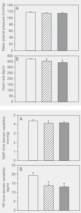
The iron status of the animals treated with iron dextran confirmed our expecta-tions regarding the effect of serum and tissue iron overload on the baroreflex response (Table 1), i.e., the SIO group presented very high serum levels of iron (29-fold) com-pared to the modest increase observed in the TIO group (1.8-fold). On the other hand, tissue accumulation (e.g., spleen and liver) of iron was much more evident in the TIO group, even though less iron was injected when compared to the SIO animals.
Both iron-treated groups showed a slight, but significant, bradycardia compared to the
sham group (SIO protocol: 341 ± 4 vs 373 ± 5
bpm; TIO protocol: 354 ± 7 vs 373 ± 5 bpm). However, baseline MAP values were not sig-nificantly different between groups (Figure 1). A slight decrease in pulse pressure was observed in TIO rats compared to sham rats
(37.9 ± 0.92 vs 40.5 ± 0.85 mmHg, P =
0.034; data not shown). SIO rats did not present alterations in pulse pressure when
compared with sham rats (37.0 ± 0.89 vs
38.8 ± 0.94 mmHg, P = 0.186; data not shown). The HR time domain variability was decreased in both iron-loaded groups compared to the sham group (SIO protocol:
14 ± 1 vs 19 ± 2 bpm; TIO protocol: 13 ± 1 vs
19 ± 2 bpm). No statistically significant dif-ferences were observed in MAP time do-main variability (Figure 2).
Baroreflex
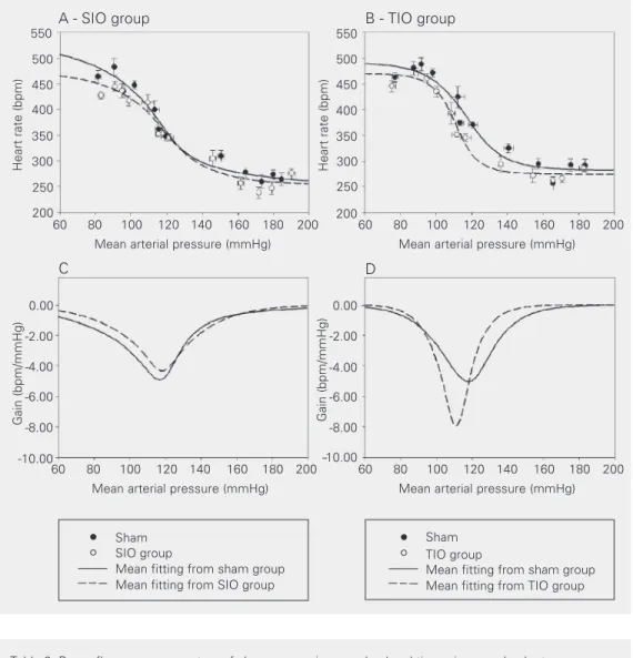
The upper panels in Figure 3 show the MAP against HR baroreflex curve, along with the mean sigmoidal fitting, for SIO (Figure 3A) and TIO (Figure 3B) rats and their respective controls. The baroreflex gain coefficient versus pressure plot is reported in the lower panels in Figure 3 for both SIO (Figure 3C) and TIO (Figure 3D) animals.
The baroreflex curve for the TIO group was shifted to the left (Figure 3B,D), closely following the changes in resting MAP (Figure
1), as indicated by the MAP50 value. No
sig-nificant changes were observed in the barore-flex curve of SIO rats compared to control (Figure 3A,C). Maximal reflex tachycardia (upper plateau) and bradycardia (lower pla-teau), and consequently HR range, for the two iron-treated groups did not differ statistically from control (Table 2). However, a tendency to a reduction in both upper plateau and HR range was noted in SIO rats when compared to sham rats (Table 2, Figure 3A).
TIO rats presented an increased barore-flex gain-coefficient (1.6-fold over control) with a concomitant narrowing of the curve accompanied by a shift to lower MAP values (Table 2, Figure 3B,D).
MAP time domain variability
(mmHg) 5 4 3 2 1
HR time domain variability
(bpm) 25 20 15 10 5 0 0 12345 12345 12345 12345 12345 12345 12345 12345 12345 12345 12345 12345 12345 12345 12345 12345 12345 12345 12345 12345 12345 12345 12345 12345 12345 12345 12345 12345 12345 * *
Figure 2. Time domain variabili-ty for mean arterial pressure (A) and heart rate (B) in sham (open bar, N = 24), tissue iron over-load (hatched bar, N = 19) and serum iron overload (filled bar, N = 12) rats over a period of 10 min at the beginning of the re-cordings. Data are reported as means ± SEM. MAP = mean arterial pressure; HR = heart rate. *P < 0.05 compared to the sham group (Student t -test).
Table 1. Iron status of sham, serum iron overload and tissue iron overload rats.
Parameter Sham SIO TIO
Serum iron (µmol/l) 35.6 ± 1.97 1032.3 ± 209.68* 64.9 ± 3.94*+ Liver iron (µmol/organ) 46.6 ± 9.49 91.3 ± 15.8* 164.7 ± 10.74*+ Spleen iron (µmol/organ) 20.6 ± 1.20 19.7 ± 2.00 30.1 ± 1.20*+
Data are reported as means ± SEM for 8 rats in each group. SIO = serum iron overload; TIO = tissue iron overload.
*P < 0.05 compared to the sham group; +P < 0.05 compared to the SIO group (Student t-test).
Mean arterial pressure (mmHg)
140 120 100 80 60 40 20 0
Heart rate (bpm)
400 350 300 250 200 150 100 50 0 12345 12345 12345 12345 12345 12345 12345 12345 12345 12345 12345 12345 12345 12345 12345 12345 12345 12345 12345 12345 12345 12345 12345 12345 12345 12345 12345 12345 12345 12345 12345 12345 12345 12345 12345 A * *
Figure 1. Baseline mean arterial pressure (A) and heart rate (B) of sham (open bar, N = 24), tis-sue iron overload (hatched bar, N = 19) and serum iron over-load (filled bar, N = 12) rats over a period of 10 min at the begin-ning of the recordings. Data are reported as mean ± SEM. *P < 0.05 compared to the sham group (Student t-test).
B
A
Heart rate (bpm)
550
500
450
400
350
300
250
-10.00 200
60 80 100 120 140 160 180 200
Mean arterial pressure (mmHg)
60 80 100 120 140 160 180 200
Mean arterial pressure (mmHg)
B - TIO group
Heart rate (bpm)
550
500
450
400
350
300
250
200
Gain (bpm/mmHg)
0.00
-2.00
-4.00
-6.00
-8.00
-10.00
Gain (bpm/mmHg)
0.00
-2.00
-4.00
-6.00
-8.00
60 80 100 120 140 160 180 200
Mean arterial pressure (mmHg)
60 80 100 120 140 160 180 200
Mean arterial pressure (mmHg)
A - SIO group
D C
Sham SIO group
Mean fitting from sham group Mean fitting from SIO group
Sham TIO group
Mean fitting from sham group Mean fitting from TIO group Figure 3. The upper panels (A
and B) show peak values of mean arterial pressure and heart rate after baroreflex stimulation with phenylephrine and sodium nitruprusside. The broken lines and open circles represent iron-treated rats and the continuous line and filled circles represent sham rats. The data for serum iron overload (SIO) rats are illus-trated in the panels on the left (A and C) and the data for tissue iron overload (TIO) rats are illus-trated in the panels on the right (B and D). The logistic sigmoi-dal-fitting baroreceptor curve is the mean of ten sigmoids fitted from the data for conscious SIO rats (N = 12), TIO rats (N = 12) and respective sham (N = 12) rats. The lower panels indicate baroreflex gain, e.g., the first de-rivative of the sigmoid functions, at specific mean arterial pres-sure values.
Table 2. Baroreflex curve parameters of sham, serum iron overload and tissue iron overload rats.
Parameter SIO TIO
Sham Iron treated Sham Iron treated
Upper plateau (bpm) 528 ± 27 473 ± 11 492 ± 11 469 ± 9
Lower plateau (bpm) 253 ± 10 254 ± 5 282 ± 5 274 ± 5
Heart rate range (bpm) 275 ± 27 218 ± 9 210 ± 13 196 ± 9
MAP50 (mmHg) 114 ± 4 117 ± 3 117 ± 1 111 ± 1*
Gain (bpm/mmHg) -4.91 ± 0.69 -4.34 ± 0.74 -5.03 ± 0.39 -7.93 ± 1.08*
Data are reported as means ± SEM for 12 rats in each group. SIO = serum iron overload; TIO = tissue iron overload; MAP50 = mean arterial pressure at the midpoint of the heart rate range.
*P < 0.05 compared to the sham group (Student t-test).
Effects of acute deferoximine treatment
The upper panel in Figure 4 represents the gain coefficient of the baroreflex
(Figure 4, lower panel; copied from Figure 3B indicating the MAP range).
Infusion of the free iron chelator (DFO) reduced the baroreflex gain values of the TIO rats (Table 3) to control levels (5.28 ±
0.86 vs 3.57 ± 0.27 bpm/mmHg) but did not
affect the baroreflex gain coefficient in sham
animals (3.77 ± 0.40 vs 3.66 ± 0.41 bpm/
mmHg) when baroreflex was evaluated by sodium nitroprusside injection.
Discussion
In this study, we sought to determine whether iron overload produced by intraper-itoneal injections of iron dextran affects baro-reflex function in conscious rats. The iron profile of SIO and TIO rats confirmed our expectation (Table 1) of an experimental model that would compare the effect of high serum iron levels with the long-term effects of iron accumulated in tissue. The results demonstrated that the baroreflex gain was in-creased in TIO rats (Figure 3), whereas no differences in baroreflex function were ob-served in SIO rats. Furthermore, the data indi-cate a direct effect of iron in tissue, since DFO, a chelator, was able to reverse the phenome-non (Figure 4). Therefore, the present data support the view that iron overload plays a role in the functional changes regarding the effec-tor components of baroreflex function, as suggested previously by others (22).
Gain (bpm/mmHg)
0.00
-1.00
-2.00
-3.00
-4.00
-5.00
-6.00
-7.00
12345 12345 12345 12345 12345 12345 12345 12345 12345 12345 12345 12345 12345 12345 12345 12345 12345 12345 12345
12345 12345 12345 12345 12345 12345 12345 12345 12345 12345 12345 12345 12345 12345 12345 12345 12345 12345
* **
550
500
450
400
350
300
250
200
Heart rate (bpm)
60 80 100 120 140 160 180 200
Mean arterial pressure (mmHg) Sham
(MAP range: 90 a 115 mmHg)
TIO rats
(MAP range: 90 a 119 mmHg)
Figure 4. General baroreflex gain elicited before (filled bar) and after (hatched bar) deferox-amine infusion (20 mg/kg, over 20 min). Left bars indicate sham rats and right bars indicate tissue iron overload (TIO) rats. The bottom panel is a copy of Figure 3B with the marked area showing the linear region of the baroreflex curve in which baroreflex was tested in the deferoxamine experiment. *P < 0.05 compared to the sham group; **P < 0.05 compared to the TIO rats before deferoxamine infusion (Student t-test).
Table 3. Changes in heart rate, mean arterial pressure and mean gain values of baroreflex evaluated by sodium nitroprusside before and after deferoxamine (20 mg/kg) infusion in sham and tissue iron overload rats.
Group Before DFO After DFO
1.0 µg/kg NP 4.0 µg/kg NP 1.0 µg/kg NP 4.0 µg/kg NP
∆HR ∆MAP Gain ∆HR ∆MAP Gain ∆HR ∆MAP Gain ∆HR ∆MAP Gain
Sham 79 ± 6 -20 ± 2 4.24 ± 0.43 94 ± 5 -29 ± 2 3.30 ± 0.29 81 ± 5 -21 ± 2 4.02 ± 0.45 89 ± 7 -28 ± 2 3.30 ± 0.34 TIO 86 ± 11 -14 ± 1* 6.53 ± 1.01 89 ± 9 -22 ± 2 4.08 ± 0.24 85 ± 8 -26 ± 3 3.44 ± 0.32 99 ± 11 -27 ± 2 3.71 ± 0.26
Data are reported as means ± SEM for 7 rats in each group. HR = heart rate; MAP = mean arterial pressure; DFO = deferoxamine mesylate; NP = sodium nitroprusside; TIO = tissue iron overload.
Chronic iron overload produces heart dysfunction in a dose-dependent manner (8), probably resulting from the time lag neces-sary to deposit iron in tissues. In the heart, iron, initially deposited in the epicardium, has a long term-effect on transmural wall thickness (23,24). Such long-term effects do not seem to have occurred in TIO rats since DFO was able to acutely reverse the effect of iron overload on the baroreflex gain coeffi-cient. The interpretation of the short-term and long-term effects of iron in the present study is compromised by the lack of pub-lished information concerning acceptable levels of iron in the rat. Neither the border-line levels of iron nor the exposure time needed in order to produce iron-related dys-functions in the cardiovascular system have been reported. Nevertheless, several reports have demonstrated the possible role of car-diac iron deposition in the development of cardiomyopathy and heart failure within the iron overload paradigm (23,25).
On the basis of the Haber-Weiss and Fenton reactions, iron can produce hydroxyl radicals that might lead to oxidative stress. Extensive reviews about the effect of iron, even at low concentrations, on free radicals and calcium homeostasis are available (26-29). A dose-dependent effect of oxidative damage in rats submitted to the iron over-load paradigm (30) agrees with our findings of the stronger cardiovascular effects en-countered in TIO rats. Since deferoxamine chelates iron ions and limits the Haber-Weiss and Fenton reactions, preventing the forma-tion of hydroxyl radicals, it becomes impos-sible to dissociate a direct effect of iron on cardiac tissue from that of free radicals. Thus, further studies using specific free radical scavengers should be carried out to elucidate the mechanism by which DFO was able to reverse the effect of the TIO protocol on the baroreflex gain coefficient.
Factors that modulate calcium homeo-stasis in cardiomyocytes affect both contrac-tility and cardiac rhythm. The proper control
of calcium inward current in myocytes is essential for normal electrical rhythms, while its dysfunction may generate life-threaten-ing heart arrhythmias (31) along with other pathological conditions. Both ferrous ions
(Fe2+) and free radicals have been associated
with the impairment and breakdown of Ca2+
-ATPase proteins in the sarcoplasmic reticu-lum of the rat heart (26). Oxidative stress has been suggested to enhance calcium currents
through neuronal Ca2+ channels (27). The
heart has a high density of L-type Ca2+
chan-nels, which are directly modulated by Fe2+
ions (28). In addition, myocardial Fe2+
up-take has been reported to occur via L-type
Ca2+ channels (29), indicating a complex
interaction between calcium, iron and free radical generation in the heart and neuronal tissue. In synthesis, both excess of iron and free radicals have been implicated in the disruption of intracellular calcium homeo-stasis (32,33) not only in cardiomyocytes but also in neurons (34,35). Taken together, these reports agree with the hypothesis that the increase in baroreflex tachycardia (Figure 3) could represent an adjustment process to compensate for iron-induced failure in myo-cardial function, sustaining, at least in part, blood pressure within normal levels. In fact, the narrowing of the baroreflex curve, the increase in baroreflex gain coefficient and
the left shift of the MAP50 (Figure 3B and D)
(for the SIO and TIO protocols; Figure 3) and those of 1- and 30-day infarcted animals (18). Nevertheless, the effects of iron over-load on other factors that modulate calcium homeostasis in cardiomyocytes cannot be
ruled out, such as Ca2+-ATPase pump
in-volvement, low levels of Ca2+ in the reticular
sarcoplasm, Ca2+ voltage-dependent
chan-nel sensitivity, or ryanodine receptor expres-sion, among others (36,37).
Our results support the idea of a periph-eral action of iron affecting baroreflex func-tion, e.g., an iron-dependent impairment of myocardial function associated with heart failure, since DFO was able to reverse the effects of altered baroreflex gain observed in Figure 3. Bartfay and collaborators (6) re-ported finding a decrease of baseline HR using the Langendorff isolated heart perfu-sion technique in iron dextran-treated mice. Our in vivo findings and Bartfay’s ex vivo
findings for baseline HR could be due to the
same intrinsic mechanisms and could result, at least in part, from impairment of calcium channels due to the action of free radicals or to a direct action of ferrous iron. The de-crease in pulse pressure observed in TIO rats can be interpreted as a possible consequence of the reduction of heart strength by iron-induced heart failure.
Our results support the hypothesis of iron-induced heart failure as an important factor for the development of the observed adjust-ment in baroreflex function. The data also indicate that the iron overload model is an
interesting in vivo model for heart failure
that is both gradual and, to some extent, reversible. Although we do not exclude the possibility of an involvement of free radicals and/or iron in the adjustments of the afferent baroreflex pathway, the data strongly indi-cate that the baroreflex curve change is the neural response to myocardial incapacity to properly respond to central control.
References
1. Machado BH, Mauad H, Chianca-Jr DA, Haibara AS & Colombari E (1997). Autonomic processing of the cardiovascular reflexes in the nucleus tractus solitarii. Brazilian Journal of Medical and Biological Research, 30: 533-543.
2. Head GA (1994). Cardiac baroreflexes and hypertension. Clinical and Experimental Pharmacology and Physiology, 21: 791-802. 3. Weinberg ED (1990). Cellular iron metabolism in health and disease.
Drug Metabolism Reviews, 22: 531-579.
4. Beard J (2002). Dietary iron intakes and elevated iron stores in the elderly: is it time to abandon the set-point hypothesis of regulation of iron absorption? American Journal of Clinical Nutrition, 76: 1189-1190.
5. Asimakis GK, Inners KF & Ethridge RT (1997). Role of low molecular weight iron in functional preconditioning of the isolated rat heart.
Journal of Molecular and Cellular Cardiology, 29: 1087-1096. 6. Bartfay WJ, Dawood F, Wen WH, Lehotay DC, Hou D, Bartfay E,
Luo X, Backx PH & Liu PP (1999). Cardiac function and cytotoxic aldehyde production in a murine model of chronic iron overload.
Cardiovascular Research, 43: 892-900.
7. Crawford RD (1995). Proposed role for a combination of citric acid and ascorbic acid in the production of dietary iron overload: a fundamental cause of disease. Biochemistry and Molecular Medi-cine, 54: 1-11.
8. Bartfay WJ & Bartfay E (2000). Iron overload cardiomyopathy: evi-dence for a free radical-mediated mechanism of injury and dysfunc-tion in a murine model. Biological Research for Nursing, 2: 49-59. 9. Chen L, Wang Y, Kairaitis LK, Wang Y, Zhang BH & Harris DC
(2001). Molecular mechanisms by which iron induces nitric oxide synthesis in cultured proximal tubule cells. Experimental Nephrol-ogy, 9: 198-204.
10. Takenaka K, Suzuki S, Sakai N, Kassell NF & Yamada H (1995). Transferrin induces nitric oxide synthase mRNA in rat cultured aortic smooth muscle cells. Biochemical and Biophysical Research Communications, 213: 608-615.
11. Zhou XJ, Laszik Z, Wang XQ, Silva FG & Vaziri ND (2000). Associa-tion of renal injury with increased oxygen free radical activity and altered nitric oxide metabolism in chronic experimental hemosid-erosis. Laboratory Investigation, 80: 1905-1914.
12. Anning PB, Chen Y, Lamb NJ, Mumby S, Quinlan GJ, Evans TW & Gutteridge JM (1999). Iron overload upregulates haem oxygenase 1 in the lung more rapidly than in other tissues. FEBS Letters, 447: 111-114.
13. Johnson RA, Kozma F & Colombari E (1999). Carbon monoxide: from toxin to endogenous modulator of cardiovascular functions.
Brazilian Journal of Medical and Biological Research, 32: 1-14. 14. Silva CC, Almeida VA, Haibara AS, Johnson RA & Colombari E
(1999). Role of carbon monoxide in L-glutamate-induced cardiovas-cular responses in nucleus tractus solitarius of conscious rats. Brain Research, 824: 147-152.
15. Chapleau MW & Abboud FM (1994). Modulation of baroreceptor activity by ionic and paracrine mechanisms: an overview. Brazilian Journal of Medical and Biological Research, 27: 1001-1015. 16. Li Z, Mao HZ, Abboud FM & Chapleau MW (1996). Oxygen-derived
atheroscle-rotic rabbits. Circulation Research, 79: 802-811.
17. Mortara A & Tavazzi L (1996). Prognostic implications of autonomic nervous system analysis in chronic heart failure: Role of heart rate variability and baroreflex sensitivity. Archives of Gerontology and Geriatrics, 23: 265-275.
18. Meyrelles SS, Mill JG, Cabral AM & Vasquez EC (1996). Cardiac baroreflex properties in myocardial infarcted rats. Journal of the Autonomic Nervous System, 60: 163-168.
19. Sack S, Auricchio A, Baumann L, Kadhiresan V, Maarse A, Pochet T & Kramer A (2000). Baseline baroreflex sensitivity can identify heart failure patients who can benefit from ventricular resynchronization therapy. European Journal of Heart Failure, 2: 13.
20. Turbino-Ribeiro SM, Silva ME, Chianca Jr DA, De Paula H, Cardoso LM, Colombari E & Pedrosa ML (2003). Iron overload in hypercho-lesterolemic rats affects iron homeostasis and serum lipids but not blood pressure. Journal of Nutrition, 133: 15-20.
21. Association of Official Analytical Chemists (1980). Official Methods of Analysis. 0-492. AOAC, Washington, DC, USA.
22. Wang W, Han HY & Zucker IH (1996). Depressed baroreflex in heart failure is not due to structural change in carotid sinus nerve fibers.
Journal of the Autonomic Nervous System, 57: 101-108.
23. Liu P & Olivieri N (1994). Iron overload cardiomyopathies: new insights into an old disease. Cardiovascular Drugs and Therapy, 8: 101-110.
24. Liu P, Henkelman M, Joshi J et al. (1998). Quantification of cardiac and tissue iron by nuclear magnetic resonance relaxometry in a novel murine thalassemia-cardiac iron overload. Canadian Journal of Cardiology, 12: 155-164.
25. Aldouri MA, Wonke B, Hoffbrand AV, Flynn DM, Ward SE, Agnew JE & Hilson AJ (1990). High incidence of cardiomyopathy in beta-thalassaemia patients receiving transfusion and iron chelation: re-versal by intensified chelation. Acta Haematologica, 80: 113-117. 26. Moreau VH, Castilho RF, Ferreira ST & Carvalho-Alves PC (1998).
Oxidative damage to sarcoplasmic reticulum Ca2+-ATPase at submicromolar iron concentrations: evidence for metal-catalyzed oxidation. Free Radicals in Biology and Medicine, 25: 554-560. 27. Li A, Segui J, Heinemann SH & Hoshi T (1998). Oxidation regulates
cloned neuronal voltage-dependent Ca2+ channels expressed in
Xenopus oocytes. Journal of Neuroscience, 18: 6740-6747. 28. Winegar BD, Kelly R & Lansman JB (1991). Block of current through
single calcium channels by Fe, Co, and Ni. Location of the transition metal binding site in the pore. Journal of General Physiology, 97: 351-367.
29. Tsushima RG, Wickenden AD, Bouchard RA, Oudit GY, Liu PP & Backx PH (1999). Modulation of iron uptake in heart by L-type Ca2+ channel modifiers: possible implications in iron overload. Circulation Research, 84: 1302-1309.
30. Lucesoli F, Caligiuri M, Roberti MF, Perazzo JC & Fraga CG (1999). Dose-dependent increase of oxidative damage in the testes of rats subjected to acute iron overload. Archives of Biochemistry and Biophysics, 372: 37-43.
31. Bers DM (2002). Calcium and cardiac rhythms: physiological and pathophysiological. Circulation Research, 90: 14-17.
32. Burlando B, Panfoli I, Viarengo A & Marchi B (2001). Free radical-dependent Ca2+ signaling: role of Ca2+-induced Ca2+ release.
Anti-oxidants and Redox Signalling, 3: 525-530.
33. Kim E, Giri SN & Pessah IN (1995). Iron(II) is a modulator of ryano-dine-sensitive calcium channels of cardiac muscle sarcoplasmic reticulum. Toxicology and Applied Pharmacology, 130: 57-66. 34. Lu C, Chan SL, Fu W & Mattson MP (2002). The lipid peroxidation
product 4-hydroxynonenal facilitates opening of voltage-dependent Ca2+ channels in neurons by increasing protein tyrosine phosphory-lation. Journal of Biological Chemistry, 277: 24368-24375. 35. Shirotani K, Katsura M, Higo A, Takesue M, Mohri Y, Shuto K,
Tarumi C & Ohkuma S (2001). Suppression of Ca2+ influx through L-type voltage-dependent calcium channels by hydroxyl radical in mouse cerebral cortical neurons. Molecular Brain Research, 92: 12-18.
36 Yasumura Y, Takemura K, Sakamoto A, Kitakazem M & Miyatakek K (2003). Changes in myocardial gene expression associated with b-blocker therapy in patients with chronic heart failure. Journal of Cardiac Failure, 9: 469-473.