Camp. Biochem. Phy siol. Vol. 106A, No. 4, pp. 813- 821, 1993 Printed in Great Britain
0300- 9629/93 $6.00 + 0.00
0 1993 Pergamon Press Ltd
zyxwvutsrqponmlkjihgfedcbaZYXWVUTSRQPONMLKJIHGFEDCBA
THE EFFECT OF IRON NUTRITIONAL STATUS ON
2-R YPANOSOMA CRUZI INFECTION IN GERMFREE
AND CONVENTIONAL MICE
MARIA L. PEDROSA,* JACQUFS R. NICOLI,~ MARCELO E. SILVA,~ MARCIO E. SILVA,~
MARC~LIO E. C. SnvA,t LEDA Q. VrEtr&t EDUARDO A. BAMBIRI@ and ENIO C. VIEIRAt
11
*Departamento de CiSncias Biolcgicas, Instituto de Ciincias Exatas e Biolbgicas, Universidade Federal de Ouro Preto, Ouro Preto, MG, Brazil; tlaboratcrio de Gnotobiologia, Departamento de Bioqulmica e Imunologia, Instituto de Ci&ncias Biolbgicas, Universidade Federal de Minas Gerais, C.P. 2486, 30161-960 Be10 Horizonte, MG, Brazil (Fax: 55-31-441-5963); SDepartamento de Nutrk$o, Universidade Federal de Ouro Preto, Ouro Preto, MG, Brazil; §Departamento de Anatomia Patologica, Faculdade deMedicina, Universidade Federal de Minas Gerais, Belo Horizonte, MG, Brazil
(Received 12 January 1993; accepted 17 February 1993) zyxwvutsrqponmlkjihgfedcbaZYXWVUTSRQPONMLKJIHGFEDCBA
Ab stra c - 1. Conventional (CV) and gnotobiotic (GN) female CFW mice were infected with the Y strain of Trypanosoma cruzi.
2. After infection, both CV and GN groups received injections of iron-dextran or desferrioxamine. Non-injected mice served as controls.
3. The parasitemia was more intense in iron-dextran-treated mice.
4. The iron levels in serum, liver, and spleen were: (a) not decreased by desferrioxamine and (b) increased by iron-dextran treatments.
5. An increase in leukocyte numbers was observed in all GN and CV groups after infection. 6. There was no difference in total iron binding capacity (TIBC) and iron saturation transferrin (IST) between GN and CV mice before infection.
7. In CV groups, after infection, TIBC was decreased whereas the levels of IST were increased; in GN the opposite occurred.
8. Trypanosome-specific IgG and IgM antibody levels were raised in the GN group but not in the CV group.
INTRODUCTION
The nutritional status of the host may affect its relationship with the parasite (Keusch and Farthing, 1986). Iron, among other specific nutrients, plays an important role in the pathogenesis of many diseases. Some evidence favors the hypothesis that mild iron deficiency would limit the proliferation of the patho- gen thus protecting the host against infection. On the other hand, some authors suggest that iron deficiency would predispose the host to infection by interfering with the immune system (Weinberg, 1984, 1990; Kent et al., 1990).
The course of experimental infection with T. cruzi is affected by nutritional factors. In a series of papers in the 196Os, Yaeger and Miller studied the effect of deficiencies of thiamine, pantothenate, pyridoxine, vitamin A, protein and zinc on the evolution of American trypanosomiasis in rats. Those papers were reviewed by Scrimshaw et al. (1968).
Recently, a relative protection against infection with T. cruzi in mice submitted to essential fatty acid deficiency was reported (Santos et al., 1992). Almeida et al. (1989) showed that the mortality index was higher in animals either deficient or overloaded with
1lTo whom correspondence should be addressed.
vitamin E as compared to controls fed on a diet containing normal levels of a-tocopherol.
In relation to iron nutriture, an increase in the cellular iron stocks through administration of iron- dextran led to higher mortality in mice infected with the Brazil strain of Trypanosoma cruzi; administration of desferrioxamine with the conse- quent decrease of iron stores led to a reduction of pathogenicity (Lalonde and Holbein, 1984). Results from this laboratory showed that treatment of mice with iron-dextran and further infection with T. cruzi
resulted in: (1) higher parasitemia and mortality with the Y strain; (2) more pronounced parasitemia with the CL strain; (3) no effect with the YuYu strain. Treatment with desferrioxamine led to: (1) milder disease with the YuYu strain; (2) no effect with the Y and CL strains (Pedrosa et al., 1990a).
The germfree (GF) animal is a good model for studies of the host-parasite relationship. The lack of any antigenic challenge by living organisms allows a specific response to any invading agent. American trypanosomiasis is much more severe in GF than in conventional (CV) mice and rats (Silva et al., 1987) whereas cutaneous leishmaniasis is much more severe in CV mice (Vieira et al., 1987).
In the present work, the effects of iron deprivation and supplementation in CV and GF mice infected
814
zyxwvutsrqponmlkjihgfedcbaZYXWVUTSRQPONMLKJIHGFEDCBA
MARIA L. PEDROSA ef al.with the Y strain of 7: cruzi were evaluated through the determination of parasitological, biochemical, hematological, immunological and histopathological parameters. A preliminary report of this work has been published (Pedrosa et al., 1990b).
absorbance was read at 492nm with an ELISA- reader.
E~~r~mentai design
zyxwvutsrqponmlkjihgfedcbaZYXWVUTSRQPONMLKJIHGFEDCBA
MATERIALS AND METHODS
Mice
CV and GF CFW 60-day-old female mice were housed in flexibte plastic isolators frrexler, 1959), handled according to established procedures (Pleas- ants, 1974) and fed on a commercial diet (Nuvilab, Nuvital, PR, Brazil). The animals were derived from a breeding nucleus of GF mice kindly supplied by Dr Morris Pollard (Lobund Laboratory, University of Notre Dame, U.S.A.). The CV animals used were derived from the GF colony after many generations in an open animal room.
Parasites
Eighty CV and 50 GF female mice were inoculated with 1700 blood forms of T. cruzi. The infected animals were divided into three groups. One group remained as controls. The remaining two groups were injected intraperitoneally either with 5 mg of iron- dextran (ferric hydroxide dextran complex, Sigma) or with 10 mg of desferrioxamine mesithylate (Desferal, Ciba, Brazil) on days 5 and 6 after infection. These drugs were previously filter-sterilized and introduced into the isolators in sealed ampoules. On days 6, 10 and 15 after infection, some of the animals from all groups were weighed and killed under ether anesthe- sia. Blood was collected from the axillary plexus. Liver and spleen were removed and weighed. Frag- ments of these organs were used for iron assays and the remaining of the viscera and carcass were used for histopathological evaluation.
T. cruzi, Y strain, was maintained by successive transfers in CFW mice. The infection was carried out as described previously (Silva et at., 1987).
~~t~path5l~gi~a~ examination
Parasitemia
Mice were bled daily from the tail, starting at the 5th day after infection, for the evaluation of the number of circulating parasites, according to Brener (1962).
Tissue samples were fixed in 4% formaldehyde and processed for paraffin embedding. The histological sections (3-5 pm) were stained with hematoxylin- eosin and the slides examined by only one person who did not have access to the codification of the animal, whose identification was done only after each report had already been written.
Assays Statistical analysis
The number of red blood cells and total leukocytes were evaluated. Serum iron and total iron-binding capacity (TIBC) were determined with a commercial kit (Labtest, Be10 Horizonte, MG, Brazil) based on the method of Goodwin et al. (1966). Spleen and liver iron levels were determined by the phenanthroline method after incineration according to AOAC (1980). Iron saturation transferrin (IST) was deter- mined from the ratio: serum ironfTBIC.
The results were compared through analysis of variance followed by the determination of the mini- mal si~ifi~nt difference (Snedecor and Cochran, 1980). The calculations were made using the EPIS- TAT computer program (T.L. Gustafson, Round Rock, TX, U.S.A.).
IgG and IgM &termination
Efleet of treatment with iron-dextr~ or desferrio~am- ine on parasitemia and histopathoiogy
Trypanosome-specific antibodies in serum samples were identified by an enzyme-linked immunosorbent assay (ELISA) in which the antigen was 4.0 x lo4 clean epimas~gotes, kindly provided by Centro de Pesquisas Rene Rachou, Fundago Oswald0 Cruz, Belo Horizonte, MG, Brazil. The parasites were fixed to the ELISA plate (Hemobag, Campinas, SP, Brazil) by drying a suspension of cells in carbon- ate/bicarbonate buffer, pH 9.6, onto the wells. The wells were blocked with bovine serum albumin (Biobms, Montes Claros, MG, Brazil). Hor~radish peroxidase conjugated goat anti-mice immuno- globulins (Sigma, St. Louis, MO, U.S.A.) served as conjugate and the peroxidase substrate was o-phenylenediamine. The procedure followed was that described by McLaren et al. (1980). Optical
Parasitemia in CV and in gnotobiotic (GN) mice (i.e. monoassociated with T. cruzi) are shown in Fig. 1. Control animals showed detectable para- sitemia from day 7 of inf~tion and peak parasitemia occurred on day 9. Parasitemia on both GN and CV mice was intensified when these animals were treated with iron-dextran. These results suggest that the increase in iron availability stimulates the prolifer- ation of T. cruzi in GN and CV mice. The parasitemia of both CV or GN mice treated with desferrioxamine did not differ from the non-treated control group.
On histopathologicai examination, no difference between iron-dextran or desferrioxamine-treated mice and their controls could be detected, either in CV or GN animals. However, the disease was more severe in GN than in CV mice as revealed by a more
Iron nutriture and Chagas’ disease in gnotobiotic mice 815
mice (Fig. 2B) on days 15 and 10 after infection, respectively.
The iron contents in spleen are shown in Fig. 2C. Before infection these contents were the same in CV and GF animals. After infection, these levels were significantly raised in CV but not in GN mice. Treat- ment with desferrioxamine did not produce any alter- ation in iron levels when compared to infected controls, both for CV and GN mice. Gn treatment with iron-dextran, there was an elevation of iron levels in spleens on day 15 after infection in CV mice and on days 10 and 15 in GN mice.
E#ect of the infection with T. cruxi on serum iron, total iron binding capacity (TIBC), and iron saturation transferrin (IST)
Days after infection
Fig. 1. Parasitemia in control (O), desferrioxamine-treated (O), and iron-dextran-treated (A) conventional (-) and gnotobiotic (- - -) mice inoculated with 1700 Trypunosoma cruzi (blood forms). No. of animals: from 7 to 10 day, 10-14 and 9-10, from day 11 to I5,4-8 and 4-6 for conventional
and gnotobiotic mice, respectively.
Table 1 shows that the levels of serum iron, TIBC and IST were the same in GF and CV controls before infection. In the course of the disease, serum iron levels were not altered in the CV group; however, in GN mice a rise in this parameter was observed. In the CV group, TIBC levels were decreased 15 days after infection whereas IST levels increased after day 10 of infection. In GN animals, TIBC increased sharply after 10 day of infection and the IST decreased at day 15.
Effect of the infection with T. cruzi on the levels of ZgG and ZgM
intense cell and tissue parasitism, confirming previous results (Silva et al., 1987).
Effect of desferrioxamine or iron-akxtran on the iron status of the host after infection with T. cruxi
The effect of the treatments with desferrioxamine and iron-dextran on serum levels of iron of infected animals were evaluated (Fig. 2A). CV and GN ani- mals treated with desferrioxamine did not show sig- nificant differences on serum iron when compared with untreated controls at the same day after infec- tion. Treatment of CV animals with iron-dextran slightly elevated the levels of serum iron; this increase was only detected on day 15 post-infection. In GN animals the levels of iron were higher in dextran- treated mice than in controls reaching a maximum on day 6 post-infection.
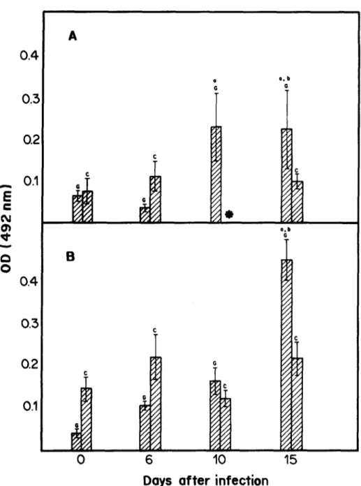
The evolution of antibody levels in each group is shown in Fig. 3. The basal levels of antibodies were the same for both GN and CV groups. After infection, the levels of these immunoglobulins were significantly raised in the GN group, this did not occur in the CV group. When compared with their CV counterparts, the GN mice had higher titers of IgG and IgM, 15 days after infection.
Efect of desferrioxamine, iron-dextran and the infec- tion with Trypanosoma cruxi on the number of red blood cells and leukocytes
Before infection and on day 6 after inoculation with T. cruzi, the iron content in liver of GF controls was higher than of their CV counterparts. Desferrioxam- ine-treated GN mice showed lower levels of iron in liver than GN controls 10 days after infection but not on day 15 when there was an increase in the iron levels. No such alterations were observed in the CV group. Treatment with iron-dextran enhanced the levels of iron in livers of GN and CV infected
Infection by T. cruzi caused a reduction in numbers of red blood cells in CV control mice, 15 days after infection (Table 2). This difference was not observed in animals treated with desferrioxamine or iron- dextran. There was an earlier and more intense re- duction of the red blood cell number in control GN mice. Desferrioxamine- or iron-dextran-treated GN animals also showed a marked reduction in red blood cell number in the course of infection.
816 MARIA L. PWXWW et nl.
C
A 0,m
zyxwvutsrqponmlkjihgfedcbaZYXWVUTSRQPONMLKJIHGFEDCBA
A 800-
t0
I
2 6Od
E
a 200
3
zyxwvutsrqponmlkjihgfedcbaZYXWVUTSRQPONMLKJIHGFEDCBA
C!z
‘;
100
f
a
0
6
IO
Days after infection
Fig. 2. Mean levels of iron in serum (&dl) (A), liver (~g/or~n) (B), and spleen (~g/organ) (Cc) in ~oto~otic (0) and inventions (C) controt (a), d~fe~o~ne-tram (m) or iron-dextran-treaty mice (0). Assays were performed before infection or on days 6, IO, and 15 after infection. Statistical differences (P < 0.05) are indicated as follows: a, in relation to day 0 within the same group; e, in relation to their conventional counterpart at day 0; m, in relation to their control counterpart at the same day of infection. The number of animals in each group are inside the vertical bar. Vertical lines represent the
standard deviations of the means. *Not done.
DiSCUSSION
Our results demonstrate that an increase in avail- able iron stimulates the parasitemia in CV and GN mice (Fig. I), confirming previous results (Pedrosa et al., 199Ob). However, the higher rate of prolifer- ation of the parasite with the increase in iron levels was not followed by a more intense tissue parasitism. The short period of experimentation could explain this apparent paradox.
Non-infected GF and CV mice presented the same values for serum iron and TIBC conlirming the results
obtained by Donati et al. (1969). After infection with
T. cruzi, the levels of serum iron were not altered and increased in CV and GN animals, respectively (Table 1). However, TIBC levels were reduced and elevated in CV and GN mice, respectively, in the course of infection. Therefore, there is a difference in iron meta~~sm between GN and CV animals. It is known that T. crud is capable of suppressing the host
Iron nutriture and Chagas’ disease in gnotobiotic mice 817
Table I. Levels of serum iron, total iron binding capacity (TIBC) and transfertin saturation index (TSI) in conventional (CV) and gnotobiotic (GN) mice infected with
1700 blood forms of ~rypanosoma CJWZ~, Y strain
Days after Serum iron TIBC TSI
Group infection (rgldl) @g/d0 W)
cv
cv
cv
cv zyxwvutsrqponmlkjihgfedcbaZYXWVUTSRQPONMLKJIHGFEDCBA
GN
zyxwvutsrqponmlkjihgfedcbaZYXWVUTSRQPONMLKJIHGFEDCBA
GN
GN
GN
zyxwvutsrqponmlkjihgfedcbaZYXWVUTSRQPONMLKJIHGFEDCBA
0
6
IO
15
0
6
10
15
155.7 * 93.9 (7)
132.3 f 60.1 309.3 f 122.7 43.3 f 23.8
(6) (3) (3)
183.9 _+ 89.9 294.3 f 152.7 72.8 5 14.4’
(7) (4) (4)
102.6 f 45.8 199.1 f 71.3’ 52.7 f 14.6’
(7) (7) (7)
104.8 + 22.2 318.3 + 97.6 35.8 k 14.0
(6) (6) (6)
73.8 & 10.9 272.0 + 35.8 28.5 f 4.5
(7) (4) (4)
205.1 f 34.5’ 545.3 * 55.YJ 36.0 f 4.7’
(5) (4) (4)
202.9 f 39.9”; 1129.0 + 218P 18.5 f 4.0”” 378.3 It 84.9 33.0 f 16.2
(6) (6)
(4) (4) (4)
Statistical differences (P < 0.05) are indicated as superscripts: a, in relation to day 0 within the same group; e, in relation to the CV counterpart on the same day. The number of mice is indicated in parentheses.
GN mice. Therefore, these lymphocytes became more reactive and synthesized more transferrin.
Differences in the immune response between GN and CV animals were earlier reported by Rogers and Balish (1978): when CV and GF rats were infected with Candida albicans and later challenged with phy- tohemagglutinin and concanavalin A, they observed: (1) suppression of splenocyte response in CV rat; (2) a five-fold increase in blastogenesis in GN rat. These results suggest that disseminated candidiasis is capable of suppressing blastogenesis in CV rat. The absence of antigenic stimuli derived from intestinal microbial flora could affect the immune response to infection with T. cruzi. Such differences may be evidenced by observing the levels of T. cruzi-antigen bound IgG and IgM in CV and GN mice. The levels of immunoglobulins in the latter were higher than in the former at day 15 after infection (Fig. 3). The GN group, therefore, produced more IgG and IgM than their CV counterparts. Higher titers of immunoglob- ulins were observed in GN rats infected with T. lewisi (Giannini, 1987) and in GN mice infected with T. cruzi (Furarah et al., 1991) than in their CV counterparts. This confirms the hypothesis that the difference in levels of TIBC between CV and GN mice may be due to differences in immunological responses.
Desferrioxamine, an iron chelator, has been widely used to evaluate the relationship between infection and iron nutriture (Duhr et al., 1989). Two mechan- isms are proposed for its action: (1) induction of iron deficiency by limiting its availability within the cell, (2) direct interference with the acquisition
of iron by the parasite (Hershko and Peto, 1988). In vitro, this chelating agent is able to remove iron from ferritin, hemosiderin and, to some extent, from transferrin (Keberle, 1964). Desferrioxamine did not affect the levels of liver, spleen and serum iron in either CV or GN mice after infection with T. cruzi (Fig. 2), except for GN mice at day 10 after infection. Desferrioxamine did not affect the parasitemia (Fig. 1). These results confirm the ones previously obtained with the Y strain in CV mice even though this strain was more sensitive to iron deficiency (Pedrosa et al., 1990a). Pedrosa et al. (1990a) also reported that 40 days after infection, there was a decrease in the levels of iron in spleen and liver of desferrioxamine-treated mice. It is possible that the removal of iron by the chelating agent may be occurring after the period of high proliferation of the parasite which explains the results presented here.
818 MARIA L. ~DROSA et d.
0.4
zyxwvutsrqponmlkjihgfedcbaZYXWVUTSRQPONMLKJIHGFEDCBA
0.3
0.2
A
0. b
G
0.b
i
‘13
Days of ter infection
Fig. 3. Mean levels of IgM (A) and IgG (B) determined by ELISA in control gnotobiotic (G) and conventional (C) mice. Assays were perform& before infection and on days 6, 10 and 15 after infection. Each bar represents an average of three to sii samples. Statistical differences (P < 0.05) are indicated as follows: a, in relation to day 0 within the same group; b, in relation to the conventional counterpart at
the same day of infection. Vertical lines represent the standard error of the means. *Not done.
(Table 1). It seems, therefore., that the uptake of iron from irondextran is more efficient in GN than in CV animals after intraperitoneal injection. On the other hand, the removal of serum iron by the reticulo- endothelial system seems to be less efficient in the former group of animals. This corroborates the data of Donati ef al. (1969) who observed that the clear- ance of radio-iron from plasma was markedly slower in the GF mouse, suggestive of a direct or indirect effect of the flora on the reticulo-endothelial system which could explain the difference in serum iron levels
between GN and CV mice. Treatment with iron- dextran promoted an elevation of the levels of spleen iron in CV and GN mice, 15 and 10 days after the infection, respectively (Fig. 2C). An increase in liver iron content in both groups was also observed (Fig. 2B). The increase in iron content in these organs positively affected the proliferation of the parasites (Fig. 1).
Iron nutriture and Chagas’ disease in gnotobiotic mice 819
Table 2. Number of red blood cells/mm’ in conventional (CV) and gnotobiotic (GN) mice treated with desferrioxamine (D), iron dextran (F) or control groups (C)
Days after infection
Grow 0 6 10 15
cv-c
1590 * 147 I183 + 956(5) (5)
CV-D 1605 * 174
(4)
CV-F 1137 * 461
(5)
zyxwvutsrqponmlkjihgfedcbaZYXWVUTSRQPONMLKJIHGFEDCBA
GN-C I772 f 225 949 f 35wc
(5) (7)
GN-D 1040 + 91s”
(5i
GN-F 1521 f 556
(51
1280 + 540 (5)
1429 f 231 (5)
1562k319
(4)
684 f 133’*
(5)
613 + 184’c (6)
949 + 3 16” (4)
1090+448*
(3)
1147 + 120 (4)
1365 f 171
(4)
619 f 268’.=
(4)
456 f 268” (5)
521 f 159l.C (41
Assays were performed before infection with 1700 blood forms of Tryponosoma crux, Y strain, and on days 6, 10 and 15 after infection.
Statistical differences (P < 0.05) are indicated as superscripts; a, in relation to day 0 within the same group; e, in relation to the CV counterpart on the same day. The number of mice is indicated in parentheses.
numbers in both groups (Table 2). Transitory hemato- logical alterations have been described in various infections with trypanosomes which have been at- tributed to different factors: hemodilution, bone marrow injury, destruction of red blood cells and alteration of their turnover (Cardoso and Brener, 1980). In iron dextran-treated GN mice, a reduction in the number of red blood cells was observed after day 10 of infection. In control and desferrioxamine-treated GN mice the red blood cell numbers were altered after day 6 of infection. A marked anemia in the GN group could be explained by the findings of Hashimoto and
Hashimoto (1968) who observed a smaller hematopoi- etic activity in GN than in the CV rat. Furthermore, the low IST in GN mice may have contributed to the decrease of the red blood cell count in these animals as compared with their CV counterparts.
An elevation of the number of leukocytes was ob- served in all CV and GN groups as infection pro- gressed (Table 3). These results contradict the ones obtained by Cardoso and Brener (1980) who reported leukopenia in CV albino mice infected with lo4 trypomastigotes. This discrepancy may be explained by the different inoculum size or by the difference
Table 3. Number of leukocytes/mm’ x lo-’ of blood in conventional (CV) and gnotobiotic (GN) control mice (C) or in mice treated with desferrioxamine (D) or
iron d&ran (Fj
Days afier infection
Group 0 6 10 12
cv-c 8.2652.12 22.6 + 0.8” 37.1 * 15.7’ 35.4 *4.1a
(5) (5) (5) (4)
CV-D 35.2 + 14.5’ 34.5 f 13.op 56.4 f 9.5’
(5) (5) (3)
CF-F 25.3 i 17.9’ 51.9 f 22.4’.m 54.8 + 4.5’,m
(5) (5) (4)
GN-C 4.6 f 1.7’ 6.3 f 2.4’ 12.2 rt 3.8” 33.5 + 8.3’
0 (7) (5) (4)
GN-D 16.2 f 4.9= 10.8 f 3.4’ 13.3 f 3.5a.m
(5) (5) (5)
GN-F 6.4 k 1.5 17.3 + 7.0a 20.0 f 6.61.”
(5) (4) (5)
Assays were performed before infection with 1700 blood forms of
zyxwvutsrqponmlkjihgfedcbaZYXWVUTSRQPONMLKJIHGFEDCBA
Trypmosoma cruri, Y strain, or on days 6, 10 and 15 after infection820 M AW L. I ‘EDROSA et al.
in mice strains. In GN control, desferrioxamine- and iron-dextran-treated mice, the rise in leukocyte number was less pronounced that in their CV counterparts, except for the GN control group on day 15 after infection. Anosa and Kaneko (1983) reported leukocytosis with lymphocytosis, monocytosis, eosinopenia and basophilia in T.
brucei-infected mice. One may suppose that in GN mice, the leukocyte proliferation occurs at a slower rate. On day 15 after infection the levels of leukocytes in GN mice reached the ones of CV animals but treatment with desferrioxamine or iron-dextran bin- dered this recovery. In CV mice there was a rise in leukocyte numbers from 3- to ‘I-fold in all groups.
We have shown that GN mice differ from their CV counterparts in: (1) their response to infection by
T. cruzi, (2) iron metabolism in different conditions
of iron nutriture and (3) red blood cell and leukocyte numbers. These results demonstrate that, under the conditions described, desferrioxamine was not capable of producing alterations in the iron status of both GN and CV mice, and therefore, did not interfere with the uptake of iron by T.
cruzi. Iron-dextran, on the other hand, did elevate the
levels of iron in the host and, therefore, its avail- ability, what facilitated the proliferation of the para- site.
Acknowledgements- This work was supported by Finan-
ciadora de Estudos e Projetos (FINEP), Conselho National de Desenvolvimento Cientifico e Tecnologico (CNPq), Fun- da@io de Amparo a Pesquisa do Estado de Minas Gerais (FAPEMIG), and Pro-Reitoria de Pesquisa da Universi- dade Federal de Minas Gerais (PRPQ-UFMG). The authors are greatly indebted to Ronilda Maria de Paula and Maria Cristina Lego Monteiro de Barros for skilful techni-
cal help.
zyxwvutsrqponmlkjihgfedcbaZYXWVUTSRQPONMLKJIHGFEDCBA
REFERENCES
Almeida M. R., Silva M. E., Soares F. M. L. and Vieira E. C. (1989) Influence of vitamin E nutriture on the evolution of Chagas’ disease in mice. M em. Inst. Oswald0 Cruz 84 (Suppl. II), 28.
Anosa V. 0. and Kaneko J. J. (1983) Pathogcnesis of
Trypanosoma brucei infection in deer mice (Peromy scus
maniculatus): hematologic, erythrocyte biochemical, and
iron metabolic aspects. Am. J. uet. Res. 44, 639- 644. A.O.A.C. (Association of Official Analytical Chemists)
(1980) O fficial M eth& of Analy ses (Edited by Horowitz E.), ‘13th Edn. Assoc:ation- of ‘Ofhcial _ Analytical Chemists, Washington, DC.
Brener Z. (1962) Therapeutic activity and criterion of cure on mice experimentally infected with Trypanosoma cruzi. Rev. Inst. M ed. Trop. St70 Paul0 4, 380- 396.
Cardoso J. E. and Brener Z. (1980) Hematological changes
in mice experimentally infected with Trypanosoma cruzi.
M em. Inst. Oswald0 Cruz 75, 97-104.
Dhur A., Galan P. and Her&erg S. (1989) Iron status, immune capacity and resistance to infections. Comp.
Biochem. Phy siol. 94A, 11-19.
Donati R. M., McLaughlin M. M., Levri E. A., Berman A. R. and Stromberg L. W. R. (1969) The response of
iron metabolism to the microbial flora: studies on 8crmfree. mice. Proc. Sot. exp. biol. M ed. 130, 920-922.
Furarah A. M.. Crocco-Afonso L. C.. Silva Marcel0 E.. Silva Mdrcio E., Silva M. E. C., Nicoli J. R., Vieria E. C: and Bambirra E. A. (1991) Immune responses in germfree and conventional mice infected with Trypanosoma cruzi. Braz. J. med. biol. Res. 24, 12233 1.
Giannini M. S. H. (1987) The specific anti-parasite immune responses of germ-free and conventional rats infected with Trypanosoma lew isi. J. Parasitol. 73, 144- 148.
Goodwin J. F., Murphy B. and Guillemette M. (1966)
Direct measurement of serum iron and bindina capacity. __ - Clin. Chem. 12, 47- 57.
Hashimoto M. and Hashimoto N. (1968) Histological \ , study of bone marrow in breeding rats. In Adoanc~s in
Germfree Research and Gnotobiology (Edited by
Miyakawa M. and Luckey T. D.), pp. 149-161, Illife Books, London.
Hershko C. and Peto T. E. A. (1988) Deferoxamine inhi- bition of malaria is independent of host iron status. J. exp. M ed. 168, 375- 387.
Holbcin B. E. (1980) Iron-controlled infection with
Neisseria meningitidis in mice. Infect. Immun. 29, 886-
891.
Kebcrle H. (1964) The biochemistry of desferrioxamine and its relation to iron metabolism. Ann. N. Y. Acad. Sci. 119, 758-768.
Kent S., Weinberg E. D. and Stuart-MacAdam P. (1990) Dietary and prophylactic iron supplements: helpful and harmful. Human Nature 1, 55-81.
Keusch G. T. and Farthing M. J. G. (1986) Nutrition and infection. Ann. Reu. Nutr. 6, 131-154.
Kierzenbaum F. and Sztein M. B. (1990) Mechanisms underlying immunosuppression induced by Trypanosoma cruzi. Parasitol. Today 6, 261- 263.
Lalonde R. G. and Holbein B. E. (1984) Role of iron in Trypanosoma cruzi infection of mice. J. clin. Invest. 73, 470- 476.
McLarcn M. L., Lillywhite J. E. and Sirr S. (1980) A
Laboratory M ethod for IFAT and ELISA. The Ross
Institute of Tropical Hygiene, London School of Hygiene and Tropical Medicine, London.
Pedrosa M. L., Silva Marcel0 E., Silva Mdrcio E., Silva M. E. C., Nicoli J. R. and Vieria E. C. (199Oa) The effect of iron deficiency and iron overload on the evolution of Chagas’ disease produced by three strains of Try-
panosoma cruzi in CFW mice. Comp. Biochem. Phy siol.
VA, 235-243.
Pedrosa M. L., Silva M. E., Silva M. E., Silva M. E. C., Vieria L. Q., Vieira E. C. and Bambirra E. A. (199Ob) The effect of iron nutritonal status on experimental Chagas’ disease in conventional and germfree mice. M icroecol. Ther. 20, 453- 457.
Pleasants, J. R. (1974). Gnotobiotics. In Handbook
of Laboratory Animal Science (Edited by Melby,
Jr. E. C. and Altman N. H.), pp. 119-174. CRC Press, Cleveland.
Rogers T. J. and Balish E. (1978) Effect of systemic can- didiasis on blastogenesis of lymphocytes from germfree and conventional rats. Infect. Immun. 20, 142- 150. Santos C. F., Silva M. E., Evangelista E. A., Nicoli
J. R., Santos J. E., Bambirra E. A. and Vieira E. C. (1992) Effect of an essential fatty acid deficient diet on experimental infection with Trypanosoma cruzi in germfree and conventional mice. Braz. J. med. biol. Res. 25, 795- 803.
Scrimshaw N. S., Taylor C. E. and Gordon J. E. (1968)
Interactions of Nutrition and Infection. World Health
Organization, Geneva.
Iron nutriture and Chagas’ disease in gnotobiotic mice 821 Snedecor G. W. and Cochran W. G. (1980) Statistical Cutaneous leishmaniasis in germfree, gnotobiotic, and
Methods, pp. 215-237. New York University Press, New York.
conventional mice. Rev. Ins~. Med. Trop. S&o Paul0 29, 385-387.
Trexler P. C. (1959) The use of plastics in the design of Weinberg E. D. (1984) Iron withholding: a defense against isolator sytems. Ann. N.Y. Acad. Sci. 78, 29-36. infection and neoplasia. Physiol. Rev. 64, 65-102.