FACULDADE DE FARMÁCIA
DEPARTAMENTO DE MICROBIOLOGIA E IMUNOLOGIA
B
ACTERIOPHAGE
L
YTIC
E
NZYMES AND THEIR
ENGINEERING TOWARDS IMPROVED
A
NTIBACTERIAL
E
FFICACY
Daniela Sofia Moreira Proença
D
OUTORAMENTO EMF
ARMÁCIAE
SPECIALIDADEM
ICROBIOLOGIAUNIVERSIDADE DE LISBOA
FACULDADE DE FARMÁCIA
DEPARTAMENTO DE MICROBIOLOGIA E IMUNOLOGIA
BACTERIOPHAGE LYTIC ENZYMES AND THEIR
ENGINEERING TOWARDS IMPROVED
ANTIBACTERIAL EFFICACY
Daniela Sofia Moreira Proença
Tese orientada pelos Professores Doutores Carlos Jorge Sousa de São-José
(orientador universitário) e Miguel Ângelo da Costa Garcia (orientador
empresarial), elaborada para a obtenção do grau de doutor em Farmácia
(Microbiologia)
Financial support to Daniela Sofia Moreira Proença was provided in equal parts by the Biotech company TechnoPhage, SA and by a PhD fellowship (SFRH/BDE/51076/2010) from Fundação para a Ciência e a Tecnologia (FCT, MCTES, Portugal) under the funding program “Programa Operacional Potencial Humano do QREN Portugal 2007-2013”.
De acordo com o disposto no ponto 1 do artigo n° 45 do Regulamento de Estudos Pós-Graduados da Universidade de Lisboa, deliberação n° 4624/2012, publicado em Diário da Républica – II Série n° 65 – 30 de Março de 2012, a Autora desta dissertação declara que participou na conceção e execução do trabalho experimental, interpretação dos resultados obtidos e redação dos manuscritos.
Sempre chegamos ao sítio aonde nos esperam.
A
CKNOWLEDGMENTS
A
GRADECIMENTOS
O meu primeiro agradecimento é dirigido ao meu orientador científico Professor Carlos
São-José e ao meu co-orientador e coordenador empresarial Doutor Miguel Garcia. Carlos, obrigada
por teres acreditado que eu conseguia e que valeria a pena fazer esta viagem, agradeço por teres
entrado neste barco comigo e por o teres levado a bom porto sempre que a tempestade teimava
em não passar. Obrigada por todas as palavras de força e ânimo, nas alturas mais difíceis.
Agradeço por me teres apoiado incondicionalmente em TODOS esses momentos, toda a tua
incansável dedicação a mim, à minha formação e ao meu projeto. Agradeço todos os teus
ensinamentos que me fizeram crescer como investigadora com sede de respostas, de
conhecimento, que fizeram de mim uma melhor profissional. Agradeço-te também pela
paciência e pelos conselhos sábios, sem os quais não teria chegado aqui. Agradeço ao Doutor
Miguel por ter aceitado co-orientar este projeto e por me ter proporcionado todas as condições
e meios materiais para a execução dos trabalhos que me permitiram atingir os objetivos a que
me propus.
Agradeço a toda a equipa da TechnoPhage, SA, por me ter acolhido tão bem e por me ter
apoiado no meu trabalho ao longo destes 4 anos. Em especial agradeço às minhas queridas
colegas, amigas e companheiras Clara Leandro e Raquel Barbosa por todo o apoio profissional e
pessoal. Por me emprestarem o vosso ombro para eu me apoiar nas alturas mais difíceis, por
me emprestarem os vossos ouvidos quando precisei de desabafar, por lutarem ao meu lado nos
meus objetivos. Clara, obrigada por me ajudares a crescer como profissional, por teres
contribuído para que fosse uma pessoa mais atenta, responsável e tolerante. Raquel, obrigada
por me teres brindado todos os dias com a tua boa disposição, o teu sorriso e o teu positivismo.
Agradeço à Sofia Corte-Real por ter coordenado este projeto, por toda a amizade e apoio
sempre disponibilizados. Agradeço ao Frederico Aires da Silva por todas as dicas preciosas que
me deu ao longo destes anos, pela amizade e pelo apoio. Agradeço à minha crew: Soraia
Oliveira (Castanha ); Joana Ministro; Rita Vaz e Pedro Canhão (Pedrito), por todos os
momentos de descontração, pelas loooooongas conversas (e debates também!), pelas saídas à
noite e jantares bem regados de boa disposição, alegria e companheirismo. Obrigada por, à
disponibilidade e por se preocuparem comigo. Pela amizade.
Agradeço à Professora Madalena Pimentel, pelos inúmeros conselhos práticos que me foi dando
nas nossas reuniões semanais de segunda-feira, ao longo destes 4 anos. Pelas palavras de ânimo
e pelo carinho.
Agradeço às meninas do lado de lá da estrada: a companheira de congressos Sofia Fernandes e à
Catarina Baptista; por me “desenrascarem” sempre que precisei de algum material à última da
hora. Agradeço também a paciência que disponibilizaram para ouvir os meus lamentos,
frustrações e alegrias também! Obrigada pela amizade.
Agradeço ao meu namorado Marcelo, por me ter acompanhado e apoiado em todas as minhas
decisões. Obrigada pela tua paciência e companheirismo, obrigada por conseguires alegrar-me
nas alturas mais complicadas com o teu bom humor, com a tua alegria, com a tua magia.
Agradeço à minha segunda mãe, Paula Pires, por ter estado ao meu lado desde que me mudei
para Lisboa. Por me ajudar a ultrapassar todos os obstáculos e todas as dificuldades que
encontrei nesta viagem. Por ter estado ao meu lado sempre que precisei. Por ter sido o meu
porto seguro dos últimos anos.
Agradeço aos meus queridos pais, pelo apoio incondicional, por estarem sempre presentes,
sempre dispostos, por me darem a força necessária para continuar em frente e terminar este
projeto. Agradeço-vos por estarem aí, por me estenderem a mão sempre que eu preciso! Por
serem o meu porto seguro, por serem os melhores pais. À minha irmã Cátia por todos aqueles
artigos que me “sacou” sempre que eu não tinha acesso! Foram preciosos! Agradeço-te por,
mesmo longe, me teres apoiado com bons conselhos, com as chamadas de atenção, com as
correções de inglês. Por seres a minha alma gémea, a minha consciência, a minha guia.
Obrigada por seres a melhor irmã do mundo!!!
T
ABLE OF CONTENTS
ABBREVIATIONS
... i
SUMMARY
... iii
RESUMO
... v
THESIS OUTPUTS
... ix
C
HAPTER1
GENERAL INTRODUCTION
... 1
Bacteriophages: the viruses of bacteria... 3
The bacterial cell envelope: a barrier to phage entry and exit from host cells ... 7
Phage release from infected cells: lysis-mechanisms of dsDNA bacteriophages ... 10
Phage-encoded peptidoglycan hydrolases ... 14
Endolysins ... 17
Virion-associated lysins of dsDNA bacteriophages ... 20
The use of phage-based products to control pathogenic bacteria ... 23
Phage therapy ... 23
Endolysins as antibacterials ... 25
Engineering of phage-lytic proteins ... 27
Other applications of phage lytic proteins ... 30
Phage-encoded lytic proteins with activity against Enterococcus sp ... 32
C
HAPTER2
PHAGE ENDOLYSINS WITH BROAD ANTIMICROBIAL ACTIVITY AGAINST
E
NTEROCOCCUS FAECALIS CLINICAL STRAINS... 53
Abstract ... 57
Introduction ... 58
Cloning of Lys168 and Lys170 endolysin genes ...62
Production and purification of the endolysins Lys168 and Lys170 ...63
Evaluation of endolysin lytic action against bacterial pathogens ...64
Identification of bacterial species ...64
Results ...65
Bioinformatics of enterococcal phage endolysins Lys168 and Lys170 ...65
Heterologous production and purification of endolysins Lys168 and Lys170 ...68
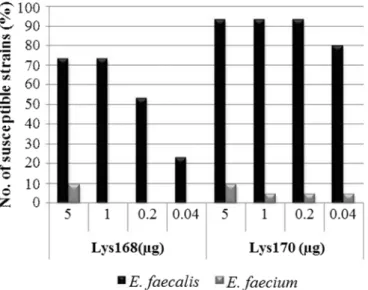
Lytic action of Lys168 and Lys170 against enterococcal clinical strains ...69
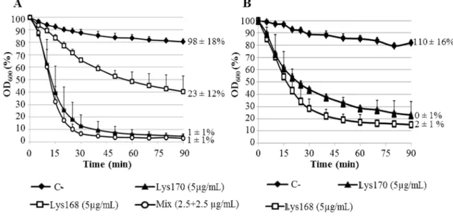
Lytic action of Lys168 and Lys170 against E. faecalis in liquid medium ...71
Activity of enterococcal endolysins against other Gram-positive pathogenic bacteria ...72 Discussion ...72 Acknowledgements ...77 References ...77 Supplementary material ...83
C
HAPTER3
A
TWO-
COMPONENT,
MULTIMERIC ENDOLYSIN ENCODED BY A SINGLE GENE... 95
Abstract ...98
Introduction ...99
Results ... 100
Expression of endolysin gene lys170 results in two stable polypeptides ... 100
Lys170FL and CWB170 polypeptides are required for full endolysin lytic activity in vitro ... 104
The two endolysin polypeptides are produced in the phage infection context ... 106
CWB170 promotes endolysin binding to target cells ... 107
Composition of the Lys170 complex ... 109
Cross-linking of endolysin multimers ... 112
Materials and Methods ... 120
Bacteria, plasmids, phage and growth conditions ... 120
General DNA techniques ... 120
General protein techniques... 121
Construction of lys170 derivatives ... 121
Production and purification of endolysin polypeptides ... 122
Protein N-terminal sequencing ... 122
Rabbit immunization with purified Lys170 ... 122
Lytic activity of Lys170 and its derivatives ... 123
Time course of endolysin production during phage infection ... 123
Binding of endolysin polypeptides to E. faecalis cells ... 124
Size-Exclusion Chromatography with Multi-Angle Light Scattering (SEC-MALS) 124 Protein Cross-linking experiments ... 125
Bioinformatics analysis ... 125
Acknowledgments ... 125
References ... 126
C
HAPTER4
EC300: A PHAGE-BASED, BACTERIOLYSIN-LIKE PROTEIN WITH ENHANCED
ANTIBACTERIAL ACTIVITY AGAINST ENTEROCOCCUS FAECALIS... 133
Abstract ... 136
Introduction ... 137
Materials and Methods ... 139
Bacteria, phage and growth conditions ... 139
General DNA techniques ... 141
General protein techniques... 142
Construction and cloning of EC300 chimeric gene and its derivatives ... 142
Protein Production and purification ... 143
Lytic activity in liquid media ... 144
Evaluation of EC300 antibacterial activity in solid medium ... 144
Bioinformatics tools ... 144
EC300 has superior lytic activity when compared to Lys170 ... 149
EC300 spectrum of activity against enterococcal clinical strains ... 150
Discussion ... 152
Acknowledgements ... 154
References ... 155
Supplementary material ... 164
CONCLUDING REMARKS AND FUTURE PRESPECTIVES
...
169
FIGURES AND TABLES
C
HAPTER1
Fig. 1. Schematic representation of the major bacteriophage families……….. 5
Fig. 2. Bacteriophage life styles……… 7
Fig. 3. Bacterial cell envelopes……….. 8
Fig. 4. Models for export and activation of endolysins………. 12
Fig. 5. Types of enzymatic domains found in phage PG hydrolases……… 15
Fig. 6. Domain architecture of Gram-negative and Gram-positive endolysins……… 18
Fig. 7. VALs domain organization and diversity of PG cleavage specificities……….. 21
Fig. 8. Schematic representation of the mode of action of a virion-associated lysin (VAL)… 22 Table 1. Major characteristics of bacteriophage families………. 4
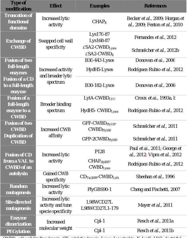
Table 2. Molecular engineering and its effects on phage PG hydrolases properties………… 28
C
HAPTER2
Fig. 1. Domain architecture and sequence relatedness of Lys170……… 66Fig. 2. Domain architecture and sequence relatedness of Lys168……….... 67
Fig. 3. Analysis of endolysins Lys168 and Lys170 purification………..……… 69
Fig. 4. Lytic action of Lys168 and Lys170……….……….. 70
Fig. 5. Lytic action of Lys168 and Lys170 in a turbidity assay………...………. 71
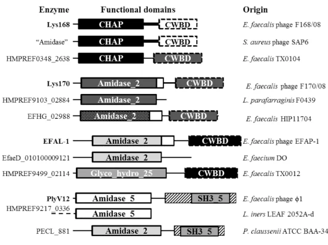
Fig. 6. Nature organization and sequence relatedness of E. faecalis phage endolysin functional domains………...……….. 74
Fig. S1. Bioinformatics analysis of endolysins Lys168 and Lys170 primary sequence…….. 83
Fig. S2. “TritonX-100-induced lysis halo assay”………..…… 83
Fig. S3. Representative lysis halos resulting from endolysin lytic action……….……… 84
Fig. S4. Lys168 and Lys170 lytic action against 73 E. faecalis and 26 E. faecium non-typed clinical isolates……….………. 84
Table 1. Typed enterococcal clinical strains used in this study…….………. 60
Table S3. Detailed characterization of the typed enterococcal clinical strains used in this
study……….………. 89
Table S4. Lytic action of Lys168 and Lys170 against the panel of typed enterococcal clinical strains……….……….. 91
Table S5. Non-typed clinical isolates from other Gram-positive pathogenic cocci used in this study……….….. 93
C
HAPTER3
Fig. 1. The two polypeptides of endolysin Lys170………..……. 101Fig. 2. Size-exclusion chromatography of the Lys170, mLys170 and CWB170 proteins….... 104
Fig. 3. Impact of CWB170 polypeptide in endolysin activity………...…… 105
Fig. 4. Time course of Lys170FL and CWB170 synthesis during E. faecalis infection by phage F170/08……….…….. 107
Fig. 5. Binding of purified mLys170, Lys170 and CWB170 to E. faecalis cells………..…… 108
Fig. 6. SEC-MALS analysis of Lys170, mLys170 and CWB170………. 110
Fig. 7. Cross-linking analysis of endolysin multimers……….. 113
Fig. 8. PG hydrolases with CWB170-like domains………..……… 117
Table 1. Analysis of the UV280nm extinction coefficient (ep) of Lys170 multimer models. 111
CHAPTER 4
Fig. 1. Rationale behind the construction of the lytic chimera EC300……….. 146Fig. 2. EC300 and mEC300 purification………... 147
Fig. 3. Impact of CWB170 polypeptide in EC300 activity………... 148
Fig. 4. Comparison of EC300 and Lys170 lytic activities……….... 150
Fig. 5. Susceptibility of a panel of typed E. faecalis strains to the EC300 growth inhibition activity………..…………. 151
Fig. 6. Evaluation of EC300 capacity to inhibit growth of four vancomycin-resistant E. faecalis strains………...……… 152
Fig. S1. Primary sequence details and domain architecture of the VAL Orf73 the endolysin
Lys170 and of the chimera EC300……….………... 164
Fig. S2. “TritonX-100-induced lysis halo assay”……….. 165
Table 1. Typed enterococcal clinical strains used in this work………..….. 140
Table S1. Growth inhibition of typed E. faecalis clinical strains by EC300... 166
Concluding Remarks and Future Prespectives Fig. 1. Pipeline of bacteriolysins-like proteins developed during this thesis……….... 173
A
BBREVIATIONS
GENERAL
CFU Colony Forming Units
DNA Deoxyribonucleic Acid
dsDNA double stranded DNA
dsRNA double stranded RNA
EFS Enterococcus faecalis strain
His Histidine tag
NCBI National Center for Biotechnology Information
OD Optical Density
CDD Conserved Domain Database
RNA Ribonucleic Acid
ssDNA single stranded DNA
Pfam Protein family
TECHNIQUES
ELISA Enzyme-linked immunosorbent assay
PCR Polymerase Chain Reaction
PFGE Pulse-Field Gel Electrophoresis
SDS-PAGE Sodium Dodecyl Sulphate - Poli-Acrilamide Gel
Electrophoresis
SEC Size-Exclusion Chromatography
SEC-MALS SEC-Multi Angle Light Scatering
REAGENTS
BHI Brain Heart Infusion
BS3 bis(sulfosuccinimidyl)suberate
DTT Ditiotreitol
HEPES 4-(2-Hydroxyethyl)piperazine-1-ethanesulfonic acid
HRP Horseradish peroxidase
UNITS
µg microgram µL microliter µM micromolar µmol micromol kb kilo basekDa kilo Dalton
LB Lysogeny broth
mg miligram
mL mililiter
pmol picomol
S
UMMARY
Increasing antibiotic resistance among bacterial pathogens has been promoting the study of bacteriophage (phage) lytic enzymes (bacterial cell wall hydrolases) as alternatives/complements to antibiotics. Phages can employ two types of these enzymes during their life cycle: i) virion-associated lysins (VALs), which promote a local cleavage of cell wall bonds to facilitate phage genome entry into the host cell; and ii) endolysins that destroy the wall at the end of infection, leading to cell burst and release of virion progeny. We studied the lytic activity of two enterococcal endolysins, Lys168 and Lys170, towards clinical isolates of different Gram-positive bacterial pathogens. In the conditions tested, both enzymes showed broad antimicrobial activity against E. faecalis, including vancomycin-resistant strains, and to less extent against E. faecium.We show that lys170 expression results in the production of the expected full length polypeptide (Lys170FL, 32.6 kDa) and of a C-terminal fragment of the enzyme (CWB170, 12 kDa), with both proteins co-eluting in the purification steps. Further analysis revealed that CWB170 corresponded to the Lys170 cell wall binding domain, which is independently produced from an in-frame, secondary translational start site. Biochemical and biophysical analysis indicated that the fully active Lys170 is a complex most likely corresponding to one subunit of Lys170FL associated to three of CWB170. Study of Lys170 has thus uncovered a new strategy of increasing the number of CWB domains in this type of enzymes.
A frequently reported problem when working with phage lytic enzymes is their propensity to become insoluble. Further, the activity of endolysins is rarely studied in conditions that promote robust growth of target bacteria. With the goal of supplanting these limitations we engineered a chimerical lysin, EC300, aimed at lysing E. faecalis growing in rich culture media. EC300 resulted from the fusion of a M23 endopeptidase domain of a VAL to the CWB170 domain of Lys170. The bacteriolysin-like protein exhibited a clear enhanced lytic activity when compared to the parental endolysin, particularly when assayed in a rich culture medium, thus having the potential to be used as an anti-E. faecalis therapy.
R
ESUMO
A emergência de bactérias patogénicas resistentes a antibióticos e a consequente limitação de antibióticos eficazes na eliminação destes microrganismos tem sido o motor para a pesquisa de alternativas ao uso da terapia antimicrobiana convencional. Nesse sentido, tem sido intensamente estudado o potencial das hidrolases do peptidoglicano da parede celular bacteriana produzidas por vírus que infetam bactérias (bacteriófagos ou, mais simplesmente, fagos), como alternativas e/ou complementos aos antibióticos. Existem dois tipos de enzimas líticas que participam em etapas distintas do ciclo de infeção bacteriofágico: i) as lisinas associadas ao virião (VALs), responsáveis por uma clivagem controlada e não-letal do peptidoglicano (PG) para facilitar a entrada do genoma viral na célula bacteriana hospedeira; e ii) as endolisinas, que destroem a camada de PG no final do ciclo de reprodução do fago, o que leva à rutura (lise) da célula hospedeira com consequente libertação da descendência viral. As endolisinas em particular têm sido muito estudadas e exploradas como terapêutica antimicrobiana, uma vez que têm a capacidade de lisar rapidamente bactérias alvo Gram-positivas quando aplicadas exogenamente na forma de enzimas recombinantes.Este trabalho iniciou-se com a identificação e estudo da actividade lítica de duas endolisinas produzidas pelos fagos de Enterococcus faecalis F168/08 e F170/08. A endolisina Lys168 apresenta um domínio catalítico (CD) da família das amidohidrolases / peptidases dependentes de cisteína-histidina (CHAP), enquanto Lys170 apresenta um CD da família Amidase_2. Ambas as proteínas foram heterologamente produzidas em fusão com uma extensão C-terminal de 6 histidinas e subsequentemente purificadas na forma de proteínas solúveis. A atividade lítica destas proteínas foi testada contra uma vasta coleção de isolados clínicos, que incluía diferentes espécies bacterianas Gram-positivas. Ambas as enzimas mostraram uma elevada especificidade contra isolados de E. faecalis, ainda que com capacidade de atuação em alguns isolados de E. faecium. Numa primeira fase, Lys168 e Lys170 foram testadas numa coleção não tipada de isolados clínicos e exibiram capacidade lítica em 81% e 97% das estirpes de E. faecalis (n = 73) e 42% e 54% das estirpes de E. faecium (n = 26), respetivamente. Numa segunda coleção de estirpes geneticamente caracterizadas composta por 30 estirpes clínicas de E. faecalis e 21 de E. faecium, incluindo enterococos resistentes à vancomicina (VRE), as lisinas Lys170 e
de E. faecium foram sensíveis à ação de ambas as enzimas. Contrastando com o espectro de ação de endolisinas de fagos de E. faecalis reportado anteriormente, Lys168 e Lys170 apresentaram uma atividade quase exclusiva sobre E. faecalis. Num ensaio controlado em meio líquido, ambas as lisinas foram eficazes na eliminação de células da estirpe VRE modelo E. faecalis V583.
Durante os ensaios de expressão heteróloga da endolisina Lys170 observou-se sistematicamente a produção de um fragmento C-terminal de Lys170 com cerca de 12 kDa, para além do polipéptido esperado correspondente à totalidade da proteína (Lys170FL, 32,6 kDa). Ambas as proteínas foram co-purificadas através de cromatografia de afinidade em colunas de níquel e subsquentemente submetidas a uma cromatografia de exclusão molecular (SEC) com o objetivo de as separar. Inesperadamente, os dois polipéptidos foram co-eluídos durante a SEC, sugerindo uma associação entre Lys170FL e o polipeptídeo de 12kDa. Análises genéticas e bioquímicas provaram que o polipéptido de menor dimensão correspondia essencialmente ao domínio que se previa mediar a ligação de Lys170 à parede celular (domínio CWB170). Demonstrou-se que este é produzido de forma independente a partir de um segundo sinal de tradução interno ao gene lys170. A eliminação deste sinal resultou na produção de uma única proteína (mLys170) de tamanho idêntico ao de Lys170FL, mas com a metionina de iniciação interna substituída por uma leucina. Surpreendentemente, a atividade lítica de mLys170 revelou ser muito reduzida quando comparada com a de Lys170 nativa (Lys170FL + CWB170). Notavelmente, a incubação de mLys170 com quantidades crescentes de CWB170 purificada permitiu melhorar progressivamente a atividade lítica de mLys170. Observou-se que CWB170 per se não produziu atividade lítica detetável contra E. faecalis, apesar de se ter demonstrado a sua afinidade para a superfície bacteriana. Análises bioquímicas e biofísicas suportam um modelo em que a forma ativa de Lys170 corresponde a um complexo constituído por uma subunidade de Lys170FL associada a três de CWB170. Complementarmente, ensaios de infeção com o fago F170/08 revelaram que os polipéptidos Lys170FL e CWB170 são igualmente produzidos neste contexto, descartando a possibilidade de produção artificial durante a sua expressão heteróloga. A endolisina Lys170 define assim uma nova família estrutural de hidrolases de PG, até à
data desconhecida, revelando uma nova estratégia de aumento do número de subunidades de ligação à parede neste tipo de enzimas.
Um problema frequentemente relatado quando se trabalha com este tipo de enzimas é a sua baixa solubilidade e/ou propensão para precipitarem durante a produção em larga escala, concentração ou armazenamento. Além disso, e com base nos estudos publicados atualmente, pode-se concluir que a atividade das hidrolases do PG de origem fágica é raramente estudada em condições que promovem o crescimento ativo da bactéria alvo. Com base nesta observação, construiu-se uma lisina quimérica, designada por EC300, com capacidade para eliminar células de E. faecalis em fase de crescimento activo em meios ricos em nutrientes. EC300 resultou da fusão de um domínio com atividade de endopeptidase do tipo M23 da VAL Orf73, também codificada pelo fago F170/08, com o domínio de ligação à parede CWB170 da endolisina Lys170. A estrutura hétero-oligomérica descrita para a endolisina Lys170 foi também observada para a quimera EC300, ou seja, a forma ativa desta proteína também corresponde a um complexo multimérico entre EC300FL e CWB170. Além de demonstrar uma elevada solubilidade, esta proteína, que apresenta uma organização de domínios funcionais semelhante a uma bacteriolisina, exibiu uma atividade lítica bastante superior à exibida pela endolisina parental, particularmente quando ambas são testadas em condições que permitem o crescimento robusto de E. faecalis. Em contraste com a Lys170, a lisina quimérica demonstrou ter a capacidade de eliminar eficazmente um painel de estirpes de E. faecalis geneticamente caracterizadas e com elevado nível de resistência a antibióticos, quando estas se encontravam em fase ativa de crescimento. A EC300 é a primeira enzima semelhante a uma bacteriolisina construída a partir de proteínas fágicas com elevada atividade antimicrobiana, constituindo assim um potencial agente terapêutico para a eliminação de infecções causadas por E. faecalis.
Palavras-chave: endolisina, peptidase M23, lisina quimérica, domínio de ligação à
T
HESIS
O
UTPUTS
The research work described in this thesis was performed in TechnoPhage SA laboratories, headquartered at Instituto de Medicina Molecular, Lisbon, Portugal, from October 2010 until November 2014, under the supervision of Dr. Carlos Jorge Sousa São-José from Faculdade de Farmácia da Universidade de Lisboa, Lisbon, Portugal, and Dr. Miguel Ângelo da Costa Garcia, president and CEO of Technophage, SA, Lisbon, Portugal.The results described in this thesis are included in published or submitted manuscripts and/or patents:
Proença, D., Fernandes, S., Leandro, C., Silva, F., Santos, S., Pimentel, M., Lopes, F., Mato, R., Garcia, M., Cavaco-Silva, P. and São-José, C. (2012) Phage endolysins with broad antimicrobial activity against Enterococcus faecalis clinical strains. Microb Drug Resist 18: 322-332.
Proença, D., Velours, C., Leandro, C., Garcia, M., Pimentel, M., and São-José, C. (2014) A two-component, multimeric endolysin encoded by a single gene. Mol Microbiol Accepted for publication.
Proença, D., Leandro, C., Garcia, M., Pimentel, M., and São-José, C. (2015) EC300: a phage-based, bacteriolysin-like protein with enhanced antibacterial activity against Enterococcus faecalis. Submitted to Applied Microbiology and Biotechnology.
Proença, D., Garcia, M., Pimentel, M., and São-José, C. (2014) EC300: a phage-based, bacteriolysin-like protein with enhanced antibacterial activity against Enterococcus faecalis. Provisional national application patent No. 20141000060398.
C
HAPTER
1
BACTERIOPHAGES: THE VIRUSES OF BACTERIA
Bacteriophages, or phages, are viruses that infect bacteria. Phages are frequently described as the most abundant and diverse biological entity on earth and they are estimated to outnumber bacteria by a factor of ten (Hendrix, 2003; Pedulla et al., 2003). Phages were first discovered by Twort (1915) and d´Herelle (1917) in independent experiments and it was soon realized that these viruses could be explored as antibacterial agents (Chanishvili, 2012). Yet, the decisive impetus to the role of phages in Biology came up with M. Delbrück, that together with other scientists such S. Luria and A. Hershey, formed a research group that went by the name "phage group". The research conducted by this group and its followers on realizing the mechanisms of phage infection and bacterial lysis are at the very foundations of the field that later came to be known as Molecular Biology (Pennazio, 2006). Bacteriophages are ubiquitous forms, found wherever bacteria reside, but they are most frequently isolated from aquatic environments. Phages are not able to infect eukaryotic cells, requiring specific target bacterial cells for replication. This specificity can be highly refined, with each phage attacking just one bacterial species and, in some cases, a few strains of a given species (Hanlon, 2007).
The International Committee for Taxonomy of Viruses (ICTV) presently classifies viruses into 7 orders, 103 families, 455 genera and 77 families with unassigned order (http://ictvonline.org/taxonomyReleases.asp). Bacteriophages presently constitute 20 families (Table 1).
Table 1. Major characteristics of bacteriophage families.
Family Description Examples
Double-stranded (ds) DNA phages
Myoviridae Contractile long tail T4
Siphoviridae Non-contractile long tail
Podoviridae Short tail T7
Corticoviridae Lipid-containing phages with icosahedral capsid PM2
Rudiviridae Non-enveloped, straight rod-shaped phages SIRV-1
Tectiviridae
Phages with internal lipoprotein vesicle icosahedral capsid
PRD1
SH1, group* SH1
STV1 group* Icosahedral with protruding vertices STV1
Fuselloviridae Lipid-containing with lemon-shape phages SSV1
Globuloviridae Enveloped, lipid-containing, spherical phages PSV
Plasmaviridae Enveloped, lipid-containing, no capsid phages L2
Guttaviridae Droplet-shaped phages SNDV
Lipothrixiviridae Enveloped, filamentous or rod-shaped phages TTV1
Ampullaviridae Bottled-shaped phages with helical nucleocapsid ABV
Bicaudaviridae Two-tailed, oval phages with helical nucleocapsid ATV
Salteprovirus** Short-tails, spindle-shaped phages His1
Single-stranded (ss) DNA phages
Inoviridae Non-enveloped Fd, MVL1
Microviridae Non-enveloped, icosahedral phages X174
Double-stranded (ds) RNA phages
Cystoviridae Enveloped icosahedral phages 6
Single-stranded (ss) RNA phages
Leviviridae Non-enveloped icosahedral phages MS2 *Preliminary designation
**No family assigned
There is a variety of bacteriophage morphological types (Table 1 and Fig. 1A), although about 96% of those reported in the literature belong to the order Caudovirales (tailed phages, Fig. 1B). Phages from this order are composed by a double-stranded (ds)
DNA-containing icosahedral head, which is attached to a tail involved in the phage DNA delivery to host cells. Figure 1 and Table 1 illustrate the morphological diversity of phages and highlight some of their most typical features. The capsid is a protein shell that contains the viral nucleic acid; when present, the tail may or may not be a contractile structure, and connected to this are usually fibers or analogous structures involved in the recognition of specific receptors of the bacterial cell surface (Fig. 1B) (Hanlon, 2007). Tailed phages are classified into three families according to the morphological features of the tail: Myoviridae, Siphoviridae and Podoviridae (Table1 and Fig. 1A). These three families comprise the order Caudovirales (Ackermann, 2007; Maniloff, 2012). The other 4% of phages are distributed into 17 families, that comprise the polyheadral, filamentous, and pleomorphic phages. The nucleic acid material of phages can be made of ds or single-stranded (ss) DNA or RNA.
Fig. 1. (A) Schematic representation of the major bacteriophage families. (B) Caudovirales prototype here
illustrated by the typical myovirus morphology.
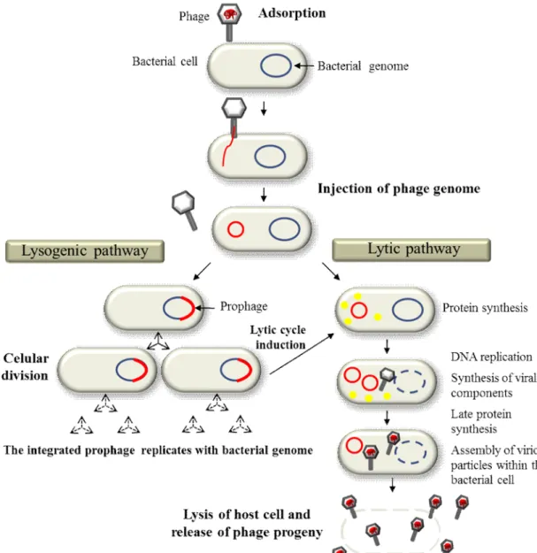
Bacteriophages, like all obligate parasites, cannot complete their life cycle by themselves and depend on host bacterial cells to replicate and maintain. The phage extracellular form, the virion, is a supramolecular structure that has evolved to maximize viral propagation by protecting the phage genome and by promoting its efficient delivery to host bacteria. When phages encounter suitable bacterial cells during random motion, they adsorb to their cell surface (Fig. 2, adsorption step) via specific receptor sites. These may be a wide
variety of cell surface components, such as proteins, oligosaccharide, teichoic acids, peptidoglycan, lipopolysaccharides, or even bacterial structures like cell capsule, flagella or conjugative pilli (Hanlon, 2007; Rakhuba et al., 2010). After adsorbing, the phage injects its genome into the bacterial cell. This step can be mediated by different mechanisms, but in Caudovirales it usually involves major structural rearrangements of the tail and the formation of a conduit across the bacteria cell envelope (wall and membrane(s)), through which the genome is delivered to the host cell cytoplasm. After genome injection, two different lifestyles can be followed depending on whether the phage as a temperate or virulent (strictly lytic) nature: the lysogenic and/or the lytic pathways (Fig. 2). The lytic pathway, which may immediately follow viral genome entry of either temperate or virulent phages, has as major role: the multiplication and spread of the virus particle. It starts with an intense viral DNA replication and viral protein synthesis, taking advantage of bacterial synthetic machinery. Later in the infection process the viral genome is encapsidated and the virion progeny assembled within the host cell. During this process, lytic functions that include the holin and endolysin proteins (in dsDNA phages) accumulate within the infected cell and, at a specific time, both proteins cooperate in killing and disrupting the bacterial cell and consequently enabling the release of the newly formed virions (Catalão et al., 2013; Young, 2014). Alternatively, temperate phages can follow the lysogenic circuit where the viral genome normally integrates into the bacterial chromosome. In some cases though, the phage genetic material can be maintained in the host cell cytoplasm as an extrachromosomal element (e.g. plasmid). In both situations the phage genome (prophage) is perpetuated as part of that of the host bacterium, with each daughter bacterial cell inheriting the viral DNA (Fig. 2, lysogenic pathway). Eventually, and generally in response to environmental factors, the prophage can be induced to enter the lytic pathway, leading to virion production and escape from infected bacteria through cell lysis, as described above.
Fig. 2. Schematic representation of the two major bacteriophage life styles (adapted from Thiel, 2004).
THE BACTERIAL CELL ENVELOPE: A BARRIER TO PHAGE ENTRY AND EXIT
FROM HOST CELLSLike all viruses, phages need to deliver their genome to the site of replication within the host cell, in this case the bacterial cytoplasm. However, in contrast to eukaryotic viruses, the genome of the vast majority of bacteriophages enters naked, or accompanied by only a few virion proteins, to the host cell cytoplasm; the emptied virion structure remains at the cell surface (Vinga et al., 2006). This most certainly reflects the rather rigid structure of the bacterial cell wall, which basically works as tight physical barrier to the passage of
most virus particles. Phages evolved mechanisms to deliver their genome into bacteria without compromising the integrity and functions of the cell envelope (Vinga et al., 2006 and see below). In contrast, and exception made for filamentous phages, escape of the viral offspring from infected bacteria typically involves extensive disintegration of the envelope structure upon the action of phage lytic functions (Catalão et al., 2013, see also below).
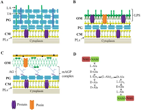
The complex and multilayer cell envelope of bacteria consists of a cytoplasmic membrane (CM), a cell wall (CW) and, an outer membrane (OM) in the case of Gram-negative bacteria and mycobacteria (Fig. 3).
Fig. 3. Bacterial cell envelopes. (A) Gram-positive bacteria, (B) Gram-negative, and (C) mycobacteria. (D)
Enterococcus faecalis peptidoglycan prototype structure (Schleifer and Kandler, 1972). OM, outer
membrane; PG, peptidoglycan; CM, cytoplasmaic membrane; LA, lipoteichoic acids; TA, teichoic acids; LPS, lipopolysaccharides; PLs, phospholipids; AG, arabinogalactan; mAGP, complex arabinogalactan-peptidoglycan; NAG, N-acetylglucosamine; NAM, N-acetylmuramic acid. Adapted from Catalão et al., 2013.
The bacterial CM is a hydrophobic phospholipid bilayer imbedded with proteins, which surrounds and contains the cytoplasm and is common to all bacteria. It is the structure responsible of energy production, lipid biosynthesis, protein secretion, and transport, and acts as a semi-permeable barrier preventing leakage of hydrophilic constituents from the cytoplasm and protecting this cell compartment from external aggressions (Silhavy et al., 2010). The CM is impermeable to protons and other ions, allowing the cell to sustain an electron-chemical gradient across the membrane and thus generating the so-called proton motive-force (PMF) (Weiner and Rothery, 2007).
Bacteria do not lyse when put into distilled water due to a rigid CW composed of peptidoglycan (PG), which protects the cells from osmotic pressure. PG is a large polymer made of repeating units of N-acetylglucosamine (NAG) and N-acetylmuramic acid (NAM), which are cross-linked by peptide side chains attached to NAM via amide bonds; due to its rigidity the CW also confers shape do the cell (Vollmer et al., 2008a). The overall variation in the PG structure of the different bacteria resides in the amino acid sequence of stem peptides and, most importantly, in that forming the interpeptide cross-bridge (Schleifer and Kandler, 1972; Vollmer et al., 2008a). Most Gram-positive bacteria have a stem peptide consisting of L-Ala-D-Glu-L-Lys-D-Ala-D-Ala (L-Lys in position 3, Lys-type PG). Stem peptides of adjacent strands are cross-linked with an interpeptide bridge from the ε-amino group of the L-Lys residue of one strand to the carboxyl group of D-Ala in position 4 of the adjacent strand. This covalent modification results in the removal of the terminal D-Ala residue at position 5 (Fig. 3D) (Hancock et al., 2014). The exact nature of these cross-bridges can be species-specific and accounts for more than 100 different PGs (Schleifer and Kandler, 1972). For most species in the genus Enterococcus, which was central to the work presented in this thesis (see next chapters), this cross-bridge is comprised of a single D-Asp residue (Kilpper-Bälz and Schleifer, 1987). Enterococcus faecalis appears to be an exception to this theme, as it possesses a cross-bridge of 2-3 L-Ala residues (Schleifer and Kandler, 1972) (Fig. 3D). In Gram-negative bacteria and some Gram-positive bacilli, peptide side chains are usually directly cross-linked, with the position 4 D-Ala of one chain being linked to the opposite meso-diaminopimelic acid (m-Dap) at position 3 (Dap-type PG) (Schleifer and Kandler, 1972; Vollmer et al., 2008a).
Gram-positive bacteria are surrounded by several layers of PG that form a cell wall thicker than that found in Gram-negative bacteria. Inside the PG mesh of Gram-positive bacteria are long anionic polymers, the teichoic (TA) and lipoteichoic acids (LTA), which
can correspond to 60% of the mass of the cell wall, making them major contributors to envelope structure and function (Neuhaus, 2003; Dramsi et al., 2008) (Fig. 3A).
In Gram-negative bacteria the thinner PG layer is surrounded by the OM, which is absent from Gram-positive organisms (Fig. 3B). This structure plays a major role in protecting Gram-negative bacteria from the environment by excluding toxic molecules and providing an additional stabilizing layer around the cell. Because the OM indirectly helps stabilize the inner membrane, the peptidoglycan mesh surrounding Gram-negative is covalently linked to the OM. The OM is a lipid bilayer composed by phospholipids (PLs) in the inner leaflet and lipophospholipids and lipopolysaccharides (LPS) in the outer leaflet (Ruiz et al., 2006).
Mycobacteria also have an OM, of distinct composition from that of Gram-negative bacteria, and, in these particular bacteria, the OM is surrounded by a capsule which is composed by proteins, polysaccharides and a small amount of lipids (Lemassu and Daffé, 1994; Lemassu et al et., 1996; Sani et al., 2010). Interestingly, in mycobacteria the PG is covalently attached to OM via arabinogalactan (AG), which is esterified to mycolic acids, forming the complex arabinogalactan-peptidoglycan (mAGP) (Brennan, 2003) (Fig. 3C).
Double-stranded DNA bacteriophages follow the most drastic strategy to overcome the host cell barriers and release their virion progeny, that is, they induce bacterial lysis. As detailed below, lysis is accomplished through specialized and regulated functions that compromise the physical integrity of the different layers composing the bacterial cell envelope.
P
HAGE RELEASE FROM INFECTED CELLS:
LYSIS-
MECHANISMS OFdsDNA
BACTERIOPHAGESThe culmination of the bacteriophage lytic cycle coincides with the lysis of the host cell to allow the release of the virion progeny. Lysis of bacterial hosts mediated by dsDNA phages seems to require at least two partners for efficient cell burst: a PG hydrolase, known as endolysin and a small hydrophobic protein designated by holin. Endolysins (see next section) are responsible for the breakdown of the PG network composing the cell wall and are essential for rapid and efficient host cell lysis. Holins are typically small
proteins (<150 amino acids) displaying 1 to 3 transmembrane domains and a hydrophilic C-terminus (Wang et al., 2000; Young 2002). Generally, holin and endolysin genes are clustered with the same transcription orientation in the phage genomes (São-José et al., 2003; Catalão et al., 2013).
The coordinated action of these two proteins in the lysis mechanism of Escherichia coli phage is, by far, the best studied and still serves as a model for most dsDNA phages employing this lysis strategy (São-José et al., 2003; Young and Wang, 2006; São-José et al., 2007). According to this model, phage endolysins accumulate in their active state in the host cell cytoplasm during phage replication, while holins are progressively embedded in CM. After reaching a critical concentration in the CM, the holins suddenly trigger to form a pore that dissipates the membrane PMF, thus killing the cell. In the case of the phage model system, this pore also constitutes the passage through which the endolysin gains access to the cell wall, which rapidly leads to its digestion, and hence to cell lysis (Young, 2013; Savva et al., 2014) (Fig. 4A).
Fig. 4. Models for export and activation of endolysins. (A) The endolysin is exported to the periplasm
through the holin pores (e.g. phage ). Holin independent, Sec-mediated export of endolysin: (B) endolysins with typical signal peptides (SP) (e.g. fOg44); (C) endolysins with signal-arrested-release sequence (SAR) (e.g. P1); and (D) mycobacteriophage Ms6 endolysin, were the endolysin export is assisted by a chaperone protein. PG, peptidoglycan; CM, cytoplasmic membrane; Cyt, cytoplasm. Adapted from Catalão et al., 2013.
The confinement of endolysins in the host cell cytoplasm during phage development was for long regarded as an imperative of any lysis strategy of dsDNA phages, simply because premature cell lysis, before the entire assembly of viral progeny, would not make biological sense. Today, however, there is an increasing awareness that phage lysis mechanisms can be diverse, with at least subtle deviations to the paradigm. For instance, it is now known that some dsDNA phages instead of making use of the holin holes to export their endolysins, they engage the host cell secretion machinery (Sec system) to carry these enzymes to the periplasm, way before the completion of the viral life cycle (São-José et al., 2000; Young, 2005; São-José et al., 2007). These phages produce endolysins with secretion signals, that is, typical signal peptides (SP) or signal-arrested-release (SAR) sequences, or synthesize chaperon-like proteins that interact with endolysins and target them to the Sec translocase (São-José et al., 2000; Xu et al., 2004; Catalão et al., 2010) (Fig 4. B, C and D).
In contrast to what was expected, it was proved that the export of these endolysins to the periplasm at early stages of virus replication had no major impact in the bacterial cell wall. This implied that the endolysins are kept inactive in the cell wall compartment, “waiting” for the exact moment for lysis to occur. An interesting observation is that the phages producing holin-independent exported endolysins also encode a holin. In fact, it has been demonstrated that even in the systems employing secreted endolysins the holins still maintain the key role of defining the lysis timing. In addition, the holin PMF-dissipating action is responsible for the activation of the pre-secreted endolysins (Nascimento et al., 2008; Young, 2013; Savva et al., 2014). It was speculated (São-José et al., 2000) and latter demonstrated (Frias et al., 2009) that the holin membrane-depolarizing function can also trigger the bacterial autolytic machinery, which contributes to fast and effective lysis of host cells. Interestingly, at least for phages relying on SAR endolysins, it has been shown that the cognate holins produce small-sized pores, as these need only to allow the passage of ions and depolarize the CM in order to fulfil their role in lysis (Park et al., 2006, 2007). These holins have been coined as ‘pinholins’ given their small-hole (pinhole)-forming character when compared to canonical holins like that of phage , which forms micron-scale holes (Park et al., 2007; Dewey et al., 2010).
In addition to the fundamental holin and endolysin players, dsDNA phages seem to have evolved auxiliary functions that contribute to the regulation and effectiveness of bacterial lysis. Well- known examples are the antiholin protein, whose role is to tune the timing of
the holin action, spanins that weaken the OM barrier of Gram-negative hosts and lipases that are thought to compromise the mycolyl-arabinogalactan external layer of the mycobacterial cell envelope (Catalão et al., 2013; Young, 2014).
Besides endolysins, dsDNA bacteriophages can also encode cell wall lytic functions that are associated with the virus particle. These often make part of multidomain, virion structural proteins that are here designated as virion-associated lysins (VALs, see next section). In addition to their role in virion morphogenesis, VALs are thought to act at the onset of phage infection by promoting a local, controlled cleavage of cell wall bonds to facilitate phage genome transference to the host bacterial cell. Since endolysins and VALs were the main object of the studies presented in this thesis, the next sections will provide a detailed description of their fundamental features.
PHAGE-ENCODED PEPTIDOGLYCAN HYDROLASES
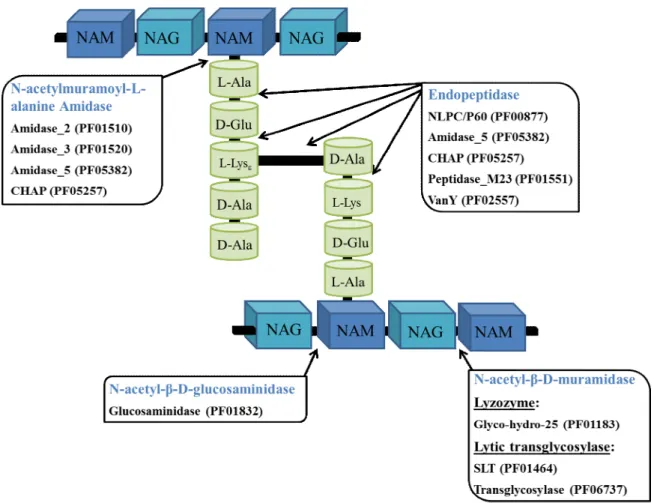
Due to the relatively conserved structure of PG, there are limited types of covalent bonds available for cleavage by phage PG hydrolases (Fig. 5). Independently of their prokaryotic or eukaryotic origin, PG hydrolases are generally classified into five major classes depending on the specific bond they attack and/or the reaction mechanism (Vollmer et al., 2008b): 1) lysozymes; 2) lytic transglycosylases; 3) N-acetyl-β-D-glucosaminidases (glucosaminidases); 4) N-acetylmuramoyl-L-alanine amidases (amidases); and 5) peptidases (carboxi- or endopeptidases, but with the latter being much more relevant in the context of phage PG hydrolases) (Fig. 5). Lysozymes and lytic transglycosylases are also collectively known as N-acetyl-β-D-muramidases (muramidases).
Fig. 5. Basic structure of peptidoglycan with indication of the bonds that are targeted by the five main types
of enzymatic domains found in phage PG hydrolases. The predominant families of catalytic domains within each class of PG hydrolases are indicated according to the Pfam database nomenclature. Note that the genesis of these families is essentially based on primary sequence relatedness; this explains why different cleavage specificities can be displayed by a single family (e.g. CHAP and Amidase_5). SLT, soluble lytic transglycosylase; CHAP, cysteine, histidine-dependent amidohydrolases/peptidases; NAM, N-acetylmuramic acid; NAG, N-acetylglucosamine.
Lysozymes and lytic transglycosylases breakdown the β-1,4-glycosidic bond between NAM and NAG. However, the latter differ from true lysozymes in that they cleave the bond with concomitant formation of an intramolecular 1,6-anhydro ring at NAM by an intramolecular transglycosylation reaction (Holtje et al., 1975; Thunnissen et al., 1994). These glycosidase activities are frequently found in phage-encoded PG hydrolases, including VALs, like the tail-associated lysin CwlP from Bacillus subtilis phage SP-β (Sudiarta et al., 2010), and endolysins, such as the pneumococcal lysozyme Cpl-1 (Garcia et al., 1987) and the phage lytic transglycosylase R (Taylor and Gorazdowska, 1974). Lysozyme and lytic transglycosylase activities are also common in PG hydrolases
produced by bacteria, either in enzymes involved in cell wall metabolism or in bacteriolysins (PG hydrolases released to the extracellular media to attack bacterial competitors, formerly class III bacteriocins, Cotter et al., 2005), and also in enzymes produced by eukaryotic cells (Scheurwater et al., 2008; Callewaert and Michiels, 2010) Glucosaminidases cleave the glycan component of the PG on the reducing side of NAG. This kind of activity is much more frequent in autolysins, such as AltA from E. faecalis (Mesnage et al., 2008), but it has also been described for the streptococcal LambdaSa2 endolysin (Pritchard et al., 2007).
The amidases target the amide bond between the C2 on the NAM and the primary L-Ala of the stem peptide. This activity is among the most frequently found in PG hydrolases and seems to be associated quite often with endolysins, maybe because this bond is highly conserved in the bacterial cell wall PG (Nelson et al., 2012). In addition, since hydrolysis of this bond separates the glycan polymer from the stem peptides, it may be more destabilizing to the PG mesh than the cleavage of other bonds and thus it may have been favored evolutionarily by bacteriophages for rapid lysis of host cells (Nelson et al., 2012). This activity has been demonstrated for the amidase domain of the staphylococcal phage 11 endolysin (Navarre et al., 1999), the phage K endolysin, LysK (Becker et al., 2009;) and the Listeria phage endolysins Ply511 (Loessner et al., 1995), just to give a few examples.
Finally, endopeptidases are the lytic enzymes that cleave any of the peptide bonds within or between the peptide stems. As referred to above, the most important variation among the bacterial cell wall PG resides in the interpeptide cross-bridges. Therefore, the activity of a given endopeptidase tends to be restricted to a particular type of PG. The listerial Ply500 and Ply118 endolysins display L-alanyl-D-glutamate endopeptidase activity (Loessner et al., 1995). The endolysin of the staphylococcal phage 11 is a bifunctional enzyme, cleaving the bond between position 4 D-Ala and the first Gly residue of the pentaglycine cross-bridge (D-alanyl-glycine endopeptidase) in addition to its amidase activity (Navarre et al., 1999). The also bifunctional endolysin of the streptococcal phage B30 exhibits D-alanine-L-alanine endopeptidase and lysozyme activities (Pritchard et al., 2004). The bacteriolysin lysostaphin from Staphylococcus simulans cleaves the S. aureus pentaglycine cross-bridge (Iversen and Grov, 1973). Besides a lytic transglycosylase activity (see above), the VAL CwlP from B. subtilis phage SP-β also harbors a peptidase domain of the M23 family (Sudiarta et al., 2010).
Phage lytic enzymes, i.e., endolysins and VALs, harbour at least one of the five muralytic activities just described but, as perceived from examples given above, some have been reported that comprise two distinct enzymatic specificities, which are generally attributed to two separate catalytic domains (CDs). In addition to these, the endolysins of phages infecting Gram-positive bacteria and mycobacteria typically harbor a C-terminal cell wall binding (CWB) domain, which mediates substrate recognition and enzyme anchoring (Nelson et al., 2012; Payne and Hatfull, 2012; Schmelcher et al., 2012; Oliveira et al., 2013). Bioinformatics and structural studies have been evidencing the diversity of catalytic and CWB domains present in PG hydrolases, when considering their primary sequence and fold, with the same PG bond being cleaved by CDs of distinct configurations. Despite this, the wealth of enzyme sequences deposited in databases has been enabling, through bioinformatics analysis, the organization of CDs and CWB motifs into different superfamilies and/or families (López and García, 2004; Firczuk and Bochtler, 2007; Layec et al., 2008a,b; Scheurwater et al., 2008; Payne and Hatfull, 2012; Oliveira et al., 2013). This, complemented with the development of sequence analysis tools (e.g. Marchler-Bauer et al., 2011), generally allows the inclusion of the functional domains of a given PG hydrolase in known superfamilies/families. This type of analysis though should be taken with caution when trying to assign the cleavage specificities of lytic enzymes as it can lead to erroneous conclusions. For example, some CHAP CDs have been shown to specify amidase activity (Nelson et al., 2006), others are endopeptidases (Navarre et al., 1999; Pritchard et al., 2004), and there is at least one example where a single CHAP displays both amidase and endopeptidase activities (Linden et al., 2014).
Endolysins
Structural diversity
Analysis of the overall structure of known phage endolysins generally leads to a distinction of those targeting Gram-positive and mycobacteria from those acting on Gram-negative bacteria, which again probably reflects the major differences in the cell wall architecture of these major bacterial groups.
In Gram-negative bacteria, the PG lies between the OM and the CM and is a relatively thin layer. Endolysins from phages that infect this type of bacteria are usually single domain, globular proteins that typically harbor a single CD and range in mass from 15 to 20 kDa (Nelson et al., 2012). Exceptions have been described, such as the Gram-negative endolysins KJ144 and EL188, both from Pseudomonas phages, which have been shown to carry a catalytic domain and an N-terminal CWB domain (Briers et al., 2007; Fokine et al., 2008) (Fig. 6).
Fig. 6. Domain architecture of Gram-negative and Gram-positive endolysins. Functional domains not
drawn to scale. Green boxes correspond to catalytic domains (CD); blue boxes represent cell wall binding domains (CWBD); N, N-terminus; and C, C-terminus.
Gram-positive organisms lack the OM and the PG is a highly cross-linked multilayer followed by the CM. As referred to above, Gram-positive endolysins show a modular structure (Diaz et al., 1991) (Fig. 6) and are usually composed by one or two N-terminal CDs connected to one to several repeats of CWB motifs at the C-terminus, which specifically recognize the host PG or other cell wall components (López and García,
2004; Nelson et al., 2012; Schmelcher et al., 2012). The two endolysin functional domains are usually linked by a flexible peptide chain (Korndorfer et al., 2006). The staphylococcal lysin LysK is an example of a bifunctional endolysin, which bears a CHAP endopeptidase and an amidase CD in the N-terminal region linked to a SH3b CWB domain (SH3_5 family, (Pfam08460 ) (O’Flaherty et al., 2005; Horgan et al., 2009).
The cell wall binding domain can have a significant impact in the activity range of endolysins. Several conserved CWB motifs have been described in the literature such as: the LysM domain (Visweswaran et al., 2011), which is the most common CWB domain in PG hydrolases and has been shown to bind to NAG residues in the sugar backbone of the PG (Ohnuma et al., 2008); the bacterial SH3b domain (Whisstock and Lesk, 1999), which is also present in some bacteriolysins; the choline-binding modules of Cpl-1 and other pneumococcal lysins (Lopez and Gracía, 2004), which specifically recognize the choline-containing theichoic acids in the cell wall of S. pneumonia; and the Cpl-7 biding domain, which binds to ethanolamine molecules present in the pneumococcal cell walls (Bustamante et al., 2010).
The recognition specificity of a CWB domain in many cases encompasses an entire bacterial genus, as observed in studies using various GFP-tagged staphylococcal SH3b binding domains (Gu et al., 2011), and is in general broader than the spectrum of the respective phage. This indicates recognition of a rather conserved ligand such as the pentaglycine interpeptide bridge shared by the most staphylococcal strains (Schleifer and Kandler, 1972). Other interesting feature about CWB motifs is that frequently they appear in multiple copies. Cpl-1 endolysin bears 6 tandem copies of the choline-binding repeats and its lytic activity depends on activation through choline binding (Garcia et al., 1990). The related pneumococcal endolysin Cpl-7 harbors 3 tandem repeats of a different CWB motif and appears to lyse bacteria both exhibiting choline and ethanolamine at the cell wall (Diaz et al., 1991).
Gram-positive endolysins are generally described as being monomeric proteins and are thus the product of a single gene. A remarkable exception is the pneumococcal endolysin PlyC, which is composed of two different subunits, PlyCA and PlyCB encoded by separate genes. PlyCA is a two CD-containing polypeptide that associates with eight PlyCB subunits with CWB activity (Nelson et al., 2006; McGowan et al., 2012) (Fig. 6).
Endolysin mode of action
The degradation of the PG layer by the action of endolysins in the context of phage infection leads to lysis of the bacterial cell. As revealed by thin-section electron microscopy, endolysins seem to display their lethal effects by forming holes in the cell wall through PG digestion. The high intracellular osmotic pressure causes extrusion of the cytoplasmic membrane, ultimately leading to hypotonic lysis (Fischetti, 2008; Fischetti, 2005). In principle, a single endolysin molecule should be sufficient to cleave several numbers of bonds. However, Loessner and collegues (2002) showed that a listerial phage endolysin had a binding affinity approaching that of an IgG molecule for its substrate, suggesting that phage proteins are one-use enzymes, probably requiring several molecules attacking the same region to efficiently weaken the cell wall.
Virion-associated lysins of dsDNA bacteriophages
As described above, bacteriophages must transport their genome across the bacterial cell envelope to initiate infection. The common obstacles to phage genome transit are the PG and CM layers but additional barriers like an OM and/or a polysaccharidic capsule may be present depending on the host. While the OM is generally traversed by puncturing (for example by a device of the tail), crossing of capsule and PG layers generally benefit from depolymerizing activities carried in the virion structure (Casjens and Molineux, 2012). Most phage particles carry at least one protein with cell wall degrading activity (the VAL) that allows access of the tail tube to the CM (Moak and Molineux, 2004). Traffic through this last barrier likely involves pore formation and/or membrane fusion events but its molecular details remain the less understood in the process of virus entry (Letellier et al., 2004). VALs are designed to promote a “surgical” lesion in the cell wall without leading to cell death. However, if a VAL-carrying phage adsorbs at very high multiplicities to a host cell, it can culminate in cell destruction. This phenomenon is denominated by “lysis from without”, as it is a lysis that does not rely on phage infection (Abedon, 2011).
VALs seem to be quite common in both Gram-negative and Gram-positive infecting phages (Moak and Molineux, 2004). These enzymes are typically associated to the phage DNA injection machinery and are most frequently incorporated in the tail structure (Fokine and Rossmann, 2014). The P7 VAL of the tail-less, dsDNA phage PRD1 which
infects various Gram-negative bacteria, such as E. coli, Salmonella enterica and Pseudomonas aeruginosa, is associated with the membrane beneath the icosahedral capsid (Rydman and Bamford. 2000). VALs are much less studied compared to endolysins. Very often they correspond to multifunctional proteins that, in addition to the PG hydrolase activity, play a role in the assembly of the phage tail. Known examples of this are the tape measure proteins (TMP), which determine the length of the tail and at the same time may display PG hydrolase activity (Piuri and Hatfull, 2006; Boulanger et al., 2008). VALs may also make part of central tail knobs, fibers or spikes (Moak and Molineux, 2000; Kanamaru et al., 2002; Kenny et al., 2004; Xiang et al., 2008). They are usually larger than cognate endolysins, present high sequence diversity and variable domain organization (Rodriguez-Rubio et al., 2012) (Fig. 7).
Fig. 7. VALs domain organization and diversity of PG cleavage specificities. Three illustrative examples
(not to scale) of known VALs targeting Gram-negative and Gram-positive bacteria are shown. CD families: SLT, soluble lytic transglycosylase; M23, peptidase M23; Lyz, lysozyme; CHAP, cysteine, histidine-dependent amidohydrolases/peptidases.
The domains of VALs responsible for PG hydrolase activity are related to those of endolysins and bacterial PG hydrolases. Yet, in contrast to the endolysins acting on Gram-positive bacteria, the VALs targeting this group of bacteria usually lack a domain responsible for cell wall binding (Rodriguez-Rubio et al., 2012). The lack of a CWB
domain is not surprising given the context of action of these proteins. Receptor binding proteins (RBP) carried in the phage tail distal end are responsible for recognition and attachment to bacterial surface receptors. This RBP/receptor interaction triggers major conformational changes in the tail structure that ultimately places the VAL it in close contact with its substrate (Fig. 8), thus becoming unnecessary the presence of a specific domain to direct the enzyme, as it happens with Gram-positive endolysins (Veesler and Cambillau, 2011; Rodriguez-Rubio et al., 2012; Fokine and Rossmann, 2014). One exception to this rule seems to be the staphylococcal phage 68 VAL P17, which shows a typical endolysin domain organization composed by an N-terminal CD and a C-terminal CWB domain (Takac et al., 2005). Interestingly, Rodriguez and collaborators (2011) showed that the two CDs of the VAL HydH5, encoded by the S. aureus phage phiIPLA88, had the ability to bind target cells. In fact, VALs acting on Gram-positive bacteria frequently display two CDs (Rodriguez-Rubio et al., 2013). To date, there is no described VAL from a Gram-negative infecting phage that harbors more than one CD.
Fig. 8. Schematic representation of the mode of action of a virion-associated lysin (VAL) of a prototype
Gram-positive Myoviridae bacteriophage.
A curious observation is the apparent abundance of VALs of Gram-positive phages carrying a CD of the peptidase M23 family in (our analysis). As far as we know, such CD has never been observed in VALs from Gram-negative phages. The M23 peptidase CD is also present in other PG hydrolases that, similarly to VALs, “attack” the bacterial cell from the outside. This is the case of the bacteriolysins lysostaphin (Shindler and