194
PARTIAL CHARACTERIZATION OF A LYTIC METHICILLIN RESISTANT-STAPHYLOCOCCUS AUREUS
BACTERIOPHAGE
Sulaiman Al-Yousef*
Address(es): University of Dammam, College of Applied Medical Science, Department of Medical Laboratory Technology, 1704, Hafr Al Batin-319 91, Saudi Arabia.
*Corresponding author: [email protected]
ABSTRACT
Keywords: MRSA, phage, concentration, characterization
INTRODUCTION
Staphylococcus aureus (S. aureus) is a versatile pathogenic bacterium (Schmidt et al., 2010) responsible for a significant number of heal thcare-associated and community-acquired infections (Kurlenda and Grinholc, 2010). It causes a broad spectrum of infections ranging from acute to chronic disease (Van den Berg et al., 2011 and Francois et al., 2010) and is a common etiological agent of opportunistic infections. It is well known that the organism acquires resistance soon after the introduction of new antibiotics (Lyon and Skurray, 1987). Methicillin was developed in 1960 for the treatment of such multidrug resistant Staphylococcus aureus. However in the same year, Jevons (1961) discovered Methicillin Resistance S. aureus (MRSA), which by 1970s has become spread all over the world. There was a new interest in the possibilities of bacteriophage therapy, which a specific kind of viruses that attack only bacteria used to kill the pathogenic microorganisms (Barrow and Soothhill, 1997). Duckworth and Gulig, 2002 showed that phage therapy would have a role against infectious diseases. Although bacteriophages had not been extensively used as antimicrobial agents since the emergence of antibiotics, the rise of multiple antibiotic resistant strains had a renewed in phage therapy (Platt et al., 2003). Amenu, (2014) examine various aspects of bacteriophage ecology, particularly focusing on phages specific to the bacterial species Staphylococcus aureus, Pseudomonas aeruginosa, and Escherichia coli. Basdew and Laing (2014) studied sensitivity of six S. aureus-phage isolates towards simulated environmental. Three of the six isolated phages performed optimally during the in vitro assays and have considerable potential for in vivo applications. Recently, some Staphylococcal phages have been isolated and characterized (Gu et al., 2012; Cui et al., 2012; Amenu, 2014; Swift and Nelson, 2014; El Haddad et al., 2014; Li and Zhang, 2014). Our study was performed to complete the previous work (Al-Yousef, 2013). Also, objectives of the present work were to study biological and partial characterize of MRSA-phage.
MATERIAL AND METHODS
Staphylococcus aureus and bacteriophage
Methicillin-Resistant S. aureus (MRSA) strain and their specific bacteriophage was obtained from previous work (Al-Yousef, 2013). Methicillin resistance of S.
aureus was determined by MRSA screening agar containing 6μg /ml oxacillin.
Confirmation for methicillin resistance was performed by determining the oxacillin-MIC by the standards of antimicrobial susceptibility testing according to CLSI, 2013 and MicroScan® instrumentation (Siemens Healthcare Diagnosis Inc, USA) (Barry, 2007). Bacteriophage stocks were generated by combining
500 μL of MRSA culture with 500 μL of each bacteriophage (1.0 x10⁶ PFU mL-1)
in a test tube, pouring the contents onto a tryptic soy agar plate, and incubating
the plate overnight at 37 °C. Following incubation, the plate’s content was
collected into a 50 mL tube by washing with tryptic soy broth (TSB), scraping with a sterile plastic loop, and aspirating with a pipette. The tube was then centrifuged for 20 minutes at 3500 rpm to pellet out the debris, and the
supernatant was filtered through 0.2 μm Millipore membrane to remove
extraneous debris. The final phage filtrate was quantified using a traditional plaque assay (Adams, 1959).
Plaque purification and lysate preparation
A single plaque was picked from the plate with a sterile tooth-pick, introduced into 3 ml of a log phase culture of the MRSA host in nutrient broth, and was
incubated at 37ºC in an environmental shaker at 120 rpm for 12 hours. This was
then centrifuged at 10.000g followed by filtration through 0.22 μm Millipore
membrane. The lysate obtained was used for double agar overlay. This procedure was repeated 6 times, until uniform sized plaques were obtained on the plate.The plates with uniform sized plaques were overlaid with 10 ml of SM buffer solution [100 mM NaCl, 10 mM MgSO4, 50 mM Tris-HCl, pH 7.5 and 0.01% (w/v)
gelatin] and were incubated overnight at 4°C, with gentle rocking so that phages
could easily diffuse into the buffer. The phage suspension was recovered after incubation from all the plates and pooled. Chloroform was added to this pooled mixture to a final concentration of 5 % (v/v), mixed well using a vortex mixer and incubated at room temperature for 15 minutes. The cell debris was then removed by centrifugation at 5000 g for 10 minutes (Sigma, 3K30, Germany) and the supernatant was transferred to sterile polypropylene tube. Chloroform was
added to a final concentration of 0.3 % (v/v) and this was stored at 4°C until use.
The titer of this lysate was noted after serial dilution according to Malke, (1990). A marked increase in the infection incidence caused by methicillin-resistant Staphylococcus aureus (MRSA) strains has been noted in medical practice in recent years. This study was conducted to study the biological and characterize of MRSA-phage. Methicillin resistance of Staphylococcus aureus was detected and confirmed by determining of the MIC of oxacillin by the standard agar dilution method. Phage was biologically purified using single plaque technique, then phage characterization were studied using host range, adsorption time, particle morphology and its structural protein. MRSA phage showing lytic nature was purified by repeated plating after picking of single isolated plaques. This phage is active against all 11 isolates either of S. aureus or MRSA tested as hosts. Phage produced clear plaques indicating their lytic nature. This phage was concentrated employing polyethylene glycol (PEG)-NaCl precipitation method. Morphologically, MRSA Phage has a hexagonal head having a long non-contractile tail, indicating his icosahedral nature. Adsorption studies showed 100% adsorption of MRSA-Phage after 35 minutes of exposure. Sodium dodecyl sulfate-polyacrylamide gel electrophoresis (SDS-PAGE) experimentation indicated that the phage particles contain one major structural protein (about 30 Kda).
ARTICLE INFO
Received 7. 9. 2014 Revised 25. 9. 2014 Accepted 25. 9. 2014 Published 1. 12. 2014
Regular article
J Microbiol Biotech Food Sci / Sulaiman Al-Yousef 2014/15 : 4 (3) 194-197
195 Host range determination
Spot tests were used for host range studies, and, in all cases, positive tests were confirmed by plaque assay. Eighteen bacterial isolates, (6 MRSA; 5 S. aureus; 2 Pseudomonas aeruginosa; 2 Klebsiella pneumonia and 3 Escherichia coli) (Table 1) were used as indicators for the determination of phage sensitivity by an agar spotting method (Adams, 1959). Ten microliters from bacteriophage samples (109 PFU/ml) were placed on each lawn of indicator cells and left to stand for 30
min. Plaque formation was examined for lysis after 24h incubation at 37°C. The
results were recorded as positive (+) and negative (-).
Phage adsorption
The first step in the growth of bacteriophage was its attachment to susceptible bacteria, this process is called adsorption. Log phase culture of host was infected
using the phage then incubated at 37°C. Aliquots of 5 ml were sampled at 0, 5,
10, 15, 20, 25, 30, 35, 40 and 45 minutes time intervals after infection. All the
samples drawn were immediately filtered through 0.22 μm membrane filter
(Millipore, USA). The phage titer was determined using double agar overlay method after appropriate dilutions. All plating was done in triplicates and appropriate controls were maintained. The percentage of phage adsorption was calculated as follows: [(control titer -residual titer)/control titer] X 100% (Durmaz et al., 1992). The phage titer observed at time zero was considered as the control titer.
Phage concentration
Phage was concentrated using Polyethylene glycol (PEG) 6000 as described by Malke, (1990). Briefly, 1% (v/v) of an overnight culture of the host bacteria was transferred to 200 ml nutrient broth, and incubated at 37°C for 3 hours in an
environmental shaker at 100 rpm. Phages were added and the incubation at 37°C
was continued at 100 rpm for 12 hours. This broth was then centrifuged at 8000 rpm for 20 minutes (Sigma, 3K30, Germany), the supernatant was collected and
filtered through 0.22 μm membrane Millipore filter. Solid NaCl was added to a
final concentration of 1 M and dissolved by stirring with a sterile glass rod. This mixture was kept in ice for 1 hour, followed by centrifugation at 9000 rpm for 10
minutes at 4°C. Solid PEG 6000 was added to the supernatant at a final
concentration of 10% (w/v), dissolved by slow stirring on a magnetic stirrer at room temperature. This was then kept in ice overnight, followed by centrifugation at 9000 rpm for 10 minutes at 4ºC. The supernatant was discarded completely, while the pellet was resuspended in 5 ml of phosphate buffered saline (PBS). PEG and cell debris were removed from the phage suspension by the addition of an equal volume of chloroform, vortexing for 30 seconds,
followed by centrifugation at 2000 rpm for 15 minutes at 4ºC. The aqueous phase
containing the phage particles were recovered and stored at -20ºC. Phage stock
was stored with chloroform (5% by volume) at 4 ºC, and an aliquot was frozen at
-80 ºC in MRS broth containing 15% glycerol.
Morphological analysis by transmission electron microscopy (TEM)
Formvar membrane-coated grids were placed on a drop of bacteriophage diluted with distilled water. The grids were then placed on a drop of 2% uranylacetate for 10–20 s, lifted and dried with filter paper, then dried in air. The grids were examined using a Hitachi 7100 transmission electron microscope (Joil, model GEM 1010).
Phage structural protein analysis by sodium dodecyl sulfate-polyacrylamide gel electrophoresis (SDS-PAGE)
SDS-PAGE was performed for determining the size and number of the proteins of phage particles. Concentrated and purified sample phage (0.1-ml) was heated
at 100°C for 5 min to ensure denaturation before loading on a gel for
electrophoresis. Electrophoresis was carried out with a 4% stacking layer and 12.5% separating gel according to Laemli (1970). Standard proteins were used as
size markers. Each well was loaded with 10 μl of sample containing
approximately 10 μg of protein. Following electrophoresis, protein bands were
stained with Coomassie blue R-250 solution, followed by destaining with 7.5% glacial acetic acid and 5% methanol, and then photographed.
RESULTS AND DISCUSSION
A marked increase in the infection incidence caused by MRSA strains has been noted in medical practice in recent years. Colonization with MRSA has been described as a risk factor for subsequent MRSA infection (Davis et al., 2004 and Khairulddin et al., 2004). The prospects of lytic phages as bio-control agents against pathogenic bacteria are being reconsidered worldwide with the surfacing of multiple antibiotic resistances (Sulakvelidze et al., 2001). S. aureus is one among many bacterial pathogens which phages are experimented as therapeutic agents (Leszczyňski et al., 2006). Thus the rationale of the present study was to search for specific lytic phages where it was most likely to be found i.e. from the
soil (the same environment where their host S. aureus is known to reside) (Al-Yousef, 2013).
Bacteriophages isolation
MRSA was the host bacterium that helped to isolate lytic bacteriophage from the soil samples in previous work. The phage was obtained only via the enrichment protocol. MRSA Phage consistently exhibited excellent bacterial cell lysis capability. MRSA Phage was purified by repeated plating and picking of single isolated plaques from the lawns of MRSA plates, and produced clear and round plaques with well-defined edges indicating their lytic nature (Figure 1).
Figure 1 Plate showing plaques formed by Staphylococcus aureus-phage on bacterial lawn of MRSA
Host range
The host range of MRSA phage was determined with 18 selected isolates from 4 genera. MRSA phage was lytic against both MRSA and S. aureus. The other bacterial isolates were not sensitive to phage (Table 1). Host range within the genus is considered to be a desirable quality of phage as a bio-control agent.
Table 1 Host range of MRSA-phage
Bacterial isolates Lysis(a) Methicillin Resistance S. aureus (MRSA)
Methicillin Resistance S. aureus (MRSA) Methicillin Resistance S. aureus (MRSA) Methicillin Resistance S. aureus (MRSA) Methicillin Resistance S. aureus (MRSA) Methicillin Resistance S. aureus (MRSA) S. aureus
S. aureus S. aureus S. aureus S. aureus
Pseudomonas aeruginosa Pseudomonas aeruginosa Klebsiella pneumonia Klebsiella pneumonia Escherichia coli Escherichia coli Escherichia coli
+ + + + + + + + + + + - - - - - - - a + = Plaques formed; - = no plaque formed
Concentration of MRSA Phage
As concentration and purification of virus particles are prerequisites for structural and functional characterization of phages (Boulanger, 2009), MRSA phage was purified and concentrated. Concentration was done employing PEG-NaCl precipitation method was as described by Malke, (1990). The efficiency of this method is almost independent of phage concentration and is therefore useful in order to concentrate even phage lysates with very low titer (Yamamoto et al.,
1970). MRSA phage was concentrated up to 1 x 10¹⁰ PFU/ml using PEG
precipitation and these phage concentrates prepared in large quantities were used for all further studies.
Characterization of isolated MRSA-phage
Morphological analysis by TEM
J Microbiol Biotech Food Sci / Sulaiman Al-Yousef 2014/15 : 4 (3) 194-197
196
hexagonal head (64 ± 2nm) having a long non-contractile tail (132 ± 2nm), which
are typical morphological features of family Myoviridae (Figure 2).
Figure 2 Transmission electron micrograph image of phage stained with 2% uranyl acetate MRSA phage.
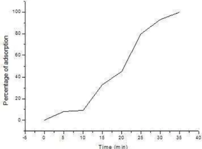
Phage adsorption
Phage adsorption on to the susceptible host is the second significant factor affecting the successful phage-host interaction. Adsorption is described as the first step of phage infection and may be defined as the attachment of phage particles to bacterial surfaces so that phage and bacteria can sediment together (Adams, 1959). The phage extracellular search is a time of free diffusion that delays the onset of virus attachment (Shao and Wang, 2008). Careful determination of the time taken by the phages to adsorb onto to the host cell is of paramount importance, as it may serve in later experiments for accurate characterization of the phage. The adsorption curve of MRSA-phage is shown in Figure 3. MRSA phage adsorption achieved 100% after 35 minutes of exposure to the host bacteria.
Figure 3 Adsorption curves of MRSA phage.
Phage protein
The structural protein composition of Staphylococcus phage was analyzed by SDS-PAGE. One major structural protein was detected during the analysis of the phage. Protein profiling can be used as a molecular signature tag of a phage, helping in identifying as well as differentiating it from other phages, as phages even within the same family tend to have different structural proteins owing to differences in phage specificity (Shivu et al., 2007) as evidenced by the lack of comparable protein profile among phages belonging to Podoviridae family. The structural protein of MRSA-phage was analyzed by SDS-PAGE (Figure 4). It showed that the virions contained one major protein with a molecular weight about of 30 kDa.
Figure 4 Sodium dodecyl sulfate-polyacrylamide gel electrophoresis of MRSA phage structural protein. Lanes: M, standard proteins; 1, phage protein, stained with Coomassie blue. The size of the phage proteins is expressed in kDa.
CONCLUSION
In this study, virulent phage with lytic activity against MRSA strain was isolated. Morphologically, the phage is member of the family Myoviridae, and exhibited an identical host range. Adsorption studies showed 100% adsorption of MRSA-Phage after 35 minutes of exposure. SDS-PAGE indicated that the phage particles contain one major structural protein (about 30 Kda). Our results indicated good efficacy for this phage, which consistent with data cited by Kaźmierczak et al., 2014. They studied the phages which able to kill S. aureus in the treatment of various human diseases, e.g., venous leg ulcers and eye infections, septicemia, staphylococcal lung infections, and others. Because there are not enough chemotherapeutics to destroy bacteria and to counteract the problem of infections in the human population, however the field of phage therapy as antibacterial agents has advanced considerably as an alternative to antibiotics (Kwiatek et al., 2012; Chhibber et al., 2008; Kim et al., 2007 and Westwater et al., 2003). Finally, phage treatment may help to reduce the frequency of potentially lethal infections in the hospital environment, with related costs that can be significantly lower than those of antibiotics.
REFERENCES
ADAMS, M. 1959. Bacteriophages, Interscience Publishers, NY Blackie Academic & Professional, London.
AL-YOUSEF, S. 2013. Methicillin-Resistant Staphylococcus aureus Phages: Isolation and Identification. Journal of pure and applied microbiology, 7(2), 939-945.
AMENU, D. 2014. Isolation of Bacteriophages Specific to Escherichia coli, Staphylococcus aureus and Pseudomonas aeruginosa. Bio-Genetics J., 2(1), 32-36.
ASWANI, V., TREMBLAY, D., MOINEAU, S., SHUKLA, S. 2011. Staphylococcus epidermidis Bacteriophages from the Anterior Nares of Humans.
Applied and environmental microbiology, 77(21), 7853–7855.
http://dx.doi.org/10.1128/aem.05367-11
BARROW, P., SOOTHILL, J. 1997. Bacteriophage therapy and prophylaxis: rediscovery and renewed assessment of potential. Trend Microbiology, 5(7), 268– 271. http://dx.doi.org/10.1016/s0966-842x(97)01054-8
BARRY, A. 2007. An Overview of the clinical and laboratory standards institute (CLSI) and its impact on antimicrobial susceptibility tests. Antimicrobial Susceptibility Testing Protocols [Internet]. CRC Press; May 22, 1-6, Available from: http://dx.doi.org/10.1201/9781420014495.ch1
BASEW, I. H., LAING, M. D. 2014. Stress sensitivity assays of bacteriophages associated with Staphylococcus aureus, causal organism of bovine mastitis. African Journal of Microbiology Research, 8(2), 200-210.
BOULANGER, P. 2009. Purification of bacteriophages and SDS-PAGE analysis of phage structural proteins from ghost particles. Methods in molecular biology, 502, 227-238. http://dx.doi.org/10.1007/978-1-60327-565-1_13
CHHIBBER, S., KAUR, S., KUMARI, S. 2008. Therapeutic potential of bacteriophage in treating Klebsiella pneumoniae B5055-meiated lobar pneumonia in mice. J Med Microbiol, 12, 1508-1513.
CLSI, 2013. Performance Standards for Antimicrobial Susceptibility Testing; Twenty-Third Informational Supplement. CLSI document M100-S23. Wayne, PA: Clinical and Laboratory Standards Institute.
CUI, Z., SONG, Z., WANG, Y., ZENG, L., SHEN, W. 2012. Complete genome sequence of wide-host-range Staphylococcus aureus phage JD007. J Virol, 86, 13880–13881.
DAVIS, K., STEWART, J., CROUCH, H., FLOREZ, C., HOSPENTHAL, D. 2004. Methicillin- resistant Staphylococcus aureus (MRSA) nares colonization at hospital admission and its effect on subsequent MRSA infection. Clinical
Infectious Diseases, 39(6), 776-782. http://dx.doi.org/10.1086/422997
DUCKWORTH, D., GULIG, P. 2002. Bacteriophage. BioDrugs, 16(1), 57–62.
http://dx.doi.org/10.2165/00063030-200216010-00006
DURMAZ, E. 1992. A fourth mechanism of bacteriophage resistance in Lactococcus lactis subsp. lactis ME2. MS Thesis. NC State University, Raleigh, NC.
EL HADDAD, L., BEN ABDALLAH, N, PLANTE,P. L., DUMARESQ, J., KATSARAVA, R., LABRIE, S., JACQUES, C., ST-GELAIS, D., MOINEAU, S. 2014. Improving the safety of Staphylococcus aureus polyvalent phages by their production on a Staphylococcus xylosus strain. PLoS ONE, 9(7), 1-10 e102600. doi:10.1371/journal.pone.0102600
FRANCOIS, P., SCHERL, A., HOCHSTRASSER, D., SCHRENZEL, J. 2010. Proteomic approaches to study Staphylococcus aureus pathogenesis. Journal of
Proteomics, 73(4), 701–708. http://dx.doi.org/10.1016/j.jprot.2009.10.007
JEVONS, M. 1961. “Celbenin” - resistant Staphylococci. Br. Med. J., 1(5219),
124–125. http://dx.doi.org/10.1136/bmj.1.5219.124-a
GU, J., LIU, X., LU,, R., LI, Y., SONG, J. 2012. Complete genome sequence of
Staphylococcus aureus bacteriophage GH15. J Virol, 86, 8914–8915.
KAŹMIERCZAK, Z., GŐRSKI, A., DABROWSKA, K. 2014. Facing antibiotic
J Microbiol Biotech Food Sci / Sulaiman Al-Yousef 2014/15 : 4 (3) 194-197
197 KHAIRULDDIN, N., BISHOP, L., LAMAGNI, T., SHARLAND, M., DUCKWORTH, G. 2004. Emergence of methicillin resistant Staphylococcus aureus (MRSA) bacteraemia among children in England and Wales. Archives of
Disease in Childhood, 89(4), 378–379.
http://dx.doi.org/10.1136/adc.2003.028712
KIM, K. P., KLUMPP, J., LOESSNER, M. J. 2007. Enterobacter sakazakii bacteriophages can prevent bacterial growth in reconstituted infant formula. Int J Food Microbiol, 115, 195-203.
KURLENDA, J., GRINHOLC, M. 2010. Current diagnostic tools for methicillin-resistant Staphylococcus aureus infections. Molecular Diagnosis and Therapy, 14, 73-80. http://dx.doi.org/10.1007/bf03256356
KWIATEK, M., PARASION, S., MIZAK, L., GRYKO, R., BARTOSZCZE, M., KOCIK, J. 2012. Characterization of a bacteriophage, isolated from a cow with mastitis, that is lytic against Staphylococcus aureus strains. Arch Virol, 157, 225– 234.
LAEMMLI, U. 1970. Cleavage of structural proteins during the assembly of the heated of bacteriophage T4. Nature, 227(5257), 680-685.
http://dx.doi.org/10.1038/227680a0
LESZCZYNSKI, P., WEBER-DABROWSKA, B., KOHUTNICKA, M., LuczakLUCZAK, M., GORSKI, A. 2006. Successful eradication of methicillin-resistant Staphylococcus aureus (MRSA) intestinal carrier status in a healthcare worker-case report. Folia Microbiolica, 51(3), 236-238.
http://dx.doi.org/10.1007/bf02932128
LI, L., ZHANG, Z. 2014. Isolation and characterization of a virulent SPW specific Staphylococcus aureus from bovine mastitis of lactating dairy cattle. Mol Biol Rep, 41, 5829-5838.
LYON B., SKURRAY, R. 1987. Antimicrobial resistance of Staphylococcus aureus: genetic basis. Microbiol. Rev., 51, 88-134.
MALKE, 1990. J. SAMBROCK, E. F. FRITSCH and T. MANIATIS, Molecular Cloning, A Laboratory Manual (Second Edition), Volumes 1, 2 and 3. 1625 S., zahlreiche Abb. und Tab. Cold Spring Harbor 1989. Cold Spring Harbor Laboratory Press. ISBN: 0-87969-309-6. Journal of Basic Microbiology, 30(8), 623–623. http://dx.doi.org/10.1002/jobm.3620300824
PLATT, R., REYNOLDS, D., PHILLIPS, G. 2003. Development of a novel method of lytic phage delivery by use of a bacteriophage P22 site-specific recombination system". FEMS Microbiology Letters, 223(2), 259-265.
http://dx.doi.org/10.1016/s0378-1097(03)00388-4
SCHMIDT, F., SCHARF, S., HILDEBRANDT, P., BURIAN, M., BERNHARDT, J., DHOPLE, V., KALINKA, J., GUTJAHR, M., HAMMER, E., VOLKER, U. 2010. Time-resolved quantitative proteome profiling of host-pathogen interactions: the response of Staphylococcus aureus RN1HG to internalization by human airway epithelial cells. Proteomics, 10(15), 2801-2811.
http://dx.doi.org/10.1002/pmic.201000045
SHAO, Y., Wang, I. 2008. Bacteriophage adsorption rate and optimal lysis time.
Genetics, 180(1), 471–482. http://dx.doi.org/10.1534/genetics.108.090100
SHIVU, M., RAJEEVA, B., GIRISHA, S., KARUNASAGAR, I., KROHNE, G., KARUNASAGAR, I. 2007. Molecular characterization of Vibrio harveyii bacteriophages isolated from aquaculture environments along the coast of India.
Environ. Microbiol., 9(2), 322-331.
http://dx.doi.org/10.1111/j.1462-2920.2006.01140.x
SULAKVELIDZE, A., ALAVIDZE, Z., MORRIS, J. 2001. Bacteriophage therapy. Antimicrobial Agents and Chemotherapy, 45(3), 649–659.
http://dx.doi.org/10.1128/aac.45.3.649-659.2001
SWIFT, S.M., NELSON, D.C. 2014. Complete Genome Sequence of Staphylococcus aureus Phage GRCS. Genome Announc, 2(2), e00209-14. doi:10.1128/genomeA.00209-14.
VAN DEN BERG, S., BOWDEN, M., BOSMA, T., BUIST, G., VAN DIJL, J., VAN WAMEL W., DE VOGEL, C., VAN BELKUM, A., BAKKER-WOUDENBERG, I. 2011. A multiplex assay for the quantification of antibody responses in Staphylococcus aureus infections in mice. Journal of Immunological
Methods, 365(1-2), 142-148. http://dx.doi.org/10.1016/j.jim.2010.12.013
WESWATER, C., KASMAN, L. M., SCHOFIELD, D. A., WERNER, P> A., DOLAN, J. W., SCHMID, M. G., NORRIS, J. S. 2003. Use of genetically engineered phage to deliver antimicrobial agents to bacteria: an alternative therapy for treatment of bacterial infections. Antimicrob Agents Chemother, 47, 1301-1307.