Faculdade de Motricidade Humana
Dancers’ Landing Strategies – Experimental and Computational Approaches
Ana Paula Morais Teixeira de Azevedo
Orientador: Professor Doutor Raul Alexandre Nunes da Silva Oliveira Co-orientadores: Professor Doutor Nelson Filipe Neves Cortes
Professor Doutor João Pedro Casaca Rocha Vaz
Tese especialmente elaborada para obtenção do grau de Doutor no ramo Motricidade Humana, na especialidade de Comportamento Motor
Júri
Presidente:
Professor Doutor Francisco José Bessone Ferreira Alves
Presidente do Conselho Científico da Faculdade de Motricidade Humana da Universidade de Lisboa
Vogais
Doutor Nelson Filipe Neves Cortes Associate Professor
Sports Medicine Assessment Research & Testing (SMART) Laboratory da George Mason University, Virginia, EUA
Doutor Orlando de Jesus Semedo Mendes Fernandes Professor Auxiliar
Departamento de Desporto e Saúde da Universidade de Évora Doutora Filipa Oliveira da Silva João
Professora Auxiliar
Faculdade de Motricidade Humana da Universidade de Lisboa Doutora Emma Redding
Professor
Trinity Laban Conservatoire of Music and Dance, Londres 2019
Nome: Ana Paula Morais Teixeira de Azevedo Endereço eletrónico: apmtazevedo@gmail.com Telefone: +351 96 44 38 793
Número do Cartão de Cidadão: 12057382
Título da Tese: Dancers’ Landing Strategies – Experimental and Computational
Approaches
Orientador: Professor Doutor Raul Alexandre Nunes da Silva Oliveira Co-orientadores: Professor Doutor Nelson Filipe Neves Cortes
Professor Doutor João Pedro Casaca Rocha Vaz
Ano de conclusão: 2019
Ramo de conhecimento do Doutoramento: Motricidade Humana, especialidade de
Comportamento Motor
NÃO É AUTORIZADA A REPRODUÇÃO INTEGRAL DESTA DISSERTAÇÃO. .
Faculdade de Motricidade Humana – Universidade de Lisboa Cruz Quebrada, 2019
Assinatura:_____________________________________ (Ana Paula Morais Teixeira de Azevedo)
GRANTS, PEER-REVIEWED PAPERS & INTERNATIONAL CONFERENCE
ABSTRACTS, AND AWARDS DURING THE PhD
During the PhD program, two outstanding visiting research scholarships were awarded to further develop and improve research skills. Additionally, the PhD findings have been disseminated (2018) and are in the process of being presented (2019) at international conferences or annual meetings. The scientific meetings focus on fields related to this PhD thesis, such as Biomechanics, Dance Science, Bioengineering, and Computational Modeling.
A. Grants
Academic year 2017-2018:
• Fulbright Visiting Research Scholarship at George Mason University (VA, USA); Mentor Dr. Nelson Cortes (Associate Professor). Duration: 9 months. Academic year 2018-2019:
• Short-term Scholar – Visiting Researcher at George Mason University (VA, USA); Mentor Dr. Nelson Cortes (Associate Professor). Duration: 6 months.
B. Peer-reviewed Papers
2019
• Published: Azevedo AM, Oliveira R, Vaz JR, & Cortes N (2019). Professional Dancers Distinct Biomechanical Pattern during Multidirectional Landings. Med Sc
Sport Exerc, 51(3): 539-547.
• Published: Azevedo AM, Oliveira R, Vaz JR, & Cortes N (2019). Foot Modeling Affects Ankle Sagittal Plane Kinematics during Jump-landing. J Biomech,
https://doi.org/10.1016/j.jbiomech.2019.109337
• Accepted – to be published in indexed IEEE Xplore and Medline/PubMed: Azevedo AM, Oliveira R, Vaz JR, & Cortes N (2019). Effect of Two Different Pose
Estimation Approaches on Lower Extremity Biomechanics in Professional Dancers, IN PRESS.
C. Peer-reviewed Conferences or Annual Meetings
2018
• June – Thematic Poster
65th American College of Sports Medicine Annual Meeting (Minneapolis, MN,
USA). The Biomechanical Pattern of Multidirectional Single-leg Landing in
Professional Dancers and Non-dancers.
• August – Poster Presentation
42nd American Society of Biomechanics Conference (Rochester, MN, USA).
Landing Biomechanics differ between Professional Dancers and Non-dancers: Experimental and Preliminary Modeling Approaches.
• September – Oral Presentation
2nd Lisbon Foot & Ankle Clinical Biomechanics Course 2018, Sports Edition
(Lisbon, Portugal). Movement Analysis in Dancers – Landing Biomechanics. • October – Oral Presentation
28th Annual Meeting of the International Association for Dance Medicine &
Science (Helsinki, Finland). Professional Dancers have Distinct
Multi-segmented Foot-ankle Biomechanics than Non-dancers during Multidirectional Landings (2018 IADMS Research Student Award).
2019
• July – Ignite Session and Poster Presentation
41st Engineering in Medicine and Biology Conference (Berlin, Germany). Effect
of Two Different Pose Estimation Approaches on Lower Extremity Biomechanics in Professional Dancers.
• August – Poster Presentation
27th Congress International Society of Biomechanics and 43rd American Society
of Biomechanics Conference (Calgary, Canada). Musculoskeletal
Computational Model Optimization: The Critical Role of Markers Error Adjustments.
• October – Poster Presentation
29th Annual Meeting of the International Association for Dance Medicine &
Science (Montreal, Canada). Professional Dancers Shock Absorption
Mechanism Differs from Non-dancers during Landings.
D. Conference Award
2018
The oral communication Professional Dancers have Distinct Multi-segmented
Foot-ankle Biomechanics than Non-dancers during Multidirectional Landings, which is
part of this PhD thesis and aforementioned, was distinguished with the Conference
Award, 2018 IADMS Research Student Award at the 28th Annual Meeting of the
TITTLE:
Dancers’ Landing Strategies – Experimental and Computational ApproachesABSTRACT
Jump-landing activities are an essential part of dance training technique, and have aesthetic and technic constrains. Previous research reported that dancers display an extended landing posture at initial ground contact, which has been considered a deleterious pattern. However, this population shows higher joint excursions of the lower extremity allowing to appropriately dissipate the impact forces. Additionally, computational modeling and simulation (e.g., OpenSim) has had limited applications in dance science. This PhD thesis main purpose was to determine lower extremity biomechanical patterns in professional dancers during single-leg landings in lateral, diagonal and forward directions and compare to non-dancers. Five studies were conducted employing experimental and computational modeling approaches: 1) a more broad study that exhibited extensive kinematics and kinetic differences between professional dancers and non-dancers in multidirectional jump-landings; 2) a study that explored the different foot modeling and highlighted lower ankle magnitudes in the multi-segmented foot model than the single rigid foot model; 3) an investigation that revealed higher kinematic magnitudes in professional dancers foot-ankle complex, providing additional data related to foot kinematics (e.g., forefoot-hindfoot); 4) an experiment that ensured that knee and ankle sagittal plane kinematics were less sensitive to the kinematic model chosen than hip joint, when comparing OpenSim and Visual 3D methods; 5) a modeling approach that sought to determine the impact of different processing phases while developing a kinematic model on the sagittal lower extremity kinematics. Overall, the findings of this PhD provided scientific data for dance science, supporting that some characteristics of professional dancers landing patterns may be appropriate for energy dissipation and therefore may contribute to the shock absorption mechanism in jump-landings. Moreover, it is important to recognize that OpenSim is a multistep process, and each stage is interdependent with potential interlinked effects, where the user responsibility is to ensure that the model represents the experimental data and the simulation is reliable. Further research is needed to decrease such subjectivity in the modeling approach. Additional investigation is also required to continue exploring jump-landings to support risk management and development of effective intervention and prevention programs.
KEYWORDS: Biomechanics; Dance Science; Single-leg landing; Multi-segmented foot
TÍTULO:
Análise Biomecânica do Landing em Bailarinos Profissionais.RESUMO
As actividades de jump-landing são parte essencial no treino dos bailarinos. Estes apresentam uma postura mais em extensão no instante do contacto inicial com o solo, após um salto. Este padrão é associado a uma menor eficiência na dissipação das forças de impacto. Contudo, os bailarinos apresentam grandes amplitudes de movimento nas articulações do membro inferior, o que poderá permitir uma absorção das forças de impacto mais eficaz. Por outro lado, investigação na área da dança tem explorado pouco os modelos e simulações computacionais (ex. OpenSim). Esta tese de Doutoramento teve como principal objectivo investigar o padrão biomecânico do membro inferior dos bailarinos profissionais, nas actividades de jump-landing em várias direcções, em apoio unipodal. Ao longo deste projecto de investigação, cinco estudos foram realizados utilizando dois métodos, experimental e computacional: 1) estudo mais abrangente que evidenciou as diferenças cinemáticas e cinéticas entre bailarinos profissionais e não-bailarinos, durante jump-landings multidirecionais; 2) estudo que explorou o modeling do pé e tornozelo, revelando que o modelo (segmento rígido vs. multissegmentado) afecta a análise cinemática do tornozelo (menores valores no modelo multissegmentado); 3) estudo que corroborou que os bailarinos profissionais apresentam maiores amplitudes articulares no complexo articular pé-tornozelo comparado aos não-bailarinos, o que contribuiu para o conhecimento mais real sobre a cinemática e participação dos vários segmentos do pé (ex.: retro-pé, médio-pé); 4) estudo onde se verificou que os ângulos articulares da anca, no plano sagital, são mais sensíveis ao modelo cinemático escolhido quando se utilizam dois software diferentes (OpenSim e Visual 3D); 5) estudo que demonstrou como os ângulos articulares do membro inferior são afectados em diferentes fases de processamento, aquando do desenvolvimento de um modelo cinemático. Os resultados deste projecto de Doutoramento providenciaram informação relevante para a dance science, enfatizando que o padrão de landing dos bailarinos profissionais pode trazer vantagens para a dissipação de energia e consequentemente poderá contribuir para um mecanismo de absorção do impacto mais eficiente. No OpenSim, o investigador é responsável por assegurar que o modelo representa os dados recolhidos. Estudos são necessários para minimizar a subjetividade inerente ao processo de desenvolvimento de modelos e simulações computacionais. Este software apresenta potencial para se investigar actividades de jump-landing e assim poder contribuir para melhorar a gestão de risco e fundamentar programas de intervenção e prevenção de lesões.
PALAVRAS-CHAVE: Biomecânica; Dance Science; Landing em apoio unipodal;
TABLE OF CONTENTS
CHAPTER I ... 1
1. Introduction ... 1
Rationale, Research Questions, and Hypotheses ... 3
Thesis Structure ... 8
Chapter II ... 9
2. Literature Review ... 9
2.1 Dance ... 9
2.1.1 Dancers’ injury epidemiology of the lower extremity ... 9
2.1.2 Modifiable risk factors related to the lower extremity... 10
2.1.3 Dancers training technique ... 11
2.2 Jump-landing biomechanics ... 12
2.2.1 Jump-landing patterns in dancers ... 15
2.2.2 Different types of jump-landings ... 17
2.2.2.1 Drop landing and drop jump tasks ... 17
2.2.2.2 Single- and double-leg landings ... 18
2.2.2.3 Jump-landing directions ... 19
2.3 Foot-ankle complex ... 21
2.3.1 Foot-ankle complex in dancers ... 22
2.3.2 Multi- vs single-segmented foot models ... 23
2.4 Computational modeling and simulation ... 25
2.4.1 OpenSim: a computational modeling and simulation approach ... 26
2.4.2 Challenges presented in computational modeling and simulation ... 27
CHAPTER III ... 29
3. Professional Dancers Distinct Biomechanical Pattern during Multidirectional Landings ... 29 Abstract ... 30 Introduction ... 30 Methods ... 32 Results ... 36 Discussion ... 39 Conclusions ... 43 Acknowledgements ... 44
References ... 44
CHAPTER IV ... 47
4. Foot Modeling Affects Ankle Sagittal Plane Kinematics during Jump-landing ... 47 Abstract ... 48 Introduction ... 49 Methods ... 50 Results ... 52 Discussion ... 53 Acknowledgments... 54 References ... 55 CHAPTER V ... 57
5. Oxford Foot Model Kinematics in Landings: a Comparison between Professional Dancers and Non-dancers ... 57
Abstract ... 58 Introduction ... 59 Methods ... 60 Results ... 63 Conclusion ... 67 References ... 68 CHAPTER VI ... 71
6. Effect of Two Different Pose Estimation Approaches on Lower Extremity Biomechanics in Professional Dancers ... 71
Abstract ... 72 Introduction ... 72 Methods ... 74 Discussion ... 78 Conclusions ... 80 Acknowledgements ... 80 References ... 80 CHAPTER VII ... 82
7. The Impact of Marker Adjustment in Musculoskeletal Modeling on Kinematic Parameters ... 82
Abstract ... 83
Introduction ... 83
Results ... 87 Discussion ... 89 Acknowledgements ... 91 References ... 91 CHAPTER VIII ... 93 8. General Discussion ... 93 8.1 Main findings... 93 8.2 Limitations ... 99
8.3 Recommendations for future research ... 100
8.4 Practical Implications ... 102
CHAPTER IX ... 104
9. Conclusions ... 104
CHAPTER X ... 105
LIST OF FIGURES
Figure 1. Sagittal view of the stance phase of a jump-landing: (A) initial contact; (B) Peak knee flexion; (C) and toe off events; (A) to (B) represents the landing phase, and (B) to (C) represents the take-off phase. ... 13
Figure 2. Example of an anterior (left) and sagittal (right) views of the extensive trunk flexion strategy during double-leg drop landing task (65). ... 14
Figure 3. Depiction of a dancer in a demi-plié position (posterior view): ankle
dorsiflexion, knee flexion, and external rotation of the lower extremities (en dehors), with the patella aligned with the second metatarsal. ... 16
Figure 4. Demonstration of the starting position of a double-leg drop landing task (18). ... 18
Figure 5. Representation of the three directions (A) lateral, (B) diagonal, (C) forward of the jump-landings performed in the studies of this PhD thesis. ... 20
Figure 6. Depiction of the single-leg jump-landing in the lateral direction, followed by a vertical jump-landing; marker set on the trunk and left dominant lower extremity. The highlighted image represents the landing phase analyzed in the studies of this PhD research... 20
Figure 7. Representation of the three-segment foot of the Oxford Foot Model.
Segments: TB = Tibia; HF = Hindfoot; FF = Forefoot; HX = Hallux (86). ... 24
Figure 8. Anterior view of the foot and ankle markers placement according to the Oxford Foot Model. ... 25
Figure 9. Illustration of the sagittal view experimental (blue) and virtual (pink) markers, after running OpenSim inverse kinematics. ... 28
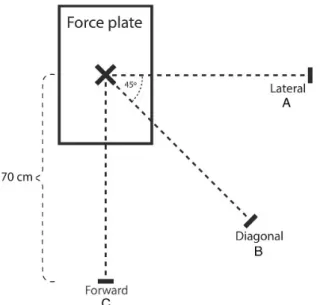
Figure 10. Representation of the jump-landing directions: (A) lateral (LJ); (B) diagonal (DJ); (C) forward (FJ). Distance between starting point and center of force place for each jump was 70cm. ... 34
Figure 11. Visual illustration of the task (e.g., lateral direction): single-leg jump-landing, followed by vertical jump. ... 34
Figure 12. Comparisons of mean (SD) of hip, knee and ankle sagittal and frontal plane angles observed between professional dancers and non-dancers. Left column
illustrates aggregated sagittal plane data, and the right column depicts ensemble frontal plane data. ... 36
Figure 13. Comparisons of mean (SD) of hip, knee and ankle sagittal and frontal plane angles observed among multidirectional jumps. Left column illustrates aggregated sagittal plane data, whereas the right column depicts ensemble frontal plane data. .... 38
Figure 14. Sagittal plane of the single- (ankle) and multi-segmented (hindfoot-tibia) foot models kinematics (angles in degrees), during forward and lateral jump-landing
directions, from initial contact to toe off (stance phase). ... 52
Figure 15. Professional dancers and non-dancers hindfoot-tibia and forefoot-hindfoot angles (in degrees), in the sagittal plane, from initial contact to toe off (stance phase). ... 64
Figure 16. Professional dancers and non-dancers hindfoot-tibia and forefoot-hindfoot excursions (angles in degrees) in the sagittal plane. ... 64
Figure 17. Example of a professional dancer single-leg forward jump-landing at initial contact (A & D); peak knee flexion (B & E); toe off phases (C & F) in Visual 3D (A, B, & C) and OpenSim (D, E, & F). ... 76
Figure 18. Offset (mean and standard deviation) of 6 professional dancers’ lower extremity joint angles (º) in the sagittal plane during a single-leg forward jump-landing. ... 77
Figure 20. Example of 1 participant markers errors (m) during forward jump-landing stance phase, in the three processing phases: i) before markers adjustment, ii) after
single max marker adjusted, iii) after all markers with max errors adjusted. Markers: m8 = right acromion, m2 = left acromion, m6 = left scapula, m1 = C7. ... 88
Figure 21. Example of 1 participant lower extremity sagittal joint angles (in degrees) differences during forward jump-landing stance phase, in the three processing phases: i) before markers adjustment (solid green line), ii) after single max marker adjusted (dashed black line), iii) after all markers with max errors adjusted (dashed red line). .. 89
Figure 22. Representation of the sagittal plane dominant lower extremity of a
professional dancer (left), and a non-dancer (right) at initial contact during a forward single-leg jump-landing. ... 95
LIST OF TABLES
Table 1. Descriptive table (mean ± standard deviation) of the kinematic (angles in degrees) and kinetics (joint moments in Nm/Kgm) variables at IC, PvGRF and PKF. . 37 Table 2. Descriptive table (mean ± standard deviation) of the multi- (hindfoot-tibia) and single-segmented (ankle) foot models kinematics (angles in degrees) at IC, PvGRF and PKF, in forward and lateral directions. ... 52 Table 3. Descriptive table (mean ± SD) of the multi-segmented foot model (HFTBA: hindfoot-tibia; FFHFA: forefoot-hindfoot; HXFFA: hallux-forefoot) kinematics (angles in degrees), during lateral and forward directions, at IC, PvGRF and PKF. ... 63
LIST OF ABBREVIATIONS
DJ DOF FJ IC LE LJ Max PKF PvGRF RMS SD Diagonal Jump-landing Degrees of Freedom Forward Jump-landing Initial Contact Lower Extremity Lateral Jump-landing MaximumPeak Knee Flexion
Peak vertical Ground Reaction Force Root Mean Square
CHAPTER I
1. Introduction
Dance movements result from a set of coordinated biomechanical degrees of freedom, ruled by technical and aesthetic requirements (1). The development of dance aptitudes requests continuous and progressive work, beginning at an early age and undertaken over years (2, 3). Professional dancers represent the elite that achieve the higher levels of the profession (2). In dance, as in other athletic activities, the injury rates have been a hot topic, resulting in several studies. In these two populations, the lower extremity has an expressively higher number of musculoskeletal injuries compared to other anatomical structures (4-10). Interestingly, it has been reported that acute lower extremity injury rate is lower in dancers, compared to other athletic populations (4-6, 10). Furthermore, dancers are frequently exposed to jump-landings that are usually performed with horizontal displacement of the body with single- or double-leg support, in many directions, as part of their training technique to use a wide range of the stage or classroom. Jump-landing proficiency is thereby a request for dancers to progress in the professional career (11). They are not only exposed to the strenuous physical demands placed on their bodies, but also to the consequences of repetitive jump-landing performances, constrained by technical and aesthetic requirements (12, 13).
Several studies have examined jump-landings, predominantly using drop landing tasks, to propose strategies to target biomechanical modifiable risk factors (13-21). Extended landing posture is, for instance, one of the examples. It has been associated with less ability of the lower extremity to mitigate the landings forces, and consequently produces a higher peak vertical ground reaction force (PvGRF) (17, 18, 22). Despite many studies proposing injury prevention programs, the injury rates remain steady across populations and years (7-9, 23), which undoubtedly is a key-problem in athletic activities. Based on previous research, dancers display an extended landing posture at initial contact (IC) of the landing phase (13, 15, 24). Yet, most likely this should not be seen as a deleterious factor, since dancers are encouraged to use more lower extremity joint excursions (15), particularly of the knee and foot-ankle joints, with longer landing phase that may suggest impact forces been absorbed over a longer period of time. Injury risk factors still need a thorough biomechanical
investigation to develop and implement effective intervention and injury prevention programs.
Motion analysis in dancers and the effect of dance training on dancers’ body is evolving. Still, few research has been examining dancers jump-landings in multiple directions with a single-leg support. The landing phase of a jump-landing is a demanding task that involves rapid deceleration to “brake” the movement, and where considerable amount of forces are acting on the lower extremity. This becomes more challenging when the base of support is just one limb (25, 26), and the motion is taken in different directions (27), which have a great effect on the biomechanical profiles. Also, research incorporating a multi-segmented foot model during landing is required to better understand the intricate structure of the foot and ankle joints, particularly due to its influence on other lower extremity joints during landing (19, 21, 28).
Additionally, computational modeling and simulation (e.g., OpenSim) is an impressive tool to study variables difficult to obtain experimentally (e.g., muscle and joint contact forces) (29-32). It is a field that has not been yet sufficiently explored in dance science, and can be valuable to further study the mechanisms underlying dance movement, particularly lower extremity joint functions during jump-landings. However, despite the considerable value to gain insights into human movement using OpenSim, it is a complex and multistep process (31, 33). The implementation of this approach not only involves a subjective process, but also is dependent on users’ skills and experience (34, 35). During that process, manual adjustments of the markers are needed to achieve effective marker set optimization. Therefore, the development of a musculoskeletal model can produce errors that can propagate through the workflow, affecting interpretation of the outputs (34, 36).
Hence, in this PhD research, an experimental and computational modeling and simulation approaches were conducted. The purpose was to investigate whether professional dancers landing pattern would possibly exhibited alternative strategies to implement in future intervention and injury prevention programs. The leading focus of this PhD research involved the study of the lower extremity biomechanical patterns of professional dancers and non-dancers, while performing multidirectional single-leg landings. For that, 3D motion analysis, force plate, and computational modeling and simulation were used. Accordingly, this PhD research sought to contribute not only to dance science, but also to provide further evidence-based information regarding jump-landing performances, that might be beneficial for sports sciences to potentially contribute to decrease the risk of injury rate in jump-landings.
Rationale, Research Questions, and Hypotheses
The main purpose of this PhD thesis was to investigate professional dancers’ lower extremity biomechanical patterns during single-leg multidirectional jump-landings. To accomplish this aim, a scientific research process was conducted employing a single-session descriptive group comparison design. Each study had its specific research question and respective hypothesis based on a scientific rationale. This is presented through the five scientific papers that have been published, accepted or are currently under review to international peer-reviewed journals. Paper 5 (Chapter VII) is still in preparation to be submitted in a near future.
• Paper 1
Title: Professional Dancers Distinct Biomechanical Pattern during Multidirectional Landings.
Status: Published.
Rationale: It is well reported that dancers have a significantly high overuse injury rate,
mainly on the lower extremity. Conversely, the lower extremity acute injury rate is considerably lower in dancers when compared to other athletic populations. Previous research has proposed optimal jump-landing biomechanical patterns to attenuate the deleterious factors during landing (e.g. high impact forces). It is also established that injury prevention programs have targeted modifiable risk factors to decrease injury rates in jump-landing activities. Still, overall lower extremity injury rates in many athletic populations have remained steady over the past decade. Dance, as other athletic activities, is characterized by a significant volume of jump-landing tasks. Thus, dancers are exposed to jump-landing training technique since young age, with required aesthetic and technique constraints. Due to insufficient scientific evidence related to dancers single-leg multidirectional landings, this study presented the findings of the lower extremity biomechanical landing patterns in professional dancers and respective comparison to non-dancers’. Additionally, this study can provide further understanding of the importance of the jump-landing directions to implement in prevention and intervention programs.
Research question: Are there differences in the lower extremity biomechanical parameters
between professional dancers and non-dancers during single-leg multidirectional jump-landings?
Hypothesis:
i) Professional dancers would demonstrate a more extended landing posture (lower hip and knee flexion, and higher plantarflexion), higher ankle excursion, and PvGRF during single-leg jump-landings in multidirectional jump-landing activities, compared to non-dancers.
ii) Lateral jump-landing direction would have higher knee and lower hip flexion angles than the other two directions (forward and diagonal).
• Paper 2
Title: Foot Modeling Affects Ankle Sagittal Plane Kinematics during Jump-landing. Status: Published.
Rationale: Based on the findings of paper 1, a natural progression was to focus on the
foot-ankle complex. It is a noteworthy anatomical structure that initially attenuate the impact forces and is of utmost importance in dancers. It is a key-element in jump-landing activities and also has influence on the other joints of the lower extremity. Considerable biomechanical studies have employed the single-segmented foot to analyze this intricate structure. However, differences in the ankle kinematics have been reported when using the multi- or the single-segmented foot models, which may lead to inaccuracies on ankle joint kinematics. Furthermore, the single foot model does not consider the other foot joints. Still, there are limited studies investigating ankle kinematics between multi- and single-segmented foot models during single-leg multidirectional jump-landings. Thus, this study compared ankle kinematics between the two foot models (multi-segmented and single-segmented) in jump-landing tasks.
Research question: Are there differences in ankle joint kinematics computed by a
multi-segmented foot model (Oxford Foot Model) and a single-multi-segmented foot model (conventional ankle) in multidirectional single-leg landings?
Hypothesis:
The hindfoot-tibia angles and excursion magnitudes would be lower in the multi-segmented foot model compared to the ankle of the single-segmented foot model.
• Paper 3
Title: Oxford Foot Model Kinematics in Landings: a Comparison between Professional Dancers and Non-dancers.
Status: Under second revisions.
Rationale: When quantifying the ankle kinematics, in paper 2, differences were found
between the multi- and single-segmented foot models. The foot-ankle complex has a remarkable importance in jump-landing tasks; it allows to initially mitigate the landing forces, to support the body, and has an influence on the other lower extremity joints. Dancers not only often perform jump-landings, but also commonly rely on this anatomical structure while dancing. Most biomechanical studies in dancers jump-landings have been using the single foot model. However, the foot-ankle complex is an intricate and frequently required structure in dance; and limited research exists regarding dancers multi-segmented foot model during single-leg jump-landings. Hence, this study took into consideration the different foot segments (hindfoot, forefoot, hallux), using the Oxford Foot Model, and compared dancers’ and non-dancers’ kinematics of the foot-ankle complex.
Research question: Are there differences in the multi-segmented foot (Oxford Foot Model)
kinematics between professional dancers and non-dancers, during lateral and forward single-leg landings?
Hypothesis:
Professional dancers would have higher plantarflexion and eversion angles and excursions in the foot and ankle joints, using the multi-segmented foot model, than non-dancers.
• Paper 4
Title: Effect of Two Different Pose Estimation Approaches on Lower Extremity Biomechanics in Professional Dancers.
Status: Accepted for publication.
Rationale – Musculoskeletal modeling and simulation of dynamic activities permit the
analysis and estimation of athletic performance. Different modeling approaches have been used to explore lower extremity biomechanics in jump-landing tasks. While computational modeling has been employed in many fields, it is significantly lacking in the dance science field. Visual 3D and OpenSim are commonly used software in biomechanical studies. The
main difference between these two approaches is the algorithm used to estimate the position and orientation of musculoskeletal models. Visual 3D uses segment optimization, whereas OpenSim uses global optimization. For both approaches, pros and cons have been reported in the literature. During the earlier phases of the PhD work, Visual 3D was the software chosen to compute joint angles, and moments. However, to further explore and apply computational modeling into this project, OpenSim software was also selected with the purpose to conduct inverse kinematics, inverse dynamics, muscle analysis, and to compute lower extremity joint loads. The differences in these two approaches’ algorithms merit further investigation. Thus, this preliminary study was pertinent to understand whether the different approaches were providing similar outputs of the studied variables, using the same data set of a single-leg forward jump-landing. This comparison is considerably important for the next steps, after the PhD, concerning muscle analyses and joint reaction forces estimations on the lower extremity joints.
Research question – Do different pose estimation approaches (Visual 3D and OpenSim)
have an effect on the hip, knee, ankle joint angles and moments in professional dancers, during a single-leg forward jump-landing?
• Paper 5
Title: The Impact of Marker Adjustment in Musculoskeletal Modeling on Kinematic Parameters.
Status: In preparation to be submitted.
Rationale – The accuracy level of a musculoskeletal model depends on the application and
not only on the model itself. During the process of biomechanical studies to create a musculoskeletal model, errors in early stages of the model development, such as marker errors, can propagate and affect the results leading to biomechanical inaccuracies. Currently, in computational modeling using OpenSim software, it is essential to compute a musculoskeletal model. However, it is a subjective process that relies on the user skills and experience. In inverse kinematics, for effective marker set optimization it is essential to identify, examine and adjust all markers with max error above the recommended threshold. This is important to ensure that the model is adequately representing the musculoskeletal system that is simulating. Understanding the importance of this step is critical, since a lack of emphasis has been given on the sensitivity of the models and dependency on user skills, which may influence the final biomechanical outputs.
Research question – Are there differences in the sagittal lower extremity joint angles in
three difference processing phases of developing a musculoskeletal model i) before markers adjustment, ii) after single max marker error adjustment, and iii) after all makers with max errors adjusted?
Thesis Structure
The PhD thesis structure is organized in X chapters, assembling information needed to understand the conducted research:
● Chapter I – Introduction: introduces the main research topics of the PhD thesis, including the significance of the conducted research. It also displays the rationale behind all the research done, as well as, the research questions, and hypotheses for each study;
● Chapter II – Literature Review: summarizes the state of the art of the main research topics of the PhD thesis. It contributes to address different and fundamental topics concerning research in multidirectional single-leg jump-landings, such as dance, jump-landing biomechanics, foot-ankle complex and computational modeling and simulation.
● Chapter III to VII – Research Studies: presents the five scientific studies written during the PhD process. Each of them was organized following the structure of a scientific paper format: Introduction, Methods, Results, Discussion, Conclusions, and References; and each one was reformatted for consistency with the thesis. The original paper already published can be seen in Appendix.
● Chapters VIII to IX – General Discussion and Conclusions: provides the main findings of chapters III to VII, and the overall conclusion of this PhD thesis. Additionally, based on the main findings it also includes limitations, recommendations for future research, and practical implications.
● Chapter X – List of References: enumerates the used references in the chapters Introduction, Literature Review, General Discussion, and Conclusions. This chapter does not include the references of each study, since they are already included in their own chapter.
Chapter II
2. Literature Review
2.1 Dance
Dance is a demanding and elaborate form of body movement that requires development and proficiency of specific skills over manifold years. Due to the physical and aesthetic demands during dancers’ careers, they acquire and develop singular movement patterns of the musculoskeletal system. This is also a result of the unique anatomical positions often performed, particularly noticeable in the lower extremities, leading dancers to a distinctive biomechanical pattern. Since a young age, they are exposed to high physical demands placed on their bodies while performing dance technique (5, 11, 13, 37). This daily occurrence can lead to a high risk of musculoskeletal injuries (4, 5, 38) that can have considerable repercussions throughout their lifespan. On the other hand, dancers combine technique with body alignment and postural control while dancing, increasing awareness of their body position and orientation compared to other athletic populations.
In an effort to better understand the underlying movement performance, numerous biomechanical studies have been conducted in athletes (7-9, 14, 17-20, 22, 23, 26, 27, 39-53). Though, in dance science, there is still a lack of biomechanical research. Due to the distinctive and singular technique, undertaken through many years of intensive training, more extensive studies are needed. Furthermore, it is well-established the importance of studying movement biomechanics to determine and assess injury risk factors. This can eventually contribute to determine strategies that may be implemented in intervention and injury prevention programs (54).
2.1.1 Dancers’ injury epidemiology of the lower extremity
Dancers have a high injury rate with more than 60% of all injuries occurring to the lower extremity (4-6); which is similar to other athletic populations (7-10). In dance, the most common anatomical locations of injuries in the lower extremity are: the knee (7% to 15%) (4-6), lower leg (9% to 18%) (4, 5), ankle (14% to 33%) (4-6), and foot (9% to 20%) (4-6). Dancers’ injuries are commonly linked to stress-related factors, involving tendons, ligaments and/or muscles (4). These injuries are frequently attributed to overtraining,
repetitive dance movements and the demanding extreme ranges of motion. Consequently, high physical demands are placed on the lower extremity with excessive microtrauma in the anatomical structures (5, 38). Accordingly, 66% to 72% of the injuries in dancers are classified as overuse (4-6), resulting from cumulative microtrauma or repetitive use and stress (10, 55). Those injuries in dancers have a greater incidence and severity than traumatic injuries (5). Subsequently, the remaining 28% to 34% of the injuries have been reported as acute (4-6), which is about 2.5 times lower compared to other athletic populations – i.e., approximately 70% of the injuries are acute in nature (10).
Acute and overuse injuries often limit unrestricted athletic practice (23). The injuries can lead to short and long-term health consequences, affecting performance and quality of life with potential for extended disability (54) in dancers and athletes’ lifespan. Due to their technique, dancers are exposed to similar overuse injuries as other athletic activities (56), such as tendinopathies or stress fractures (4, 55). Nonetheless, dancers have unique dance-related injuries (56), such as foot strain (5) or snapping hip (4). Additionally, there are movement patterns commonly seen in dancers’ technique that are not observed in other athletic populations. Few examples are the knee flexion immediately before and after a jump (known as demi-plié) or rolling the feet in landing phase (12, 13, 57). Interestingly, these dance features have been associated to biomechanical improvements in jump-landing tasks, to reduce the risk of injuries. These dance-related characteristics will be further analyzed in Chapter II – section 2.2.1.
2.1.2 Modifiable risk factors related to the lower extremity
Lower extremity musculoskeletal injuries have a multifactorial etiology (58). Injury risk factors must be clearly established, so prevention and intervention programs can be devised to adequately and successfully target them (58). The risk factors interact in multiple ways. The modifiable risk factors can be subject to intervention by physical training or behavioral approaches (54). Due to an extensive high injury rate in the lower extremity, a particular emphasis should be given on this anatomical region concerning prevention and intervention programs (7). Deficits in muscle strength and/or muscle imbalance (54, 59), altered neuromuscular control (22, 42, 59, 60), extended landing posture (17, 18), decreased ankle excursion in landings (51, 61, 62), and poor technique (38, 58, 63) are some of the well-established modifiable risk factors commonly associated to lower extremity injuries. It is noticeable that in the past decades, considerable research has been conducted to identify those factors, particularly during jump-landing activities (13, 15-19, 22, 26, 27,
39, 41, 42, 48, 64). Still, tracking and managing risk factors in biomechanics research remains a challenge. Injury prevention programs in athletic populations have been shown to be effective in changing some of the proposed modifiable risk factors and decreasing injury rates in the specific study sample (49, 50, 65). Nevertheless, despite extensive research and development of prevention programs, lower extremity injury rates have remained steady across multiple populations and years (7-9, 23). Additionally, the implementation, adoption, and maintenance of these programs in real life remain a challenge. Injury prevention programs should involve wider, appropriate and engaging implementations to achieve its goals. Therefore, it is important to identify and understand the movement behind jump-landing activities to adequately target the modifiable musculoskeletal risk factors linked to the lower extremity injuries.
2.1.3 Dancers training technique
Professional dancers emerge as a result of a demanding selection process, representing the elite of dance hierarchy (2, 3). Those dancers undertake a rigorous dance training over numerous years. High levels of performance in dance require several specific athletic skills, such as muscular strength and endurance, anaerobic and aerobic energy consumption, speed, agility, coordination, motor control and psychological readiness (63). The development of those skills requires a continuous and progressive work and is critical to dancer’s performance. Moreover, for functional and aesthetics purposes, the technique requirements of dance performance not only involve considerable amount of en dehors (external rotation of the lower extremities), joint mobility, and muscle flexibility and strength, but also within extreme ranges of motion in several joints. Since early ages of dance training there is a strong emphasis on body alignment, particularly in ballet dancers. These dancers are trained to be in an upright upper body and neutral pelvis position. Additionally, both lower extremities are externally rotated (en dehors), while keeping the patella aligned with the second metatarsal. It is further expected to have neutral position of the hindfoot, associated with strong feet arches (2, 12, 13). Subsequently, all the aforementioned attributes are considerably important in training technique, making dancers a unique population, with distinctive biomechanical pattern compared to other athletic populations. Some of the dance training effects have been reported in previous literature, particularly in jump-landing activities.
Dancers are characterized by a smooth landing sequence, a request in their technique, for the audience to not visualize the movement effort (11). Also, they are encouraged in their
training process to initially contact the ground with the toes in a fully plantarflexed position, followed by lowering through the foot, until a controlled heel contact (13, 24, 57). This increases the ankle range of motion, the peak ankle dorsiflexion and the knee flexion angles during landing, producing a deeper demi-plié, while controlling eccentric loads (13, 24). These dance-related features will be further examined in Chapter II – section 2.2.1. Hence, the lower injury rate of severe traumas (e.g., anterior cruciate ligament injury) in dancers when compared to athletes (11), may be explained by the combination of repetitive and specialized training that dancers undergo to obtain jump-landing and balance skills, along with longer time to achieve fatigue (15, 16, 24). Further, the relatively predictability nature of dance environment, which does not involve ball-handling as in other athletic populations (11), may contribute to the lower injury rate. Thus, observational and measurement of performance variables in dance through biomechanical analysis is important to enhance the understanding and provide thorough information of dance movements and skills in jump-landing activities.
2.2 Jump-landing biomechanics
For the development of prevention and intervention strategies, it is imperative to identify and understand injury mechanisms (45, 54). Jump-landing tasks are fundamental features of many athletic and dance activities and have received substantial research attention in the last decades (11, 13-16, 19, 20, 22, 24, 26, 27, 39, 40, 42-44, 47, 53, 66). The study of jump-landing biomechanics allows to investigate the demands placed on the body while performing these tasks (66). Jump-landing sequence can be divided into two phases: i) the flight phase in which the foot is off the ground, and ii) the stance phase when it is contacting the ground. Additionally, the stance phase (Figure 1) can be divided into: a) landing phase, which is corresponding to the deceleration phase, defined as the time period from IC with the ground to the peak knee flexion (PKF) (13, 51); and b) take-off phase, defined as the time period between the PKF and the toe off, when the foot stop contact with the ground. In this PhD research the prime focus was in the landing phase. It corresponds to the period of the greatest loading; when considerable large forces are applied to the human body (14, 41, 52, 65).
Figure 1. Sagittal view of the stance phase of a jump-landing: (A) initial contact; (B) Peak
knee flexion; (C) and toe off events; (A) to (B) represents the landing phase, and (B) to (C) represents the take-off phase.
The lower extremity absorbs the body’s energy during landing (53). A primary goal in this phase is to stabilize the body and maintain the center of mass within the base of support. For that, the lower extremity plays an important role, to reduce and control the downward momentum acquired during the flight phase; and also, to dissipate that kinetic energy generated (19, 44, 67). Altogether, it places strenuous demands on the lower extremity (67). During jump-landings, injuries may occur from a multifactorial etiology, including repetitive malalignment of the lower extremity segments or from inadequate absorption of impact forces (61). A more flexed landing pattern, with higher trunk, hip and knee flexion during the landing phase (Figure 2) is associated with higher shock absorption mechanism, important contributor to energy dissipation, and consequently lower ground reaction forces (17, 18, 22). Thus, when employing an active trunk flexion strategy during landing, decreased PvGRF, peak knee extensor moment, higher hip and knee flexion angles and hip extensor moment have been reported (17, 18, 46). Though, despite the magnitudes of the attained hip and knee flexion (51º and 82º, respectively) being suggested to be protective of
excessive joint loads (17, 18), it is plausible that such magnitudes are most likely incompatible with athletic performance, due to the concomitant excessive active trunk flexion (i.e., 96º of active trunk flexion), as depicted in Figure 2.
Figure 2. Example of an anterior (left) and sagittal (right) views of large trunk flexion
strategy during double-leg drop landing task (65).
Being at a nearly fully extended position at IC it maximizes the range of motion (known as excursion in Chapters III to V) over which flexion can occur to dissipate energy (67). The lower extremity flexion during landing, controlled by extensor musculature eccentric work, contributes to the shock absorption mechanism (67). This is further beneficial as the increased sagittal joint excursions are related to concurrently reduced frontal plane joint excursions (14). There is also strong evidence suggesting that a decreased weightbearing ankle sagittal excursion in landing influences lower extremity mechanics and may be related to injuries due to a compensatory mechanical pattern in the lower extremity (37, 62). As an example, excessive foot pronation or compromised medial knee alignment (37). Additionally, the foot landing technique (e.g., toe-to-heel versus heel-to-toe landing) has noteworthy implications regarding the forces transmitted to the body and the body’s ability to dissipate these forces (53, 67, 68). It has been suggested that a toe-to-heel landing technique leads to a considerably lower hip flexion angle at IC compared to the rearfoot (defined in this PhD thesis as hindfoot) and self-preferred landing techniques (53). Biomechanically, in the toe-to-heel strategy, a relatively large lever arm exists between the point of force application and the ankle joint, increasing the deceleration distance and thus decreasing the impact experienced (67). Increased plantarflexion angle at IC also provides a mechanical advantage, since it permits a subsequent increased ankle excursion during landing, established as an important contributor to shock absorption (19, 21, 41, 67). Accordingly, jump-landing technique influences the lower extremity ability to mitigate the
impact forces. The landing technique therefore plays an important role, as it may influence the likelihood of injury (67) .
2.2.1 Jump-landing patterns in dancers
Biomechanical research is an emerging field in dance science. Dancers training places a significant focus on specific motor organization patterns to develop the performance and technique requirements (1). While substantial research studying the biomechanical responses of the lower extremity during landings exist, there is a paucity of these studies in dancers. Jump-landings are an important aspect of many dance styles (24). Accomplishment of aesthetically precise balance and jump-landing skills are necessary for dancers to advance in the professional careers (11). As in other athletic populations, dancers are often exposed to jump-landing activities; being part of their training technique throughout the years of practice (11, 69); a ballet dancer can perform up to 300 jumps per hour (69). Besides, dancers are not only exposed to the effects of repetitive jump-landings, but also have to be aware and develop the aesthetic requirements during their performances that other athletic populations do not have (11-13, 15, 70). Some of those aesthetic requirements are the alignment of the lower extremity (e.g., patella aligned with the second metatarsal) and the foot work (e.g. toe-to-heel landing) that subsequently allows extensive plantarflexion angle in the ankle joint (11, 13, 57). Hence, there are specific characteristics of jump-landings that are unique in dance (3).
Previous research has described dancers biomechanical landing pattern. An adequate jump-landing technique in dance is defined by starting and finishing in demi-plié position; meaning knee flexion, while heels contact the ground (Figure 3). The spring action of the
demi-plié is essential in the jump-landing activities to prepare the jump and also to support
the shock absorption mechanism during the landing phase (37). It also enables the use of the required en dehors while executing these movements (37).
Figure 3. Depiction of a dancer in a demi-plié position (posterior view): ankle dorsiflexion,
knee flexion, and external rotation of the lower extremities (en dehors), with the patella aligned with the second metatarsal.
From the demi-plié position, the jump begins with hip and knee extension, and foot-ankle plantarflexion (12, 37), transferring the weight from the heel to toes (12). The last point of contact with the ground, before the flight phase, are the toes to push-off. Maximum ankle plantarflexion (pointé) and alignment of the lower leg, ankle and foot are required during the flight phase (12, 66) preventing the sickling mechanism (presented in Chapter II – section 2.3.1). Still, in this phase, dancers have to maintain the lower extremities externally rotated (en dehors), which provides a medio-lateral work of the hip to assist with the alignment of the lower extremity during jump-landings (37). At the peak of the flight phase, the knees are extended and the ankles fully plantarflexed (12, 66). They also present an erect trunk posture during the entire jump-landing (13, 37). At the initial ground contact, the foot and ankle are plantarflexed; the first part of the body contacting the ground in the landing phase are the toes, and then the ball of the foot (12, 71). After the IC, the plantarflexed foot-ankle complex quickly and forcefully moves into a dorsiflexed position after the foot entirely contacts with the ground (71). Therefore, at IC, dancers land with nearly fully extended lower extremities; demonstrated by lower hip and knee flexion compared to other athletic populations, as well as maximum use of the ankle plantarflexion (13, 15). This pattern emphasizes the use of lower extremity joints excursion and also highlights the developed ability of the foot-ankle to roll throughout the foot (13, 57); this topic will be further addressed in Chapter II – section 2.3.1. Dancers then continue the landing phase by placing the heel on the ground coupled with knee flexion (demi-plié) to prepare for the next jump (12).
It is believed that the aforementioned extended landing pattern of dancers may hinder appropriate neuromuscular control to absorb the landing forces, which can potentially result in higher ground reaction forces (18, 22, 41, 68). This mechanism may increase the load on passive structures of the lower extremity joints (20), and consequently lead to a higher risk of injury. Yet, dancers appear to compensate the erect posture at initial contact with higher knee and ankle excursions during landing (13, 24). This biomechanical strategy allows the dissipation of the impact forces over a larger range of motion, and at the same time to achieve the desired aesthetics of a smooth and seemingly effortless landing in dancers (11, 13). This suggests that dancers appropriately take advantage of their extensive training of
demi-plié movement. Also, dancers demonstrate decreased knee valgus compared to
athletes (15, 24) that may contribute to a lower incidence of injuries (e.g., anterior cruciate ligament injury) in dancers (11). Despite the substantial research conducted in jump-landings, a comprehensive overview of lower extremity kinematics and kinetics in these tasks is still needed to comprehend the underlying patterns contributing to shock absorption mechanism in dancers and other athletic populations (known in Chapters III, IV, V, and VII as non-dancers).
2.2.2 Different types of jump-landings
It is noticeable that different tasks have been used in biomechanical studies investigating the lower extremity during jump-landing activities. A limitation of the biomechanical laboratory studies is that one cannot determine how the measured movement patterns relate to the biomechanics of real athletic motions (47). Nevertheless, an effort should be made to attempt to mimic athletic movements in the laboratory, as realistic as possible, increasing the ecological validity.
2.2.2.1 Drop landing and drop jump tasks
The drop landing has been primarily and extensively used to investigate lower extremity biomechanics (13-16, 19-22, 41, 46, 72). This task isolates the landing phase from the entire jump-landing sequence and may not take into account the preparation of a landing (45). The activation of the lower extremity musculature prior to landing affects joints’ kinematics and kinetics and potentially mediate loading (67). To drop to land (Figure 4) is reasonably different to performing a preceding jump before landing, as there is no dynamic motion prior to landing. Also, it is rarely performed outside the laboratory environments, and is considered as a less complex movement task (45). The landing phase of a drop landing and a spike jump have considerably differences in kinetic, kinematic and muscle recruitment
strategies observed in volleyball players. Drop landings displayed a more extended landing pattern, most likely due to a more vertical body position (45). Thus, a drop landing does not mimic jump-landing tasks. Results from research using drop landing mechanics to model a landing phase of an entire jump-landing sequence should therefore be taken with caution (45, 47).
Additionally, previous research has primarily employed a drop vertical jump to assess injury risk (42, 68). This task is in a controlled environment, primarily vertical in nature, and may not fully represent the demands of various jumps (e.g., vertical vs. horizontal displacements) performed in many athletic and dance activities. For instance, there are substantial differences in the magnitude of knee kinematics and knee joint loadings between drop jumps and sidestep cutting tasks (47). In the sidestep cutting, athletes had lower knee flexion angle, higher knee valgus and internal rotation angles at IC, and higher knee moments (e.g., knee abduction moment was 6 times higher) (47). Sidestep cutting and drop jumps have different biomechanical motion patterns with the double-leg drop jump considered a more controlled and less complex task (47).
Figure 4. Demonstration of the starting position of a double-leg drop landing task (18).
2.2.2.2 Single- and double-leg landings
Double-leg landings can provide valuable information, however, may not represent a comprehensive view of an athletes’ high-risk biomechanics (27); injuries often occur during single-leg landings (25, 47). This injurious mechanism is most likely due to a significantly more strenuous biomechanical demand on a single-leg compared with a double-leg (27). The impact forces, during single-leg landings, are solely absorbed by one lower extremity, with a decreased base of support and increased biomechanical demands from the trunk
and pelvis (27). Moreover, single-leg landings have demonstrated lower sagittal hip and knee angles at IC and respective excursions compared to double-leg landing (20, 25).
Biomechanical magnitudes, especially joint moments, are greater during single-leg than double-leg landings, which suggests higher musculature demands needed to dissipate energy when landing in one limb (27, 44). Larger demands are required of posterolateral hip musculature in single-leg landings, to maintain a leveled pelvis and prevent excessive hip adduction and internal rotation (components of dynamic lower extremity valgus). Previous research have reported hip flexion and adduction moments in single-leg landings that were 2.2 and 6.6 times higher, respectively, than during double-leg landings (27). It is suggested that single-leg landings are challenging tasks that may enable better assessment of high-risk movement patterns (27). Also, different lower extremity energy dissipation strategies are adopted when performing a single- or double-leg landing, in sagittal and frontal planes (25). For example, in the sagittal plane, the hip and knee are the major contributors to energy dissipation in double-leg landings, whereas the hip and ankle primarily contribute during single-leg landings. Findings from previous research, exhibited that double-leg sagittal plane movements may not fully represent the neuromuscular strategies during high-risk activities (25, 27, 47). The biomechanics elicited during double-leg sagittal plane are not representative of biomechanics during more multidirectional sport-specific activities. Hence, single-leg and multidirectional tasks may complement scientific information (27).
2.2.2.3 Jump-landing directions
The different jump-landing directions meaningfully affect the lower extremity biomechanical strategies adopted to dissipate the impact forces during the landing phase (26, 27, 39, 40, 73). These biomechanical patterns suggest distinctive neuromuscular demands for each direction. For example, during the lateral jump-landing direction lower hip flexion, higher knee flexion, ankle dorsiflexion, and peak knee valgus angles were reported when compared to diagonal and forward directions (39, 40). Jump-landings in a frontal plane movement elicit more predominant signs of dynamic lower extremity valgus (27). Thus, using only the forward direction in jump-landing assessment may be misrepresenting essential aspects of neuromuscular control that are likely important to improve intervention programs of lower extremity injuries (73).
Multidirectional single-leg landings may better differentiate the biomechanical profiles in landings (26). Dancers also perform repetitive multidirectional landings, frequently supported in single-leg, with considerable horizontal displacement as part of their artistic and technical requirements. Thereby, this PhD research focused on multidirectional single-leg landings (Figures 5 and 6) of professional dancers and compared to non-dancers, to investigate the underlying biomechanical responses in different directions.
Figure 5. Representation of the three directions (A) lateral, (B) diagonal, (C) forward of
the jump-landings performed in the studies of this PhD thesis.
Figure 6. Depiction of the single-leg jump-landing in the lateral direction, followed by a
vertical jump-landing; marker set on the trunk and left dominant lower extremity. The highlighted image represents the landing phase analyzed in the studies of this PhD
2.3 Foot-ankle complex
The foot-ankle complex is a unique masterpiece of the human body. The complexity of the foot-ankle anatomy has a remarkable influence on its biomechanics (74). Unlike other segments of the lower extremity, the foot includes multiple bones and joints with a very intricate interaction (75, 76). This structure enables not only motion with a notable level of joint stability but is also a key-element in a wide variety of daily and functional activities (76). The movement of the foot-ankle complex joints is multiplanar and exceptional (37, 77, 78), which represents a challenge to define and characterize in vivo foot kinematics. Nonetheless, due to its important function, and also improvement of the accuracy of motion capture systems, the measurement of the foot kinematics has becoming more common (77).
The foot-ankle complex plays an important role to transfer forces and mechanical energy throughout the entire lower extremity, influencing its biomechanics due to the closed kinetic chain (19, 21, 28, 72). This structure provides a unique foundation to static and dynamic stability, mechanical leverage, shock absorption, and also to maintain balance (79). Additionally, it is remarkably energy efficient (79). Thus, all of the characteristics taken together demonstrates the natural resilience of the human foot (79). Thereby, the several joints of the foot and ankle have an outstanding position and function in the human body motion.
The interface between the lower extremity and the ground is the foot-ankle complex (74). The biomechanics of this structure is critical to the functionality of the lower extremity (28) in a wide variety of dynamic activities, including jump-landings. The individual joints and segments of the foot work simultaneously in a coordinated and synchronized system rather than isolated movements (76). The foot-ankle complex therefore can provide optimum forces attenuation and energy-absorbing characteristics (79) that must be investigated. Previous biomechanical studies in dancers jump-landings (13, 15, 16, 24) commonly modeled the foot as one rigid segment. This model neglects the intricate structure of the foot and ankle functional anatomy (77, 80, 81) and may attribute motion to one joint when in fact it occurs in more than one foot joints (82). Thus, biomechanical data concerning the foot-ankle complex kinematics using a multi-segmented foot model may provide further evidence related to dancers’ foot-ankle structure.
2.3.1 Foot-ankle complex in dancers
Dancers often rely on the intricate foot-ankle complex while performing jump-landings. One of the main purposes of this structure is to initially attenuate the landing forces imposed on the body, while providing dynamic stability (19, 21, 76). A well-trained dancers’ foot and ankle therefore are able to provide full support in landing tasks, and most likely work as an advantageous shock absorption mechanism (13, 19, 21, 37).
Dancers exhibit high motion in these structures due to the aesthetics and technique requirements. Professional dancers present 129% increased plantarflexion motion over the norm, which provides a total ankle range of motion 82% greater than norm (2). However, a standard goniometer was used to measure joint angles and ranges of motion (2). Additionally, dancers exhibited increased strength of the ankle plantarflexors; approximately 40% greater than the norm, which may be linked to the great and specific demands on the foot-ankle complex in dance (2). Although foot and ankle flexibility is of utmost importance to dancers aesthetics and technique, there is little evidence to support that extreme ankle range of motion is a predictor for subsequent injury (56).
There are some unique features to dancers, such as dancing en pointé (63). To dance en
pointé, female dancers should have approximately 90º to 100º of plantarflexion in the
foot-ankle complex joints (2). A considerable amount of plantarflexion is required in dancers, linked to strength and extensive mobility of different segments of the foot (1, 38, 83), and not just the ankle joint. The observed plantarflexion motion in dancers comes from the combined motion of different foot joints, such as talocrural, subtalar, and midtarsal joints of the foot-ankle complex (2, 83). Furthermore, it has been reported that during weightbearing
en pointé and demi-plié positions in female ballet dancers, the talocrural joint is the greater
contributor to plantar and dorsiflexion motions, with approximately 70% of the overall motion, measured using x-ray. Whereas the remaining 30% occurs in other foot joints (83). This suggests that when computing the foot-ankle complex using a single rigid segment to measure kinematics, it is likely providing insufficient information related to foot function as the other foot joints are also a contributor to the foot overall motion (77, 80, 81). This motion pattern in dancers is doubtless an effect of years of training technique (1, 2, 11, 13, 63) and most likely a result of some degree of skeletal molding in young dancers while bones are still maturing (2).
As previously described in Chapter II – section 2.2.1, dancers are frequently exposed to jump-landing activities with the foot-ankle complex having an important responsibility due