Dalton
Transactions
Dynamic Article Links
Cite this: DOI: 10.1039/c2dt31688a
www.rsc.org/dalton
PAPER
Decavanadate, decaniobate, tungstate and molybdate interactions with
sarcoplasmic reticulum Ca
2+-ATPase: quercetin prevents cysteine oxidation
by vanadate but does not reverse ATPase inhibition
Gil Fraqueza,
aLuís A. E. Batista de Carvalho,
bM. Paula M. Marques,
cLuisa Maia,
dC. André Ohlin,
eWilliam H. Casey
fand Manuel Aureliano*
gReceived 26th July 2012, Accepted 23rd August 2012 DOI: 10.1039/c2dt31688a
Recently we demonstrated that the decavanadate (V10) ion is a stronger Ca2+-ATPase inhibitor than other
oxometalates, such as the isoelectronic and isostructural decaniobate ion, and the tungstate and molybdate monomer ions, and that it binds to this protein with a 1 : 1 stoichiometry. The V10interaction is not
affected by any of the protein conformations that occur during the process of calcium translocation (i.e. E1, E1P, E2 and E2P) (Fraqueza et al., J. Inorg. Biochem., 2012). In the present study, we further explore this subject, and we can now show that the decaniobate ion, [Nb10= Nb10O28]6−, is a useful tool
in deducing the interaction and the non-competitive Ca2+-ATPase inhibition by the decavanadate ion [V10= V10O28]6−. Moreover, decavanadate and vanadate induce protein cysteine oxidation whereas no
effects were detected for the decaniobate, tungstate or molybdate ions. The presence of the antioxidant quercetin prevents cysteine oxidation, but not ATPase inhibition, by vanadate or decavanadate. Definitive V(IV) EPR spectra were observed for decavanadate in the presence of sarcoplasmic reticulum Ca2+
-ATPase, indicating a vanadate reduction at some stage of the protein interaction. Raman spectroscopy clearly shows that the protein conformation changes that are induced by V10, Nb10and vanadate are
different from the ones induced by molybdate and tungstate monomer ions. Here, Mo and W cause changes similar to those by phosphate, yielding changes similar to the E1P protein conformation. The putative reduction of vanadium(V) to vanadium(IV) and the non-competitive binding of the V10and Nb10
decametalates may explain the differences in the Raman spectra compared to those seen in the presence of molybdate or tungstate. Putting it all together, we suggest that the ability of V10to inhibit the Ca2+
-ATPase may be at least in part due to the process of vanadate reduction and associated protein cysteine oxidation. These results contribute to the understanding and application of these families of mono- and polyoxometalates as effective modulators of many biological processes, particularly those associated with calcium homeostasis.
1.
Introduction
Previous studies have shown that sarcoplasmic reticulum (SR) Ca2+-ATPase activity is very sensitive to the presence of several vanadium complexes such as vanadium–citrate complexes and bis(maltolato)oxovanadium(IV) (BMOV).1,2 It has been
suggested that different vanadium coordination complexes can inhibit Ca2+-ATPase through specific protein–substrate inter-actions while the oligomeric, uncomplexed oxovanadates do not interact through the same mechanism. Moreover, the different oligovanadates such as [VO4]3− (V1), [V4O12]4− (V4), or
[V10O28]6−(V10) may have different mechanisms and effects on
the calcium ATPase activity. In particular, V10 is the only
identified polyoxovanadate that inhibits calcium accumulation by the Ca2+-ATPase when coupled to ATP hydrolysis.3 In addition, V10appears to be the most potent oxometalate Ca2+
-ATPase inhibitor out of all the vanadium ions and complexes so far analysed, exhibiting an IC50 value of 15 μM.1–4 Recently,
other transition-metal ion clusters such as niobates, tungstates and molybdates were evaluated in the inhibition of SR calcium ATPase and were found to be less efficient as inhibitors.4These findings were further explored, and the effects of five transition metal species, viz. decavanadate (V10), decaniobate (Nb10O286−,
Nb10), vanadate (V1), tungstate (WO43−, W1) and molybdate
a
ISE and CCmar, University of Algarve, 8005-139 Faro, Portugal
b
Molecular Physical Chemistry R&D Unit, University of Coimbra, Portugal
cDepartment of Life Sciences, University of Coimbra, 3004-535
Coimbra, Portugal
d
REQUIMTE, Centro de Química Fina e Biotecnologia, Departamento de Química, Faculdade de Ciências e Tecnologia, Universidade Nova de Lisboa, Campus da Caparica, 2829-516 Caparica, Portugal
e
School of Chemistry, Monash University, Clayton, Vic 3800, Australia
f
Department of Chemistry, University of California, Davis, California 95616, USA
gCCmar and FCT, University of Algarve, 8005-139 Faro, Portugal.
E-mail: [email protected]; Fax: +351 289 800066; Tel: +351 289 800905
Downloaded by RSC Internal on 12 September 2012
(MoO43−, Mo1) ions, on the structure and activity of SR calcium
ATPase were evaluated and described in the present paper. V10 and Nb10 are isostructural and isovalent, but where
vanadium is redox active and labile in solution, niobium is kine-tically inert and redox stable, making Nb10an excellent tool to
explore the type of inhibition that the V10 analogue undergoes
with the calcium pump. The SR calcium pump has proven to be an excellent model for studying the effects of oxovanadates and vanadium complexes on Ca2+homeostasis, and by extension on several signalling processes in muscle and non-muscle cells.1–4 Calcium homeostasis is also known to be involved in cell death processes such as necrosis and apoptosis. Finally, ion pumps are known as potential drug targets in medicine and science.5
Decavanadate is known to modulate the activity of many pro-teins such as phosphatases,6 myosin,7,8 and calcium pump ATPase1–4,9,10 and to interact with proteins such as actin11–15 among others.16,17 Moreover, as a well-established inhibitor of tyrosine kinases and phosphatases, vanadium is suggested to be involved in the biochemical processes of several diseases, with potential uses in anticancer18–20 and anti-diabetic therapies.21 For instance, in rat adipocytes, the V10ion has been suggested to
be a more potent insulin mimetic agent than several mononuclear vanadium coordination complexes, although the precise mecha-nism of action is still unclear.22Perhaps the most notable effect of V10described so far relates to the induction of mitochondrial
membrane depolarization and the inhibition of oxygen consump-tion at nanomolar concentrations, which leads to cell necrosis.23,24
While the literature on the biological effects of mono- and polyoxometalates has been rapidly increasing over the most recent decade, the biochemical mechanisms underpinning the effects of these oxometalates are still only poorly under-stood. This is clearly a rapidly growing field, owing to the increasing interest in the use of bioavailable metal complexes in biochemistry, biology, biotechnology, pharmacology and medicine.
2.
Materials and methods
All reagents were of biochemical analysis grade and were sup-plied by BDH, Merck, and Sigma. Anhydrous ammonium meta-vanadate, sodium tungstate and sodium molybdate salts were purchased from Riedel-de Haën. Tetramethylammonium decan-iobate, [N(CH3)4]6[Nb10O28]·6H2O, was synthesised according
to published procedures.25,26ATP (vanadium free) was supplied by Sigma.
2.1. Preparation of metal solutions
Decavanadate stock solution was obtained by adjusting the pH of an ammonium metavanadate solution (50 mM, pH 6.7) to 4.0, according to literature methods,3,16,24 and the formation of the decavanadate (V10) ion is readily observed by the characteristic
orange colour of the solution.16The kinetics of decomposition of V10is determined by following the absorbance atλ 400 nm of
the reaction medium at 25 °C, as described elsewhere.16,23,27V10
decay follows afirst-order rate law, with a half-life of more than 12 h (measured with 0.1 mM decameric vanadate species, 1 mM
total vanadate), in agreement with previous work.27At the V10
concentrations and time-scales used in the enzymatic studies per-formed in the present work, the vanadium solution contains almost exclusively the decameric species. On the other hand, vanadate solutions up to 1 mM contain mostly monomeric, dimeric and tetrameric vanadate species, as determined using NMR spectroscopy.3,27Stock solutions of sodium tungstate and sodium molybdate were prepared in milliQ water. A solution of decaniobate was prepared in water as a stock concentration of 10 mM, with a self-buffered pH of 6.0. Decaniobate solutions are stable under these conditions, as shown elsewhere.25,26 Under the experimental conditions used in this study, the addition of the metals to the reaction medium, within the concen-tration ranges used in the study, did not change the pH of the medium.
2.2 Preparation of sarcoplasmic reticulum vesicles
Isolated sarcoplasmic reticulum vesicles (SRV), prepared from skeletal rabbit muscles, were suspended in 0.1 M KCl, 10 mM HEPES ( pH 7.0), diluted 1 : 1 with 2.0 M sucrose and frozen in liquid nitrogen prior to storage at−80 °C, as described elsewhere.3,11The protein concentration was determined spectro-photometrically at 595 nm, by the Bradford method, using bovine serum albumin as a standard and in the presence of 0.125% of sodium dodecyl sulphate (SDS). The percentage of each protein present in the SR-vesicle preparations was deter-mined through densitometry analysis of SDS-polyacrylamide gel electrophoresis (7.5% acrylamide). The SR Ca2+-ATPase analysed by SDS polyacrylamide gel electrophoresis comprised at least 70% of the total protein in the SR-vesicles. The SERCA-1 (sarcoplasmic, or endoplasmic reticulum Ca2+-ATPase-1) was the predominant isoform in our SR preparations.28
2.3 ATP hydrolysis by a calcium pump
Steady-state assays of the sarcoplasmic reticulum Ca2+-ATPase were measured spectrophotometrically at 25 °C using the coupled enzyme pyruvate kinase/lactate dehydrogenase assay, as described elsewhere,2,28under the following conditions: 25 mM HEPES ( pH 7.0), 100 mM KCl, 5 mM MgCl2, 50 μM CaCl2,
2.5 mM ATP, 0.42 mM phosphoenolpyruvate, 0.25 mM NADH, 18 IU lactate dehydrogenase and 7.5 IU pyruvate kinase, with or without vanadate, decavanadate, decaniobate, tungstate or moly-bdate, in the absence or in the presence of the following anti-oxidants, wherever stated: glutathione (GSH) (1 mM), quercetin (10μM) or kaempferol (10 μM). The experiments were initiated by the addition of 10μg mL−1calcium ATPase, in the presence or absence of 4% (w/w) of calcium ionophore A23187, and fol-lowed for 5 min. Metal solutions were added to the medium immediately prior to protein addition. The absorbance of the metal solutions did not interfere with the determination of the reaction rates at the chosen wavelength of 340 nm. Furthermore, within the concentration range studied, the oxometalates did not affect the coupled enzyme method used in the assays, as observed upon addition of 40μM ADP.
Downloaded by RSC Internal on 12 September 2012
2.4 Measurements of reduced cysteine residues
The titration of accessible reduced cysteine residues in sarcoplas-mic reticulum Ca2+-ATPase was done in the absence of denaturants and was performed with DTNB (5,5′-dithiobis-(2-nitrobenzoic acid)), as indicated in previous work,13,23,24,29using an extinc-tion coefficient at 412 nm of 14.150 M−1cm−1for the colored product TNB (2-nitro-5-thiobenzoate). The assays were carried out in HEPES ( pH 7.0), 100 mM KCl, 5 mM MgCl2 and
55 mM sodium-phosphate buffer ( pH 7.2), following 5 min of incubation of 0.2 mL SR (1 mg mL−1) with vanadate, decavana-date, decaniobate, tungstate or molybdecavana-date, in the absence or in the presence of the following antioxidants, wherever stated: quercetin (10 or 100μM) or kaempferol (10 or 100 μM).
2.5 Electron paramagnetic resonance spectroscopy
X-band (9.65 GHz) EPR spectra were acquired on a Bruker EMX 6/1 spectrometer equipped with a Bruker ER4116DM rectangular cavity. The samples were cooled to 77 K with an Oxford Instruments ESR900 continuous-flow cryostat (with liquid helium),fitted with a temperature controller. The acqui-sition conditions involved a field modulation frequency of 100 kHz, a 10 G modulation amplitude, a microwave power of 635μW, a time constant of 163.840 ms, with a sweep time of 335.544 s; the spectra were acquired by sweeping the magnetic field between 2400 and 4400 G. The decavanadate reduction and subsequent vanadyl(IV) formation were evaluated using the same
samples prepared for the NMR measurements (vide infra).
2.6 NMR measurements
51V NMR spectroscopy measurements were performed on a
Bruker AM-400 MHz (9.4 T; 51V 105.2 MHz) spectrometer equipped with a 5 mm multinuclear inverse probe. Spectra were acquired at 22 °C using 0.5 mL of samples containing 10% D2O
using a calibratedπ/2 pulse. 12 000–25 000 scans were acquired with recycling delays of 10 ms, acquisition times of 86 ms at a spectral width of 45.454 kHz, as described elsewhere.3
The51V-NMR chemical shifts are reported relative to an exter-nal reference of VOCl3, as described in previous work.1–4,27
NMR spectra of decavanadate (5 mM total vanadium) in a reac-tion medium containing 25 mM HEPES ( pH 7.0), 100 mM KCl, 5 mM MgCl2and 50μM CaCl2were obtained in the absence or
in the presence of sarcoplasmic reticulum vesicles (5 mg mL−1 total protein) and 2.5 mM ATP. Similarly, NMR spectra were obtained in the presence of 1 mM of decaniobate (Nb10) in the
absence or presence of 2.5 mM ATP. The reported51V-NMR line widths of the free and bound vanadate resonances represent the full widths at half-maximum (Δν1/2) height after subtraction of
the 20 Hz used for exponential line broadening during proces-sing. The concentration of each vanadate species Vx was
calcu-lated from the fractions of the total integrated areas using the following equation: [Vx] = (Ax/At) × ([Vt]/n), where Ax
corres-ponds to the area measured for the vanadate species X with n aggregation number (number of vanadium atoms), Atthe sum
of measured areas, and [Vt] the total vanadate concentration, as
described elsewhere.3,23,27
2.7 Atomic absorption analysis
Vanadium analysis was performed by atomic absorption spectro-scopy (AAS), as described elsewhere.4 Briefly, all the protein samples in a reaction medium similar to the above NMR studies, in the absence and in the presence of decavanadate, were centri-fuged at 17 000 rpm (40 000g), during 60 min at 4 °C. The supernatant solutions over the centrifuged samples were then analysed using a GBC Avanta atomic absorption spectrometer.4 The vanadium lamp was operated at 318.2 nm, with a slit width of 0.2 nm, and the instrument was calibrated against a series of solutions containing 50, 100, 150 and 200 ppb of vanadium. Calibration standards were obtained by successive dilutions of a Merck standard solution of vanadium 1002 ± 2 mg L−1. The detection and quantification limits of the instrument for these analysis conditions, determined according to ISO 8466-1, were 5 ± 1 μg L−1 of vanadium. The method was validated using a certified reference material (TORT-1’s vanadium content 1.4 ± 0.3 mg kg−1) purchased from the National Research Council of Canada.
2.8 Raman spectroscopy
The Raman spectra were obtained at room temperature, in a triple monochromator Jobin-Yvon T64000 Raman system (focal distance 0.640 m, aperture f/7.5) equipped with holographic gratings of 1800 grooves per mm. The premonochromator stage was used in the subtractive mode. The detection system was a liquid nitrogen cooled non-intensified 1024 × 256 pixel (1′′) Charge Coupled Device (CCD) chip. The 514.5 nm line of an Ar+laser (Coherent, model Innova 300-05) was used as the exci-tation radiation, providing ca. 50 mW at the sample position. A 90° geometry between the incident radiation and the collecting system was employed. The entrance slit was set to 200μm, and the slit between the premonochromator and the spectrograph was 400μm.
Samples were sealed in Kimax glass capillary tubes of 0.8 mm inner diameter. Under the above mentioned conditions, the error in wavenumbers was estimated to be within 1 cm−1.
2.9 Statistical analyses
All parameters studied are present as averages and standard devi-ations of measurements taken from triplicate measurements from three SR preparations. Student’s t-test pairwise sets were used.
3.
Results and discussion
Oxometalates, in particular decavanadate (V10), can act as
dra-matic inhibitors (IC50 = 15 μM) of the hydrolytic activity of
sarcoplasmic reticulum Ca2+-ATPase,1 a transmembrane protein involved in calcium translocation and responsible for muscle relaxation.30Vanadium is used as a functional and structural tool in many biological studies involving ion pumps and other types of enzymes, since it is well established that monomeric vanadate(V)
can mimic transition states involved in phosphoryl-transfer reac-tions, once the chemical structure is very similar to phosphate.31 However, the physiological role of vanadium and the mode of
Downloaded by RSC Internal on 12 September 2012
action in the hydrolytic activity of sarcoplasmic reticulum Ca2+ -ATPase, an important process in Ca2+ homeostasis, are not understood. In the present study, we further investigate the effects of niobate, vanadate, molybdate and tungstate oxometa-lates on the structure and function of the sarcoplasmic reticulum Ca2+-ATPase, in order to reveal new insight into metal–protein interaction processes and the effects on calcium homeostasis, by combining NMR, atomic absorption, EPR and Raman spectro-scopies with enzyme kinetic studies.
3.1. NMR and enzymatic studies
Early work using 51V-NMR to explore the interaction between oxovanadates and proteins focussed on SR Ca2+-ATPase,9 but since then the method has been applied to a variety of biomole-cules including the muscle proteins myosin and actin, among others.3,12,16,17,32–35In general, it was found that the interaction of the protein with a specific vanadate(V) species induced
changes in the intensity and shape of the correspondent vanadate NMR signals. In some studies, these changes were not observed if the interaction was prevented by the presence of the protein’s natural ligands,3,12,33 offering an insight into the preferred modes of interaction of vanadium with these substrates. In the present study, broadening of the observed V10 NMR signal
showed that V10interacts with the calcium pump, the interaction
being stronger in the presence of ATP, the natural ligand of SR ATPase, once theΔν1/2increased from 3 to 4 (Fig. 1A, Table 1).
V10typically shows a 1 : 2 : 2 ratio of 3 peaks, namely 10A, 10B
and 10C, in its 51V NMR spectrum (Fig. 1A).3,12,33,34 Similar line broadening has been described in cases where oligomeric oxovanadates were associating with a protein3,11,12,16,33or with a lipid structure.34Moreover, it was observed that Nb10prevents
the interaction of the isostructural V10with the protein, as
evi-denced by a sharpening of the decavanadate signal once the V10Aline broadening decreases from 3 to 2.5 (Fig. 1B, Table 1),
suggesting that they compete for the same binding site, a site which is different from the ATP binding site. C2 and C1 are
NMR signals ascribed to vanadate–sucrose complexes, described elsewhere,3once the SR Ca2+-ATPase samples contain sucrose.
Although V10 has been shown to be an enzyme
modu-lator,4,6,16,17,35 little is known about the type of inhibition it engages in.3,4,6 In addition, the rich solution chemistry of vanadium as evidenced by its propensity to form a wide range of species in solution coupled with the thermodynamic lability of the V10 ion specifically opens up the possibility that the
species of vanadium present in solution under the experimental conditions may not always be the one intended – or even sus-pected. This is particularly true for experiments that require long incubation times, complex solvent compositions or elevated temperatures, which increases the rates of V10
decompo-sition.16,17,23,24,27,35 Because of this it is imperative that the vanadium species present in solution are carefully characterised before attributing a particular biological effect or a biochemical mechanism to a specific ion species.3,16,17,23,24,27,35
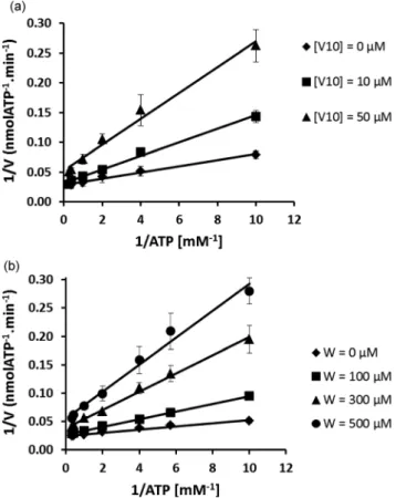
In the present study, the inhibition of the ATPase activity by V10 was found to be competitive for lower concentrations
(10μM) and non-competitive for higher concentrations (50 μM) (Fig. 2A) of V10. Previously, it was shown that the isostructural
Nb10 inhibits non-competitively at both concentrations.4
However, while Nb10 is stable and relatively inert under these
conditions, V10is not and is highly reactive. The formation and
presence of monomeric vanadate might be responsible for the processes of competitive inhibition, seen at lower V10
concen-trations (10μM). Therefore, it is suggested that while V10, like
Nb10,4interacts with calcium ATPase non-competitively, the
for-mation of monomeric vanadium(V) species is responsible for
competitive interaction. This is similar to what is seen for tung-state (Fig. 2B). Tungtung-state (W1) has a lower SR Ca2+-ATPase
inhibition capacity than decavanadate4 (IC50= 400μM for W1
and IC50= 15μM for V10), although higher than described for
Na+-K+-ATPase inhibition (W1 IC50 = 1.5 mM).36 For this
enzyme, other polyoxotungstates (W12) present a higher capacity
of inhibition, with IC50less than 15μM, the value found above
for V10.36 It thus seems that the decavanadate–calcium pump
interaction is favoured in the presence of ATP but is prevented by the iso-structural and iso-electronic decaniobate ion and V10
non-competitively inhibits the enzyme activity vis-à-vis ATP. Other studies, however, have found that the interaction of V10
with the calcium pump is competitive, it binds to two sites,9,37 and have suggested that V10will bind to the calcium pump at the
vanadate-binding site, or to the nucleotide binding site.9,37,38 This has led to the suggestion that the V10-binding site is the
same as the ATP binding site or at least close to it, since it has been observed that a covalent inhibitor that binds specifically to Lys-515 within the ATP binding site also prevents V10 from
binding to the ATPase.38
Structural studies based on V10-induced crystallization of
the calcium pump complex have indicated that the V10binding
site in the E2 conformation of the SR ATPase is located in the intersection of the three cytoplasmic protein domains that includes the nucleotide, the phosphorylation and the β-strand domains.10 It should be noticed that some ATPase enzymes include many membrane enzymes such as the E1E2-ATPases. Although many details are known with regard to the biochemical mechanisms of these ATPases, inhibitory studies assume that vanadate acts as a phosphate analogue and presumably inhibits the enzymes as a transition-state analogue for the phosphoryl group transfer. However, E1–E2 ATPases are not truly an ATPase but a phosphohydrolase, once what is hydrolyzed is the phosphate that is covalently bound to the enzyme, rather than ATP. Further studies are needed to clarify the interaction and the effects of V10on the structure and function of the calcium pump,
once it seems that it does not behave like vanadate, a phosphate analogue.
Fig. 1 105.2 MHz51V-NMR spectra, at room temperature, of 0.5 mM
decavanadate (5 mM total vanadate) in 25 mM HEPES ( pH 7.0), 100 mM KCl, 5 mM MgCl2, 50 μM CaCl2 with (A) SR calcium
ATPase, plus 0.25 mM ATP or niobate, and (B) upon niobate addition.
Downloaded by RSC Internal on 12 September 2012
3.2. Atomic absorption and EPR spectroscopy with cysteine oxidation studies
Using atomic-absorption spectroscopy (AAS) V10 has been
shown to bind to an equal extent to all protein conformations that occur during the process of calcium translocation, viz. E1, E1P, E2 and E2P.4 Thus the amount of V10that was found to
bind to each conformer of the enzyme was the same, and was independent of the medium conditions that favoured each protein conformation. A likely possibility is that the V10 binding is
always available independently of the calcium translocation-dependent conformational changes. This is in stark contrast with what has been observed for monomeric vanadate, as vanadium binding to the protein is clearly prevented in the E1 confor-mation, but is favourable when the protein is in the E2 conformation.4,10
In this study we observe that Nb10prevents the interaction of
V10with the calcium pump once the amount of vanadium bound
to the protein decreases by about 50%, whereas no effects were observed for heparin, a molecule known to interact with nucleo-tide binding sites (Fig. 3). Using NMR it was confirmed that the isostructural Nb10 prevents the V10 from interacting with the
protein. The fact that V10inhibition would be non-competitive,
as suggested above, and that the interaction with the calcium
pump is independent of the protein conformation may point to a specific mode of interaction with the calcium pump that is clearly different from the one described for vanadate alone. This observation, in agreement with previous functional studies exe-cuted at conditions very close to physiological ones, means that V10is the only vanadate species able to prevent calcium
trans-location coupled to ATP hydrolysis, whereas no effect is found for a normal vanadate solution, not containing V10, up to
con-centrations of 2 mM.3
On the other hand, more recently the IC50 value of calcium
ATPase inhibition for Nb10 (IC50 = 35 μM) was found to be
more than twice that of V10 (IC50 = 15μM).4 This shows that
V10 may have a different mode of protein interaction.
Mono-meric vanadate (V1), a product of V10 decomposition in the
medium, may also contribute to the inhibition effects. However, as indicated by AAS, monomeric vanadate interaction should not be favoured under conditions where calcium is present and the E1 conformation is dominant. In addition, the inhibition of the calcium ATPase by monomeric vanadate is less effective. There is evidence that vanadate can form a complex with ATP itself, as is expressed through broadening of the monomeric vanadate signal in the presence of ATP,3 and a similar type of complex formation has been suggested with myosin.11,12 It is also possible that the V10mediated inhibition of calcium ATPase
Table 1 Chemical shifts (δ), line widths (Δν1/2), and broadening factor of the line widths (f) of monomeric (V1), dimeric (V2), tetrameric (V4) and
decameric (V10) forms of vanadate present in 5 mM nominal decavanadate solution (0.5 mM V10) under different experimental conditions
Decavanadate (0.5 mM) V10C V10B V10A V1 V2 V4 C2 C1 Medium δ/ppm −421.0 −495.0 −512.0 −558.0 −571.0 −575.0 −537.0 −551.0 Δν1/2/Hz 1394.0 996.0 398.0 199.0 398.0 199.0 Area 2.5 6.4 10.4 4.3 1.0 5.6 [+SR Ca2+-ATPase (5 mg mL−1)] δ/ppm −421.0 −495.0 −512.0 −558.0 −571.0 −575.0 −537.0 −551.0 Δν1/2/Hz 2788.0 2390.0 1195.0 199.0 996.0 1095.0 f (2.0) (2.4) (3.0) (1.0) Area 3.6 5.4 8.5 1.2 1.0 2.8 [+SR Ca2+-ATPase (5 mg mL−1) + ATP (2.5 mM)] δ/ppm −421.0 −495.0 −512.0 −558.0 −571.0 −575.0 −537.0 −551.0 Δν1/2/Hz 3386.0 2589.0 1593.0 1195.0 1195.0 f (2.4) (2.6) (4.0) Area 2.9 5.4 6.9 0.9 2.0 [+SR Ca2+-ATPase (5 mg mL−1) + Nb10(1 mM)] δ/ppm −421.0 −495.0 −512.0 −558.0 −571.0 −575.0 −537.0 −551.0 Δν1/2/Hz 2190.0 1991.0 996.0 199.0 996.0 1095.0 f (1.6) (2.0) (2.5) (1.0) Area 2.6 5.8 9.6 0.9 0.8 2.0 [+SR Ca2+-ATPase (5 mg mL−1) + Nb10(0.5 mM)] δ/ppm −421.0 −495.0 −512.0 −558.0 −571.0 −575.0 −537.0 −551.0 Δν1/2/Hz 2788.0 2190.0 996.0 199.0 1195.0 1195.0 f (2.0) (2.2) (2.5) (1.0) Area 3.3 5.8 9.5 0.6 0.8 2.0 [+SR Ca2+-ATPase (5 mg mL−1) + ATP (2.5 mM) + Nb10(1 mM)] δ/ppm −421.0 −495.0 −512.0 −558.0 −571.0 −575.0 −537.0 −551.0 Δν1/2/Hz 2788.0 1991.0 1195.0 1195.0 1195.0 f (2.0) (2.0) (3.0) Area 2.9 4.7 8.4 0.5 1.4
Downloaded by RSC Internal on 12 September 2012
involves oxidation of the cysteine units in the protein, which would certainly affect the activity of the enzyme.39This would also lead to the reduction of vanadate(V) to vanadyl(IV), which is
an EPR active species. Such a signal was indeed found, and only for either vanadate or V10 incubation with calcium ATPase
(Fig. 4), but not in the absence of the calcium ATPase. This is strong evidence that cysteine units in the protein are oxidized in the presence of vanadium(V), as has previously been reported in
similar experiments with myosin or with actin.12,13,40
Actin was described as a potential target for decavanadate, being suggested to reduce the metal and to bind with vanadyl with a 1.1 stoichiometry.11–15This explains the difference in the inhibition caused by Nb10 vs. V10 in spite of them being
iso-structural and isovalent: while the standard reduction potential of VO2+ to VO2+ is +1.0 V, under acidic conditions niobium(V)
isn’t known to be easily reduced.25,26 This difference in redox activity means that while vanadate can reduce cysteine (E0 ∼
−0.15 V, values referred are vs. the standard hydrogen electrode and reported at pH 7), niobate won’t. In fact, only the vanadate and V10species induced protein cysteine oxidation, whereas no
evidence of oxidation was detected for the other Nb, W and Mo
oxometalates (Fig. 5). Furthermore, adding antioxidants such as quercetin or kaempferol (100μM) partially reversed the cysteine oxidation induced by vanadate (Fig. 5). With other antioxidants, such as GSH (1 mM), it was not possible to evaluate the effect, due to interferences in the method, because the com-pounds absorbed at the wavelength of the measurement or it formed complexes with the antioxidants. For the same reasons the antioxidant reversion of cysteine oxidation induced by V10could not be determined. Apparently, V10is reacting with
the antioxidant. All the data, however, point towards the oxi-dation of cysteine being a key part of the inhibitory mechanism of V10 towards calcium ATPase. On the other hand, it means
that vanadate can be reduced by thiols not only in biological conditions,41 but particularly upon interaction with specific proteins such as actin13 and also with the calcium pump, as described above.
Fig. 2 (A) Lineweaver–Burk plot of the inhibition of the sarcoplasmic reticulum calcium pump with V10. Concentrations in vanadium are
referred as decavanadate concentrations. ATPase activity was measured as indicated in the Materials and methods section. Data are plotted as means ± SD. The results shown are the average of triplicate experiments. (B) Lineweaver–Burk plot of the inhibition of the sarcoplasmic reticu-lum calcium pump with W. Concentrations in tungstenium are referred as tungstate concentrations. The ATPase activity was measured as indi-cated in the Materials and methods section. Data are plotted as means ± SD. The results shown are the average of triplicate experiments.
Fig. 3 Vanadium analysis performed by atomic absorption spec-troscopy (AAS). V10samples containing 1μM vanadate species, upon
addition of an equal concentration of sarcoplasmic reticulum (SR) calcium ATPase (1μM), in the presence of heparin or decaniobate, in the medium containing 25 mM HEPES ( pH 7.0), 100 mM KCl, 5 mM MgCl2, 50μM CaCl2and 0.25 mM ATP, were centrifuged and the
super-natant analyzed by AAS, as described in the Materials and methods section. The concentrations of vanadium, for V10solutions, were plotted
against the several medium conditions. Data are plotted as means ± SD. The results shown are the average of triplicate experiments.
Fig. 4 X-band EPR spectra of 0.5 mM decavanadate (5 mM total vanadate) in 25 mM HEPES ( pH 7.0), 100 mM KCl, 5 mM MgCl2,
50 μM CaCl2 with SR calcium ATPase, in the absence (A) and in
the presence of 0.25 mM ATP (B). The spectra were acquired as described under Materials and methods. Upon protein addition the oxidovanadium(IV) typical signals are elicited.
Downloaded by RSC Internal on 12 September 2012
It has been suggested that only one or two cysteine (Cys) residues of the SR Ca2+-ATPase are critically important for the function of the enzyme.39Modification of Cys349, located near the phosphorylation binding site, causes modulation of the SR Ca2+-ATPase activity by peroxynitrite.42 In this case the oxi-dation could be reverted by GSH, but for high concentrations of oxidizing agents the content of the SH group decreases and the effect cannot be reverted, i.e. the oxidation is irreversible.39 In fact, Ca2+-ATPase inhibitory effects promoted by reactive oxygen and nitrogen species,39,42as well as the antitumor drug 3-bromopyruvate,43 all appear to involve protein cysteine oxi-dation. To verify that glutathione (GSH) could reverse the decrease in calcium ATPase activity induced by the oxometa-lates, GSH was added to the medium just prior to oxometalate addition. Although cysteine oxidation was still observed for the two vanadate solutions, GSH could not prevent or revert the inhibition and similar IC50values were obtained with or without
GSH (Fig. 6A), for all the oxometalates. Quercetin and kaemp-ferol, antioxidants present in tea, grapes and apples, were also ineffective, suggesting that the inhibitory effects are not a conse-quence of cysteine oxidation, or that the specific cysteine oxi-dation does not contribute significantly for the modulation of the ATPase activity (Fig. 6B).
Therefore it is suggested that V10 induces oxidation of
cysteine but that only partially modulates the ATPase activity. Speculating, this cysteine unit is likely located near the nucleo-tide binding site, such as Cys675 or Cys674, not particularly involved in the modulation of the enzyme activity. Decavanadate binding to the calcium ATPase may involve all the three cyto-plasmic domains, but near the phosphorylation binding site, as described elsewhere.10,44 Decavanadatefits in the protein like a ball in a vise preventing the vise from closing.44This binding
locus would explain the inhibitory effect V10has on SR Ca2+
-ATPase activity and on calcium translocation, as described previously.3,4,16,17,35
3.3. Raman spectroscopy studies
In order to better understand the structural changes induced by the oxometalates on the calcium ATPase, the process was studied using Raman spectroscopy to explore the conformations of calcium ATPase which dominate in the presence of oxometa-lates. Raman spectroscopy is highly sensitive towards small structural changes in protein structure and has been a versatile tool in investigating protein structure modification caused by metal interactions.45,46
As is the case with infrared spectroscopy, the Raman spectrum contains several diagnostic regions which correspond to specific vibrational modes belonging to common structural motifs seen in proteins.45–48,50–52 These include a band at ca. 855 cm−1 which belongs to tyrosine residues, and a band at ca. 930 cm−1 indicative of α-helices46–48,50 (Fig. 7A). Raman spectroscopy has been used to follow peptide tyrosine phosphorylation using the two bands between 820 and 855 cm−1 48–50that are observed in the absence of phosphorylation.
Several bands that are sensitive to the average conformation of the protein can be identified in the Raman spectrum, and these can be used to evaluate the protein conformational changes
Fig. 5 Calcium ATPase cysteine (Cys) redox state, after oxometalates exposition. The accessible thiol groups, in the absence of denaturants, were measured as indicated in the Materials and methods section. Data are plotted as means ± SD. The results shown are the average of tripli-cate experiments. Titration of cysteines was carried out in HEPES ( pH 7.0), 100 mM KCl, 5 mM MgCl2and 55 mM sodium phosphate buffer
( pH 7.2), after 5 min of incubation of 0.2 mL SR (1 mg mL−1) with vanadate (1 mM), decavanadate (0.3 mM), decaniobate (0.035 mM), tungstate (8 mM) or molybdate (45 mM), in the absence or in the pres-ence of the following antioxidants, wherever stated: GSH (1 mM), quer-cetin (100μM) or kaempferol (100 μM). The increase in absorbance at 412 nm was continuously recorded over 5 min, until a steady value was reached. The results shown are the average of triplicate experiments.
Fig. 6 Effect of reduced glutathione (GSH) on sarcoplasmic reticulum (SR) Ca2+-ATPase activity in the presence of oxometalates, at IC50
con-centrations. The ATPase activity was measured as indicated in the Materials and methods section. Data are plotted as means ± SD. The results shown are the average of triplicate experiments.
Downloaded by RSC Internal on 12 September 2012
induced by external or internal factors. For instance, peptide tyrosine phosphorylation results in two bands at ca. 825 and 853 cm−1.48–50The ratio of intensities of these bands gives indi-cations about the strength of tyrosine hydrogen bonding.48–50In the present study, this Fermi resonance doublet was not observed, but instead only a single band at 837 cm−1(Fig. 7A) was found. Such an observation could be interpreted in two ways: (i) the tyrosine units are not involved in hydrogen bonding48 or (ii) the tyrosine residues are phosphorylated, leading to the collapse of the doublet into a single band.48–50 While thefirst hypothesis should be discarded, because the tyro-sines would have to be facing the interior of the protein instead of interacting with bulk water, the second one is unlikely because it is known that the Ca2+-ATPase is phosphorylated in aspartic acid.30,32
In the present study, we collected spectra on protein confor-mation E1– medium containing calcium, or on protein confor-mation E1P, in the presence of ATP (Fig. 7A). It is suggested that the more intense peak at 1650 cm−1 corresponds to the amide I CvO stretching vibration of the peptide, and the peak at 1150 cm−1 probably can be assigned to the CH2 deformation
from lipid hydrocarbon,51,52 once the sample contains not only the membranar protein but also the native lipid bilayer from the sarcoplasmic reticulum. The changes in the intensity of the peak at 1150 cm−1can be probably due to metal-induced changes to the CH2in lipid molecules,52that would suggest an effect on the
lipid-membrane structure. An increase in the peak at 1650 cm−1, ascribed to amide I (CvO stretching vibration of the peptide), is also observed (Fig. 7A), indicating changes in the secondary structure of the protein. No significant changes were detected, when the protein is susceptible to phosphorylation by inorganic phosphate, in a medium containing EGTA that complexes all the free calcium and inorganic phosphate (not shown). These ditions favour conformation E2 and by extension the E2P con-formation. Also by comparing the Raman spectra of the calcium ATPase in both conformations E1 and E2, no significant differ-ences were detected (not shown), in agreement with observations in similar studies with the Na+, K+, ATPase.52
Both molybdate and tungstate induce distinct changes in the intensities of the peaks (Fig. 8A and B), suggesting that those oxometalates are promoting different protein conformation changes. In comparison with the E1P spectra, upon addition of Mo, peaks at 837, 1075 and 1465 cm−1did not change, while the peak at 1650 cm−1is more intense and peaks at 926, 1150
and 1380 cm−1are weaker (Fig. 8A). For W, a decrease in the bands, relative to E1P, was detected for all the first six peaks found in the spectra whereas the last peak at 1650 cm−1 remained unchanged (Fig. 8B). For Mo, the increases on α-helices point to a structure-organizing effect, but clearly the Mo interaction seems to induce different calcium ATPase protein conformational changes than W, although as described above they both favour an E1 type conformation. In contrast to the former oxometalates, upon incubation of the protein with Nb10
or V10, a decrease in the intensities of all the peaks was observed
(Fig. 9). Furthermore, the decrease in the intensities of all the peaks upon V10and Nb10interaction, observed for the E1
con-formation, was also detected in the Raman spectra of the enzyme in the E2 conformation, that is, absence of free calcium due to the presence of EGTA in the medium, suggesting that those oxo-metalates affect both spectra of the E1 or E2 protein confor-mation (not shown).
In contrast to the effects induced by V and Nb on the protein Raman spectra at the E1 or E2 conformation, no significant changes were detected in the Raman spectra upon phosphate or tungstate addition to the enzyme in the E2 conformation, whereas a small decrease was observed for molybdate (not shown). Note that the presence of any natural ligand of the Ca2+ -ATPase will induce major conformational changes in the protein, thus affecting oxometalate interactions. In addition to the binding to the cytoplasmic domains, other binding sites for deca-meric vanadate have been suggested, such as the specific inter-molecular binding sites described to be essential to induce crystal formation.9,10 Therefore, although it has been used to resolve the structure of enzyme in the E2 state,10V10seems to
behave unlike a natural ligand of the enzyme.
Raman spectroscopy can also be used to characterize oxome-talate solutions,53the exploration of the viability of this approach
Fig. 9 Raman spectra of calcium ATPase upon addition of decavana-date (left) or decaniobate (right).
Fig. 7 Raman spectra of calcium ATPase at conformation states E1 upon phosphorylation (E1P).
Fig. 8 Raman spectra of calcium ATPase in the presence of molybdate (left) or tungstate (right) in the medium. Mo decreases the peak at 1150 cm−1, and it increases the peak at 1650 cm−1, belonging probably to the CH2 deformations in lipid molecules and the amide I CvO
stretching vibration of the peptide, respectively. For W, a decrease in the bands, in comparison with E1P, was detected for all thefirst six peaks, whereas the last peak at 1650 cm−1, shows the same intensity.
Downloaded by RSC Internal on 12 September 2012
to decavanadate and decaniobate solutions is currently underway. A preliminary study suggests that the more stable Nb10
oxometa-late is an excellent tool to elucidate V10 chemistry in solution
(Batista de Carvalho et al., unpublished results). Based on the data, it appears that V10and Nb10 induce similar Ca2+-ATPase
conformational changes, which are different from the known conformers E1 or E2, once similar changes in the Raman spectra were detected (Fig. 9 and not shown). W and Mo cause yet another set of conformations. It is suggested that tungstate and molybdate induce the same type of structural changes that are seen in inorganic phosphorylation, but that Nb10, vanadate and
V10 cause different changes corresponding to an as of yet
unknown type of structure. These latter oxometalates interact with both the E1 and E2 states, while this is not the case of the tungstate and molybdate. It is clear that Raman spectroscopy can be a useful diagnostic tool in providing additional information on the effect of oxometalates on calcium ATPase conformation and as such complements EPR and NMR.
4.
Conclusion
In spite of the attention paid to the role of oxometalates in bio-logical systems in recent years, their potential and modes of action are still poorly understood. For example, their influence on the activity of calcium pump ATPase, a key enzyme in homeostasis, is yet to be fully understood. Based on thefindings in this study, it is concluded that V10shows a specific interaction
with the calcium pump in comparison to other oxometalates. It induces cysteine oxidation and undergoes non-competitive inhibition. The ATPase inhibition is not reverted by GSH or quercetin, but the cysteine oxidation is partially prevented by antioxidants such as quercetin. Moreover, V10does not
discrimi-nate against any of the protein conformations that occur during the process of calcium translocation, but binds to all of them. Raman spectroscopy points to specific calcium ATPase confor-mational changes being induced by the oxometalates, but that these changes depend on what oxometalates are present. There-fore, this work confirms the fact that vanadate interaction with proteins, not only with actin but also with SR Ca-ATPase, can impact thiols and undergoing vanadium reduction. It is believed that the present observations will contribute to the understanding of oxometalate interactions with the calcium pump and the role of V10in biology, particularly the mechanisms of V10interaction
with ionic pumps.
The isostructural Nb10 is shown to be an excellent tool for
clarifying the mode of interaction and the effects of V10on the
calcium pump. Raman spectroscopy points to a new approach on the interaction of oxometalates with the calcium pump,finding distinct protein conformational changes, similar to E1P or to none of the established conformations induced by the presence of oxometalates. Putting it all together, it is suggested to be due to the putative reduction of vanadate as well as the non-competi-tive V10binding, and would explain why monomeric molybdate
and tungstate behave differently regarding the interaction with the SR calcium pump. We conclude that it is the V10or the
vana-date interaction with calcium ATPase which induces cysteine oxidation and the concomitant vanadate reduction. Therefore, many aspects of the interactions between the different
oxometalates and the calcium pump are yet to be clarified. Although some are chemically very similar they induce different effects on the structure and on the function of the protein.
Abbreviations
DTNB 5,5′-dithiobis-(2-nitrobenzoic acid) EGTA ethylene glycol tetraacetic acid GSH reduced glutathione
Nb10(Nb10O286−) decaniobate
SDS sodium dodecyl sulphate SR sarcoplasmic reticulum
51V-NMR vanadium-51 NMR
V1 monomeric vanadate
V10(V10O286−) decavanadate
Acknowledgements
MA thanks CCMAR; LAEBC and MPMM thank QFM-UC for financial support. CAO is grateful for a QEII fellowship and Discovery Project grant (DP110105530) from the Australian Research Council. WHC acknowledges support from the U.S. Department of Energy Office of Basic Energy Science via grant DE-FG02-05ER15693, the National Science Foundation via EAR-0814242 and an NSF CCI grant through the Center for Sustainable Materials Chemistry, number CHE-1102637.
References
1 M. Aureliano, T. Tiago, R. M. Gândara, A. Sousa, A. Moderno, M. Kaliva, A. Salifoglou, R. O. Duarte and J. J. J. Moura, J. Inorg. Biochem., 2005, 99, 2355–2361.
2 M. Aureliano, F. Henao, T. Tiago, R. O. Duarte, J. J. G. Moura, B. Baruah and D. C. Crans, Inorg. Chem., 2008, 47, 5677–5684. 3 M. Aureliano and V. M. C. Madeira, Biochim. Biophys. Acta, Mol. Cell
Res., 1994, 1221, 259–271.
4 G. Fraqueza, C. A. Ohlin, W. H. Casey and M. J. Aureliano, J. Inorg. Biochem., 2012, 107, 82–89.
5 B. Schiøtt, M. G. Palmgren, J. V. Møller, P. Nissen and N. Fedosova, Biochim. Biophys. Acta, 2009, 1787, 207–220.
6 T. L. Turner, V. H. Nguyen, C. C. McLauchlan, Z. Dymon, B. M. Dorsey, J. D. Hooker and M. A. Jones, J. Inorg Biochem., 2012, 108, 96–104. 7 T. Tiago, M. Aureliano and C. Gutierrez-Merino, Biochemistry, 2004, 43,
5551–5561.
8 T. Tiago, P. Martel, C. Gutiérrez-Merino and M. Aureliano, Biochim. Biophys. Acta, 2007, 1771, 474–480.
9 P. Csermely, A. Martonosi, G. C. Levy and A. J. Ejchart, Biochem. J., 1985, 230, 807–815.
10 S. Hua, G. Inesi and C. Toyoshima, J. Biol. Chem., 2000, 275, 30546– 30550.
11 S. Ramos, M. Manuel, T. Tiago, R. O. Duarte, J. Martins, C. Gutiérrez-Merino, J. J. G. Moura and M. Aureliano, J. Inorg. Biochem., 2006, 100, 1734–1743.
12 S. Ramos, J. J. G. Moura and M. Aureliano, J. Inorg. Biochem., 2010, 104, 1234–1239.
13 S. Ramos, R. O. Duarte, J. J. G. Moura and M. Aureliano, Dalton Trans., 2009, 7985–7994.
14 S. Ramos, R. M. Almeida, J. J. G. Moura and M. Aureliano, J. Inorg. Biochem., 2011, 105, 777–783.
15 S. Ramos, J. J. G. Moura and M. Aureliano, Metallomics, 2012, 4, 16–22.
16 M. Aureliano and D. C. Crans, J. Inorg. Biochem., 2009, 103, 536–546. 17 M. Aureliano, Dalton Trans., 2009, 9093–9100.
18 L. Yan-Tuan, Z. Chun-Yuan, W. Zhi-Yong, Ma. Jiang and C.-W. Yan, Transition Met. Chem., 2010, 35, 597–603.
19 F. Zhai, X. Wang, D. Li, H. Zhang, R. Li and L. Song, Biomed. Pharmacother., 2009, 63, 51–55.
Downloaded by RSC Internal on 12 September 2012
20 V. Dimitrova, K. Zhetcheva and L. P. Pavlova, J. Chem., 2011, 35, 215–223. 21 A. Zorzano, M. Palacin, L. Marti and S. Garcia-Vicente, J. Inorg.
Biochem., 2009, 103, 559–566.
22 M. J. Pereira, E. Carvalho, J. W. Eriksson, D. C. Crans and M. Aureliano, J. Inorg. Biochem., 2009, 103, 1687–1692.
23 S. S. Soares, C. Gutiérrez-Merino and M. Aureliano, J. Inorg. Biochem., 2007, 101, 789–796.
24 S. S. Soares, F. Henao, M. Aureliano and C. Gutierrez-Merino, Chem. Res. Toxicol., 2008, 21, 607–618.
25 C. A. Ohlin, E. M. Villa and W. H. Casey, Inorg. Chim. Acta, 2009, 362, 1391–1392.
26 C. A. Ohlin, E. M. Villa, J. C. Fettinger and W. H. Casey, Angew. Chem., Int. Ed., 2008, 47, 8251–8254.
27 S. S. Soares, H. Martins, J. Coucelo, C. Gutiérrez-Merino and M. Aureliano, J. Inorg. Biochem., 2007, 101, 80–88.
28 B. De Foresta, F. Henao and P. Champeil, Eur. J. Biochem., 1994, 223, 359–369. 796.
29 K. Fohr, J. Scott, G. Ahnert-Hilger and M. Gratzl, Biochem. J., 1989, 262, 83–89.
30 L. de Meis and A. L. Vianna, Annu. Rev. Biochem., 1979, 48, 275–292. 31 D. R. Davies and W. G. J. Hol, FEBS Lett., 2004, 577, 315–321. 32 G. Inesi, D. Lewis and A. J. Murphy, J. Biol. Chem., 1984, 259,
996–1003.
33 T. Tiago, M. Aureliano and J. J. G. Moura, J. Inorg. Biochem., 2004, 98, 1902–1910.
34 D. C. Crans, S. Schoeberl, E. Gaidamauskas, B. Baruah and D. A. Roess, J. Biol. Inorg. Chem., 2011, 16, 961–972.
35 M. Aureliano, World J. Biol. Chem., 2011, 2, 215–238.
36 M. B. Colović, D. V. Bajuk-Bogdanovic, N. S. Avramovic, I. D. Holclajtner-Antunovic, N. S. Bošnjaković-Pavlovic, V. M. Vasić and D. Z. Krstić, Bioorg. Med. Chem., 2011, 19, 7063–7069.
37 P. Csermely, S. Varga and A. Martonosi, Eur. J. Biochem., 1985, 150, 455–460.
38 S. Varga, P. Csermely and A. Martonosi, Eur. J. Biochem., 1984, 148, 119–126.
39 R. I. Viner, T. D. Williams and C. Schöneich, Biochemistry, 1999, 38, 12408–12415.
40 T. Tiago, C. Gutiérrez-Merino and M. Aureliano, Vanadium Biochemistry, Research Signpost, Kerala, India, 2007, pp. 75–95.
41 D. C. Crans, B. Zhang1, E. Gaidamauskas, A. D. Keramidas, G. R. Willsky and C. R. Roberts, Inorg Chem., 2010, 49, 4245–4256. 42 R. I. Viner, A. F. Huhmer, D. J. Bigelow and C. Schöneich, Free Radical
Res., 1996, 24, 243–259.
43 D. Jardim-Messeder, J. Camacho-Pereira and A. Galina, Int. J. Biochem. Cell Biol., 2012, 44, 801–807.
44 C. Toyoshima, M. Nakasako, H. Nomura and H. Ogawa, Nature, 2004, 405, 647–655.
45 P. R. Carey, Biochemical Applications of Raman and Resonance Raman spectroscopies, Academic Press, NY, 1982.
46 R. Tuma, J. Raman Spectrosc., 2005, 36, 307–319.
47 A. Tu, Raman spectroscopy in Biology, Wiley and Sons, NY, 1982. 48 Y. Xie, D. Zhang, G. K. Jarori, V. Jo Davisson and D. Ben-Amotz, Anal.
Biochem., 2004, 332, 116–121.
49 M. N. Siamwiza, R. C. Lord and M. C. Chen, Biochemistry, 1975, 14, 4870–4876.
50 Z. Arp, D. Autrey, J. Laane, S. A. Overman and G. J. Thomas, Jr., Bio-chemistry, 2001, 40, 2522–2529.
51 G. J. Thomas, Biopolymers (Biospectroscopy), 2002, 67, 214–225. 52 C. H. Nielsen, S. Abdali, J. A. Lundbaek and F. Cornelius, Spectroscopy,
2007, 22, 52–63.
53 A. Amado, M. Aureliano, P. J. Ribeiro-Claro and J. Teixeira-Dias, J. Raman Spectrosc., 1993, 24, 669–703.
Downloaded by RSC Internal on 12 September 2012