2016
UNIVERSIDADE DE LISBOA
FACULDADE DE CIÊNCIAS
DEPARTAMENTO DE BIOLOGIA VEGETAL
Characterization of Plasmodium methionine
metabolism key enzyme
Maria Inês Moreira Oliveira Leite Marreiros
MESTRADO EM BIOLOGIA MOLECULAR E GENÉTICA
Dissertação orientada pela Doutora Vanessa Alexandra Zuzarte Luís (Instituto de
Medicina Molecular, Faculdade de Medicina da Universidade de Lisboa) e pela
Professora Anabela Bernardes da Silva (Faculdade de Ciências da Universidade de
i
ACKNOWLEDGMENTS
Quero, em primeiro lugar, agradecer à Professora Doutora Maria Mota por me ter dado a oportunidade de realizar a minha tese de mestrado no seu laboratório.
À Professora Anabela Silva pelo apoio e disponibilidade demonstrados ao longo de todo este percurso. Quero agradecer, em especial, à Vanessa. Obrigada por tudo o que me ensinaste sempre com tanta paciência, dedicação e carinho. Obrigada por toda a motivação constante, toda a compreensão, por todos os conselhos e por toda a confiança que depositaste em mim e no meu trabalho. O teu entusiasmo pela ciência e o modo como a praticas são realmente inspiradores. Obrigada sobretudo pelo apoio incondicional e pela amizade, foste sem dúvida a melhor orientadora que poderia ter tido. Quero desde já agradecer a todas as pessoas do laboratório. Obrigada por me terem recebido de braços abertos e por todo o vosso apoio. Obrigada por toda a ajuda, todos os conselhos, pela alegria e boa-disposição constante e por me terem sempre guiado quando me sentia perdida. Obrigada sobretudo pela amizade e por serem uma segunda família. Embora sejam todos muito importantes quero agradecer em especial a algumas pessoas que me são mais próximas:
À minha Léninha, pela amizade e por todo o apoio e motivação constante. Obrigada por toda a ajuda com os WB, pelas inúmeras vezes que reveste a tese comigo, por todos os momentos de alegria e, principalmente, por todos os conselhos e por tudo o que me ensinaste sobre a vida.
Ao Miguel, pela amizade incondicional. Obrigada por todo o apoio, toda a paciência e compreensão. Obrigada por teres percorrido este caminho sempre ao meu lado e, sobretudo, por seres o irmão que nunca tive!
Ao Ângelo, por tudo o que me ensinaste sobre ciência e sobre a vida (e também sobre história!). Obrigada por todos os conselhos sábios nos momentos de maior indecisão, por toda a motivação, pela boa-disposição constante, pela tua amizade e principalmente por me conseguires fazer sempre sorrir (mesmo quando a vida sabia a ‘rosas de plástico’!).
Ao João, por todo o apoio, por todos os conselhos e por todas as horas que perdeste a ajudar-me no microscópio. Obrigada por toda a disponibilidade que sempre demonstraste para me ajudar e, principalmente, pela amizade.
À Ana Parreira, à Margarida Ruivo, à Inês Bento, à Maria Menezes, à Priscila e à Marta Machado por todas as palavras de força, por toda a preocupação que sempre demonstraram e pela amizade.
ii
Quero agradecer também à minha família e aos meus amigos. Sem o vosso apoio, a vossa força e motivação constante não teria conseguido. Obrigada por terem sempre acreditado em mim mesmo quando eu própria não acreditei.
Aos meus pais e à minha irmã por todo o amor e apoio. Todas as palavras parecem escassas quando tento agradecer tudo o que fizeram e fazem todos os dias por mim. Não há palavras que consigam descrever o quão importante foi ter-vos a meu lado. Obrigada por toda a paciência que tiveram comigo, por toda a compreensão, por todo o vosso carinho. Obrigada por tudo o que ensinaram sobre a vida, por todos os valores que me transmitiram e por me terem dado sempre asas para voar e para seguir os meus sonhos. São sem dúvida os melhores e tudo o que sou hoje devo-o a vocês. Adoro-vos! À minha melhor amiga, Mariana, pela amizade incondicional ao longo destes últimos 11 anos. Obrigada por todo o teu apoio, por todas as conversas, por me mostrares sempre o lado melhor da vida e por nunca me teres deixado baixar os braços. Obrigada por estares sempre ao meu lado e por me mostrares que o irás estar sempre. Adoro-te.
Ao André, por todo o amor. Não há palavras que descrevam a sorte que tenho em ter-te ao meu lado todos os dias. Obrigada pelo apoio, pela amizade, por todos os conselhos, por toda a força e motivação constante. Não há palavras que consigam agradecer toda a paciência que tiveste, todas as horas que perdeste a rever a tese comigo e toda a tua compreensão. És sem dúvida muito mais do que alguma vez esperei ter. ♥ you
iii
“There is inside you All of the potential
To be whatever you want to be; All of the energy
To do whatever you want to do.
Imagine yourself as you would like to be, Doing what you want to do,
And each day, take one step Towards your dream.
And though at times it may seem too difficult to continue,
Hold on to your dream.
One morning you will awake to find That you are the person you dreamed of, Doing what you wanted to do,
Simply because you had the courage To believe in your potential
And to hold on to your dream.”
v
ABSTRACT
Malaria is a disease caused by protozoan parasites of the genus Plasmodium that are transmitted to humans by infected female Anopheles mosquitoes. Despite countless efforts toward eradication malaria still remains one of the most prevalent infectious diseases, constituting a major public health concern. The available antimalarial drugs are insufficient to control and eradicate malaria, mostly due to the emergence of drug-resistant parasites. Thus, the development of novel intervention strategies is critical to achieve eradication. As an obligatory intracellular pathogen, Plasmodium establishes close interactions with its host that are crucial to ensure parasite development and survival, one of such is the methionine metabolism. Methionine is an essential amino acid and, as for most living organisms, Plasmodium lacks the ability to synthesize methionine de novo. During the blood-stage of infection Plasmodium obtains methionine mainly through haemoglobin digestion. However, how Plasmodium obtains methionine during the liver-stage and how the parasite modulates the host cells in order to scavenge this essential amino acid is still unknown. The first step of methionine cycle is the synthesis of S-adenosylmethionine (SAMe) through a reaction catalyzed by the enzyme SAMe synthetase (SAMS). SAMe is a key metabolite in the methionine metabolism being the main biological donor of methyl groups for transmethylation reactions. SAMe is also a key intermediate in the transsulfuration pathway generating homocysteine (Hcy) which is metabolized into glutathione (GSH), being the last step of this pathway catalysed by glutathione synthetase (GS). GSH is a powerful antioxidant that in Plasmodium acts as one of the primary lines of the defense against the damage caused by reactive oxygen species (ROS), ensuring parasite survival. In this work we have explored the role of Plasmodium enzymes responsible for SAMe and GSH synthesis throughout its life cycle and in particular during the liver-stage of infection. The liver is a particular organ in the metabolism of methionine, namely in SAMe-dependent transmethylation reactions and in glutathione synthesis and storage. Thus, we hypothesized that while replicating inside hepatocytes, Plasmodium relies on its host to ensure a sufficient supply of these crucial metabolites. The data obtained in this study suggest that: 1) Plasmodium does not rely on its own SAMS enzyme while developing inside hepatocytes; 2) that the inhibition of SAMS activity during the blood-stage of infection leads to a low parasitemia, preventing the onset of cerebral malaria and 3) the deletion of GS-encoding gene results in the arrest at the oocyst stage, preventing transmission between the mosquito vector and the mammalian host. A detailed knowledge of Plasmodium methionine pathway provides promising tools for the design and development of novel antimalarial drugs.
vi
SUMÁRIO
A malária é uma doença causada por parasitas protozoários pertencentes ao género Plasmodium que são transmitidos aos humanos por mosquitos fêmea do género Anopheles. Apesar dos inúmeros esforços realizados na tentativa de erradicar a malária esta permanece ainda uma das doenças parasíticas mais prevalentes, constituindo um problema de saúde público. Os anti-maláricos disponíveis são insuficientes no controlo e erradicação da malária, devido sobretudo ao aparecimento de parasitas resistentes. Além disso, o escasso conhecimento acerca da biologia do parasita bem como das interações que este estabelece com o hospedeiro constituem uma barreira na luta contra a malária. Assim, o desenvolvimento de novas estratégias de intervenção torna-se crucial para conseguir a erradicação. Plasmodium é um patogénio intracelular obrigatório e, como tal, as interações que estabelece com o seu hospedeiro são essenciais para garantir o seu desenvolvimento e sobrevivência, nomeadamente as que estabelece ao nível do metabolismo da metionina. A metionina é um aminoácido essencial pelo que, tal como na maioria dos organismos, Plasmodium não tem capacidade para a sintetizar de novo. Durante a fase sanguínea Plasmodium obtém metionina maioritariamente através da degradação de hemoglobina. Contudo, os mecanismos que Plasmodium utiliza para obter metionina durante a fase hepática, bem como para modular a célula hospedeira de modo a garantir um fornecimento suficiente deste aminoácido são ainda desconhecidos. O primeiro passo do ciclo da metionina consiste na síntese de S-adenosilmetionina (SAMe) numa reação catalisada pela enzima SAMe sintetase (SAMS). A SAMe é um metabolito essencial na via metabólica da metionina sendo o maior dador biológico de grupos metilo. A SAMe é ainda um importante intermediário na via da transsulfuração sendo convertida em homocisteína e subsequentemente metabolizada em glutationo, sendo o último passo desta via catalisado pela glutationo sintetase (GS). O glutationo é um antioxidante que em Plasmodium atua como uma das primeiras linhas de defesa contra espécies oxidativas. Neste trabalho explorámos o papel das enzimas de Plasmodium responsáveis pela síntese de SAMe e glutationo ao longo do seu ciclo de vida, com particular ênfase na fase hepática da infeção. O fígado tem um papel preponderante no metabolismo da metionina, nomeadamente nas reações de transmetilação dependentes de SAMe bem como na regulação da síntese e armazenamento do glutationo. Assim, a hipótese que propusemos testar é que enquanto replica no interior do hepatócito Plasmodium depende do hospedeiro para garantir a obtenção destes metabolitos essenciais. Os resultados obtidos neste estudo demonstram que: 1) durante o seu desenvolvimento no fígado Plasmodium não depende da atividade da sua enzima SAMS; 2) a inibição da atividade da enzima SAMS durante a fase sanguínea da infeção resulta numa redução da parasitémia, prevenindo o aparecimento de malária cerebral e ainda que; 3) a deleção do gene que codifica para a enzima GS inibe o desenvolvimento dos esporozoítos, bloqueando assim a transmissão entre o vetor e o hospedeiro mamífero. Assim, um conhecimento detalhado do metabolismo da metionina em Plasmodium fornece ferramentas promissoras para o desenvolvimento de novos anti-maláricos.
vii
RESUMO
A malária é a doença parasitária com maior impacto no mundo estimando-se que por ano 214 milhões de pessoas são infetadas, resultando numa taxa de mortalidade de aproximadamente meio milhão de pessoas. A África subsariana é a área geográfica com maior taxa de incidência contudo, a malária é também endémica no Sudoeste Asiático, na América Latina e ainda nas regiões Mediterrânicas Orientais. O agente infecioso causador de malária é um parasita do género Plasmodium cuja transmissão entre hospedeiros é mediada por mosquitos fêmea do género Anopheles. Em seres humanos existem 5 espécies de Plasmodium capazes de causar infeção: P. falciparum, P. vivax, P. malariae, P. ovale e P. knowlesi. Destas, P. falciparum é o parasita predominante em África sendo responsável pela manifestação mais severa da doença, a malária cerebral. Embora menos letal, P. vivax é a estirpe mais pandémica e caracteriza-se pela capacidade de gerar formas hepáticas dormentes, os hipnozoítos, que podem resultar em recidivas da doença. O ciclo de vida de Plasmodium é complexo, incluindo duas fases de desenvolvimento em hospedeiros distintos: o hospedeiro mamífero e o vetor, o mosquito Anopheles. O ciclo de vida de Plasmodium inicia-se quando um mosquito infetado se alimenta do sangue de um hospedeiro mamífero, depositando sob a sua pele centenas de esporozoítos. Os esporozoítos deslocam-se através da pele até entrarem num vaso sanguíneo migrando depois pela circulação em direção ao fígado onde, após atravessarem um determinado número de hepatócitos, estabelecem a fase hepática da infeção. Esta fase caracteriza-se por ser obrigatória e clinicamente silenciosa na qual o parasita se desenvolve rodeado por um vacúolo parasitóforo, denominado por forma exo-eritrocítica. No interior do hepatócito, o parasita inicia um ciclo de replicação assexuada denominado por esquizogonia no qual, cada parasita, origina milhares de merozoítos que são depois libertados na circulação sanguínea através de vesículas denominadas merossomas, iniciando assim a fase eritrocítica da infeção. Dentro do eritrócito os parasitas replicam mitoticamente desenvolvendo-se sequencialmente em anéis, trofozoítos e esquizontes. Os esquizontes diferenciam-se novamente em merozoítos levando à lise dos eritrócitos e consequentemente, ao aparecimento dos sintomas clínicos associados à doença. A nova descendência de merozoítos pode infetar novos eritrócitos, num ciclo que é reiniciado inúmeras vezes garantindo a progressão da infeção ou diferenciar-se nas formas sexuadas do parasita originando os gametócitos. Contudo, a divisão sexuada dos gametócitos apenas é possível no interior do mosquito. Assim, juntamente com a ingestão de sangue durante a picada do hospedeiro são também ingeridos os gametócitos. Estes migram até ao intestino médio onde se diferenciam em gâmetas masculino e feminino e sofrem fecundação, resultando na formação do zigoto. O zigoto sofre depois divisão meiótica adquirindo mobilidade sendo designado por oocineto, o qual migra pelo epitélio do intestino em direção à lamina basal onde se estabelece. Na lâmina basal o oocineto adquire uma forma globular, originando o oócisto. Este sofre um processo de maturação no qual se replica por mitoses sucessivas produzindo esporozoítos que, após rutura do oócisto, são libertados na hemolinfa. Os esporozoítos são formas móveis e alongadas, migrando depois em direção às glândulas salivares do mosquito onde estão aptos a infetar um novo hospedeiro, reiniciando assim o ciclo de vida.
viii
Plasmodium tem uma capacidade replicativa notável atingindo taxas de crescimento enormes o que implica elevadas exigências nutricionais de modo a garantir a sobrevivência, nomeadamente de metionina. A metionina é um aminoácido essencial pelo que Plasmodium não tem capacidade para a sintetizar de novo. Sendo um parasita intracelular obrigatório, Plasmodium evoluiu no sentindo de assegurar as suas necessidades nutricionais derivando nutrientes a partir da célula hospedeira. Durante a fase sanguínea Plasmodium obtém aminoácidos maioritariamente através da degradação de hemoglobina da célula hospedeira. No entanto, esta contém apenas quantidades vestigiais de metionina não sendo suficiente para satisfazer as necessidades nutricionais do parasita. Assim, Plasmodium modula a célula hospedeira induzindo na sua superfície canais que asseguram a troca direta deste metabolito do citoplasma do eritrócito para o interior do parasita. Contudo, o modo como Plasmodium modula a célula hospedeira e em que medida é o crescimento do parasita dependente do hospedeiro durante a fase hepática não é ainda conhecido. Apesar de ser assintomática, esta fase caracteriza-se por um aumento exponencial no número de parasitas sendo também marcada por inúmeras interações estabelecidas entre parasita-hospedeiro, nomeadamente a nível metabólico. Este forte tropismo do parasita para o fígado humano em conjunto com uma correlação forte entre o metabolismo do hepatócito e o do parasita tornam a fase hepática da infeção de particular interesse para o desenvolvimento de novas estratégias que atuem antes do aparecimento dos sintomas clínicos associados à doença.
O primeiro passo do ciclo da metionina é catalisado pela enzima metionina adenosiltransferase (MAT) - também conhecida por adenosilmetionina sintetase (SAMS) - e consiste na síntese de S-adenosilmetionina (SAMe). A SAMe atua como co-substrato em inúmeras reações metabólicas sendo o metabolito percursor das vias de transmetilação, aminopropilação e transsulfuração. Após ceder os seus grupos metilo a moléculas aceitadoras como DNA, RNA, lípidos e proteínas a SAMe é convertida em S-adenosilhomocisteína (SAH) que é, por sua vez, metabolizada em homocisteína. A homocisteína é então sequencial- e irreversivelmente convertida em cisteína e glutationo, sendo a última reação desta via catalisada pela enzima glutationo sintetase (GS). Dada a arquitetura do hepatócito e o papel preponderante do fígado: 1) na metabolização da metionina; 2) na mediação das reações de transmetilação dependentes de SAMe; bem como na 3) síntese e armazenamento de glutationo procurou-se neste presente trabalho testar se enquanto replica no interior do hepatócito, Plasmodium recorre ao hospedeiro para garantir um fornecimento suficiente destes metabolitos subvertendo assim esta via metabólica da célula hospedeira para garantir a sua sobrevivência. Para tal, gerou-se um parasita transgénico recorrendo à técnica do domínio destabilizador na qual a expressão de SAMS no parasita é regulada de acordo com a presença, ou não, de trimetoprima (TMP). A capacidade de Plasmodium se replicar e de estabelecer infeção na ausência de SAMS foi avaliada durante o seu desenvolvimento tanto no mosquito Anopheles bem como no hospedeiro mamífero in vivo, em modelo de roedores e in vitro, na linha celular HepG2. Os resultados obtidos in vivo sugerem que na ausência de SAMS (PbSAMS-DD – TMP) o desenvolvimento de Plasmodium durante a fase sanguínea é drasticamente afetado, resultando numa diminuição nos níveis de parasitémia. Além disso a ausência de SAMS leva a uma diminuição da virulência conferindo proteção contra a malária cerebral.
ix De seguida estudámos a importância de SAMS durante a fase hepática, in vitro. Os resultados obtidos demonstram que na ausência de SAMS os parasitas desenvolvem formas exo-eritrocíticas com igual tamanho e em igual número quando comparado com o grupo controlo (PbSAMS-DD + TMP), sugerindo assim que esta proteína não é essencial para o desenvolvimento nem para a replicação de Plasmodium durante este estadio da infeção. Os resultados obtidos in vivo estão em concordância com os obtidos in vitro, reforçando que durante a fase hepática Plasmodium não depende da sua enzima para completar o desenvolvimento e para estabelecer com sucesso a infeção. Durante o desenvolvimento de Plasmodium no mosquito tanto o número de oócistos como o de esporozoítos produzidos por parasitas que não expressam SAMS são idênticos ao obtido para o grupo controlo, sugerindo assim que esta enzima não é essencial durante esta fase do desenvolvimento.
A infeção por Plasmodium induz no hospedeiro inúmeros mecanismos de defesa que resultam na produção excessiva de espécies reativas de oxigénio, que podem resultar na morte do parasita. Em contrapartida, numa tentativa de sobreviver, o parasita recorre às suas defesas nomeadamente a antioxidantes como o glutationo. Estudos anteriores demonstraram que, na ausência de y-GCS, a enzima limitante da reação de síntese de glutationo, Plasmodium não consegue completar o seu desenvolvimento nem estabelecer com sucesso a infeção no mosquito. Tal como referido anteriormente, a síntese de glutationo envolve duas reações sendo que a GS catalisa o último passo. Assim, os resultados previamente obtidos para y-GCS permitiram-nos prever que a deleção da proteína GS em Plasmodium não iria surtir qualquer efeito no desenvolvimento do parasita durante a fase sanguínea. Para testar a nossa hipótese, gerámos um parasita transgénico que não expressa o gene que codifica a enzima GS (Pbgs-). Os resultados obtidos in vivo permitiram então comprovar que,
durante a fase sanguínea a enzima GS não é essencial para o desenvolvimento de Plasmodium. Contudo, o desenvolvimento de Plasmodium no mosquito é completamente inibido na ausência de SAMS, sendo a síntese de esporozoítos completamente bloqueada. Em suma, o conhecimento detalhado da via da metionina poderá contribuir para o desenvolvimento de novos anti-maláricos e também de novas estratégias que bloqueiem a transmissão de Plasmodium entre o vetor e o hospedeiro mamífero.
x
LIST OF FIGURES
Figure 1.1: Plasmodium life cycle: infection of the mammalian host and the mosquito vector …………. 1 Figure 1.2: Methionine metabolism in the mammalian liver ……….. 3 Figure 1.3: Schematic representation of methionine transport in Plasmodium-infected erythrocyte .. 6 Figure 4.1: Pbgs is not essential during the blood-stage of infection allowing for gene deletion ……… 13
Figure 4.2: Pbgs is essential for Plasmodium oocyst development and sporozoite production ………… 14
Figure 4.3: Inhibition of ROS production in the midgut does not rescue oocyst developmental arrest in Pbgs- parasites ……… 15
Figure 4.4: Pbsams mRNA is expressed throughout all stages of P. berghei life cycle ….……… 16 Figure 4.5: PbSAMS is a cytoplasmic protein that partially co-localises with Plasmodium endoplasmic
reticulum protein Bip during the blood-stage of infection……….……….………. 17 Figure 4.6: PbSAMS is a cytoplasmic protein during the mosquito-stage, both in oocysts and sporozoites development……….………. 18 Figure 4.7: PbSAMS is a cytoplasmic protein during the liver-stage of the development, in vitro………..……….………... 18 Figure 4.8: Pbsams gene is essential during the blood-stage of infection hindering gene deletion ….. 19 Figure 4.9: Blood-stage parasites rely on PbSAMS protein as its absence results in a developmental impairment ……….. 20 Figure 4.10: Removal of TMP leads to a PbSAMS-DD destabilisation resulting in a decrease in PbSAMS expression levels ……….. 21 Figure 4.11: Pbsams knockdown has no effect in PbSAMS-DD oocysts and sporozoites development in A. Stephensi mosquitoes ……… 22 Figure 4.12: PbSAMS destabilisation by TMP removal results in a decrease of protein levels without affecting liver-stage parasites replication and growth, in vitro ……….……… 23 Figure 4.13: PbSAMS destabilisation by TMP removal results in a decrease of protein levels during the liver-stage of infection, in vitro ……… 24
Figure 4.14: PbSAMS destabilisation by TMP removal decreases protein levels without affecting liver load infection, in vivo ………. 25 Figure 4.15: PbSAMS destabilisation by TMP removal results in a decrease of protein l during the liver-stage of infection, in vivo ………. 25
xi
LIST OF ABBREVIATIONS
BSA: Bovine Serum Albumin cDNA: Complementary DNA DD: Destabilising Domain DHFR: Dihydro-folate reductase
DMEM: Dublecco’s Modified Eagle’s Medium DMSO: Dimethylsulfoxide
DNA: Deoxyribonucleic acid
EEF: Exoerythrocytic form
ϒ-GCS: ϒ- glutamylcysteine synthetase GFP: Green fluorescent protein GS: Glutathione synthetase GSH: Glutathione
HA: Haemagglutinin Hcy: Homocysteine
HRP: Horseradish peroxidase HSP70: Heat shock protein 70 IFA: Immunofluorescence assay
i.v.: Intra-venous
MAT: Methionine adenosyltransferase NPPs: New permeability pathways PAGE: Polyacrylamide gel electrophoresis PBS: Phosphate buffer saline
PBST: Phosphate buffer saline containing
Tween 20
PCR: Polymerase chain reaction PFA: Paraformaldehyde
PV: Parasitophorous vacuole rRNA: Ribosomal RNA
RT-qPCR: Quantitative reverse transcription
PCR
RNA: Ribonucleic acid
ROS: Reactive oxygen species RT: Room Temperature SAMe: S-adenosylmethionine
SAMS: S-adenosylmethionine synthetase SDS: Sodium dodecyl sulphate
xii
TABLE OF CONTENTS
1. Malaria: a burden worldwide ... 1
Plasmodium life cycle ... 1
2. Methionine: an essential amino acid to sustain life ... 2
2.1 Methionine Metabolism in Mammals ... 3
2.2 Glutathione as a powerful tool in ROS detoxification ... 4
3. Importance of methionine during Plasmodium infection... 5
1. Ethics Statement ... 8
2. Mice and Parasites ... 8
3. Mosquitoes ... 8
3.1 Midguts dissection ... 8
3.2 Salivary glands dissection ... 9
4. Parasite infections ... 9
In vivo infections ... 9
4.2 In vitro infection... 9
5. RNA isolation and analysis by RT-qPCR ... 9
6. Immunofluorescence assays ... 10
6.1 PbSAMS localisation studies ... 10
6.2 PbSAMS-DD stabilisation by TMP treatment ... 11
7. Parasite pellet extraction for Immuno-blotting ... 11
xiii
Part I: Importance of Plasmodium berghei glutathione synthetase (PbGS) during the
different stages of the life cycle ... 12
1. Generation of a Pbgs knockout parasite line ... 12
2. GS is essential during the mosquito-stage development of Plasmodium ... 13
Part II: Importance of Plasmodium berghei SAMS enzyme during the different stages
of the life cycle ... 16
1. Expression and localisation of PbSAMS during the different developmental stages of
Plasmodium berghei life cycle ... 16
2. Effect of Pbsams knockdown in Plasmodium survival and development... 19
2.1 SAMS is essential during the blood-stage development of Plasmodium ... 20
2.2 SAMS is not essential during the mosquito-stage development of Plasmodium ... 22
INTRODUCTION | 1
INTRODUCTION
1. Malaria: a burden worldwide
Malaria is one of the most severe infectious disease in the world with an incidence of 214 million cases and a mortality rate of 438 000 individuals in 20151. Despite several efforts for prevention and control,
that greatly contributed to reduce the burden of the disease, malaria remains a public health concern.
The vast majority of deaths occur in sub-Saharan Africa (90%) however, malaria is also endemic in South-East Asia, Latin America and Eastern Mediterranean regions2.Malaria is caused by intracellular
protozoan parasites that belong to the genus Plasmodium of the phylum Apicomplexan. In humans, there are five species of Plasmodium that cause malaria, P. falciparum, P. vivax, P. malariae, P. ovale and P. knowlesi. Among them, P. falciparum is the most virulent and lethal being responsible for the severe malaria syndromes, the fatal manifestations of the disease3. In contrast, P. vivax despite being
less virulent is the most widely distributed having the ability to generate hypnozoites – dormant forms of the parasite - that can lead to relapses of the disease4.
Plasmodium life cycle
Malaria parasites have a complex life cycle that relies on two obligatory hosts: the mosquito vector in which Plasmodium completes its sexual development, and the mammalian host where Plasmodium asexual development is completed (Figure 1.1).
Figure 1.1 - Plasmodium life cycle: infection of the mammalian host and the mosquito vector. Infection of the mammalian host occurs when Plasmodium sporozoites are injected into the host skin by a female Anopheles mosquito, during a blood meal. Sporozoites enter the bloodstream and reach the liver, where each parasite invades a hepatocyte inside which grows and replicates into thousands of merozoites. Merozoites are then released in the bloodstream undergoing an asexual development inside the erythrocytes, a process that culminates with erythrocytes burst. Some parasites develop into sexual-stage gametocytes that can be ingested by a female mosquito during a blood meal. Inside the mosquito midgut, gametocytes develop to female and male gametes that fertilize and generate the zygote. In the lumen of the midgut, the zygote differentiates into the ookinete that migrates and egresses from the basal lamina. Ookinetes become sessile and develop into oocysts that grow extracellularly and burst, releasing thousands of sporozoites that migrate to the salivary glands. Sporozoites can be transmitted during the next mosquito blood meal restarting Plasmodium life cycle (Image from Su et al, 2007)5.
2 | INTRODUCTION
The infection of the mammalian host occurs during a blood meal, when an infected female Anopheles mosquito injects the Plasmodium parasite in its sporozoite form under the host skin6. Sporozoites
migrate through the dermis to enter a blood vessel and reach the liver, where they undergo an obligatory stage of development. Once in the liver sporozoites migrate through several cells before infecting a final hepatocyte7. This culminates with the development of a specialized compartment
around the sporozoite, the parasitophorous vacuole (PV). The liver-stage of infection is clinically silent and leads to a 10000-fold increase in parasite numbers8. Inside the PV each sporozoite develops into
an exoerythrocytic form (EEF) that grows as the parasite replicates. EFFs undergo several nuclear divisions by schizogony and differentiate into thousands of erythrocyte-infective merozoites that egress from hepatocytes into the bloodstream initiating the blood-stage of the infection9,10. Once in
the blood merozoites invade erythrocytes undergoing an asexual developmental process in which, each merozoite subsequently develops through ring, trophozoite and schizont-stages. Schizonts replicate into up to 32 daughter merozoites (depending on Plasmodium spp.) culminating with erythrocyte burst. The released merozoites infect new erythrocytes ensuring the progression and expansion of infection. This process is repeated umpteen times being responsible for the onset and the clinical symptoms of the disease11.
During the blood-stage of development triggered by specific environmental factors, a small subset of parasites enter a sexual phase enabling transmission from the mammalian host to the anopheline vector. In this sexual stage, parasites develop into male and female gametocytes that during a blood meal can be taken up by a female Anopheles mosquito. Within the mosquito midgut, triggered by lower temperature, higher pH levels and other mosquito-specific factors, gametocytes develop into gametes. Gametes will subsequently undergo fertilization leading to the formation of the zigote12. The
zygote develops into motile and invasive forms called ookinetes that cross the midgut epithelial cell layer, from the apical side towards the basal lamina. This step results in a reduction in ookinete numbers, owing to vector immune response. Ookinetes that survive become sessile and differentiate into oocysts. Oocysts are stationary spherical structures being the only extracellular developmental stage of Plasmodium life cycle. Through several divisions each oocyst develops into thousands of sporozoites that migrate to the mosquito salivary glands. If a Plasmodium-infected female mosquito bites a new human host, sporozoites are injected under the skin restarting the life cycle8.
2. Methionine: an essential amino acid to sustain life
Methionine is an essential amino acid for most living organisms, as such, there is no biosynthetic pathway for its de novo synthesis, meaning that it has to be obtained through the diet. As a sulphur-containing amino acid, methionine is crucial for cell survival. A sufficient supply of methionine through the diet has a massive importance for the normal growth and development of mammals13. Indeed,
genetic defects or abnormalities in the methionine metabolism are associated with cardiovascular and liver disease and with cancer14,15. After digestion of dietary proteins methionine is distributed, through
the blood, to all tissues and cells where it is used for protein synthesis or as a substrate. While all the mammalian cells have the ability to metabolize methionine, the liver is the central organ where about 50% of all dietary methionine is metabolized16. Methionine is the precursor of S-adenosylmethionine
(SAMe), the main biological methyl group donor that is synthesized in the cytosol of every cell. SAMe is mostly consumed in methyl transfer reactions and has a key role in the methionine metabolism,
INTRODUCTION | 3 pathways17,18. SAMe is a crucial regulator of multiple hepatic functions19 as such, the maintenance of
a constant hepatic content of SAMe regardless of the daily intake of methionine has an enormous importance for the normal function of the liver, being the amount of SAMe that is synthesized and catabolized strictly regulated, ensuring homeostasis20.
2.1 Methionine Metabolism in Mammals
The metabolism of methionine in mammals consists in the transmethylation, the transsulfuration and the aminopropylation pathways (detailed in Figure 1.2). Methionine cycle starts with methionine conversion into SAMe, by the enzyme methionine adenosyltransferase (MAT) – also known as S-adenosylmethionine synthetase (SAMS) - using ATP as a cofactor. In mammals, two MAT isoenzymes were described: MAT I/III, encoded by the liver-specific mat1a gene and MAT II, expressed in all tissues and encoded by the mat2a gene13. As a methyl donor, SAMe transfers its methyl groups to acceptor
molecules like DNA, RNA, proteins and phospholipids in a reaction catalyzed by specific methyltransferases (MTs). As a by-product, S-adenosylhomocysteine (SAH) is synthesized. SAH is a toxic intermediate that acts as a strong inhibitor of all transmethylation reactions by inhibiting MTs activity. The ratio between SAMe and SAH - methylation index – regulates the overall methylation capacity of the cell, being a sensitive indicator of cellular methylation status. In fact, a steep increase in SAH levels or decrease in SAMe levels can lead to disorders in the metabolism of methyl groups, which can consequently lead to cell death17. This means that the removal of excessive SAH is not only
required but also critical for normal cell growth and survival.
Figure 1.2 – Methionine metabolism in the mammalian liver. Methionine is converted to S-adenosylmethionine (SAMe) by methionine adenosyltransferase (MAT), using ATP as a co-substrate. SAMe can either be used to synthesize polyamines (aminopropylation pathway) or transfer its methyl groups to acceptor molecules leading to S-adenosylhomocysteine (SAH) synthesis (transmethylation). SAH is hydrolyzed to homocysteine (Hcy) and adenosine in a reversible reaction catalyzed by the SAH hydrolase (SAHH). Hcy can be either remethylated to salvage methionine or enter the transsulfuration pathway to be converted to cystathione and then to cysteine. Cysteine is then converted to ϒ-glutamylcysteine (ϒ-GC) and glutathione (GSH) in a two-step reaction catalyzed by ϒ-GC synthetase (ϒ-GCS) and GSH synthetase (GS), respectively.
4 | INTRODUCTION
SAH hydrolase (SAHH) is the only eukaryotic enzyme able to catalyze the reversible reaction in which SAH is hydrolyzed into adenosine and homocysteine (Hcy), completing the transmethylation pathway of the methionine metabolism21. Hcy occupies a branch point in the methionine cycle that according
to biological requirements can be either re-methylated in order to regenerate methionine or irreversibly removed from methionine cycle, entering the transsulfuration pathway allowing for cysteine synthesis – the rate-limiting amino acid for glutathione (GSH) synthesis16,22. A condensation
reaction between Hcy and serine, catalyzed by the enzyme cystathionine β-synthase (CβS), allows cystathionine synthesis. Cystathionine is then cleaved into cysteine that can be either used for protein synthesis or, due to its high reactivity, for GSH synthesis. GSH is synthesized in the cytosol of all mammalian cells in a tightly regulated manner. The synthesis rate of GSH is dependent on cysteine availability and in γ-glutamylcysteine synthetase (γ-GCS) activity – the rate-limiting enzyme - occurring via a two-step reaction23. The first step is catalyzed by the enzyme y-GCS, allowing y-glutamylcysteine
synthesis and the second step by the enzyme GSH synthetase (GS), a condensation reaction between y-glutamylcysteine and glycine that yields GSH24. While all mammalian cells are equipped with the
enzymatic machinery that allows methionine cycle, the transsulfuration pathway has a more limited distribution in mammalian tissues.
2.2 Glutathione as a powerful tool in ROS detoxification
The aerobic metabolism implies the production of ROS thus, a source of reducing equivalents is required by all cells to ensure the maintenance of an appropriate intracellular redox environment. The redox state of a cell is evaluated by the ratios between reduced and oxidized pyridine nucleotides - NAD(P)H/NAD(P)+ - and by the ratios between reduced and oxidized thiols, such as the ratio
glutathione/glutathione disulphide - GSH/GSSG 25. These ratios and the intracellular levels of reactive
oxygen species (ROS) are linked to the extent that, the activity of the enzymes involved in antioxidant processes depends on the redox state of the cell26. ROS play vital roles in cell physiology since that are
involved in life cycle regulation, induction of signaling pathways and, moreover, in the stimulation of antioxidant responses27.
However, if these antioxidant mechanisms fail to counterbalance the production of these reactive species the redox balance is lost and an oxidative stress occurs. In order to strictly regulate intracellular ROS levels aerobes evolved efficient antioxidant systems, including enzymatic scavengers of ROS - like superoxide dismutases - non-enzymatic molecules such as vitamins - vitamin E (tocopherol) and vitamin C (ascorbate) - and peptides such as the tripeptide GSH. GSH is a tripeptide of glutamate, cysteine and glycine (y-L-glutamyl-L-cysteinyl-glycine) being the most abundant low-molecular weight thiol in mammals. It is present in all mammalian tissues and cells, being particularly high concentrated in the liver28. In eukaryotic cells 90% of the cellular GSH is found in the cytoplasm but it can also be
found, at much lower levels, in the mitochondria and in the endoplasmic reticulum. This tripeptide can be found in its thiol-reduced form (GSH) or in its disulfide-oxidized form (GSSG). However, intracellularly, glutathione is maintained in its reduced form by the enzyme GSSG reductase29. GSH is
a crucial regulator of many cellular events including xenobiotics detoxification, modulation of cell proliferation, DNA and protein synthesis and it is also the most abundant endogenous antioxidant. As a powerful antioxidant GSH plays a pivotal role in the maintenance of the intracellular redox status. Furthermore, it has high stability and is a non-toxic compound which makes GSH an outstanding redox buffer30.
INTRODUCTION | 5
3. Importance of methionine during Plasmodium infection
As a rapidly multiplying organism Plasmodium has high nutritional and metabolic demands including amino acids, one of such the essential amino acid methionine. Like most living organisms Plasmodium cannot synthesize methionine, but its genome encodes all the key genes necessary for methionine catabolism, one MAT enzyme, several putative methyl transferases (MT), a SAHH, a SAMe decarboxylase (SDC) essential in polyamines synthesis and a GSH synthetase (GS)13.
During the
intraerythrocytic stages malaria parasites can acquire amino acids for protein synthesis from: 1) degradation of host haemoglobin; 2) uptake of free amino acids present in the host plasma; or 3) through de novo synthesis31. Haemoglobin degradation occurs in a specialized acidic organelle - the
food vacuole – and is the major source for amino acids acquisition during that stage of development. Once inside erythrocytes Plasmodium ingests and degrades up to 75% of the host cell haemoglobin into amino acids, detoxifying the toxic free haeme into haemozoin, a dark brown pigment32.
Nevertheless, only less than 20% of these amino acids are used for parasite protein synthesis, being the majority exported to the extracellular medium33. Thus, not all parasite amino acid requirements
are achieved through haemoglobin degradation. The amino acids isoleucine and methionine are absent, or at such low levels in human haemoglobin that an external source is required34.
Mature erythrocytes are terminally differentiated cells that are unable to synthesize proteins de novo, hence erythrocytes nutritional demands are ensured by the presence of membrane transporters, namely the L-system35. The L-system is a nutrient transporter for apolar branched or aromatic amino
acids, such as the essential amino acid methionine. However, in infected erythrocytes Plasmodium nutritional demands are not fulfilled by the narrow range and limited amount of amino acids transported by the L-system. To survive in this scarce environment Plasmodium parasites evolved mechanisms to modulate human erythrocytes permeability, in a way that the directly exchange of metabolites is allowed31. An outcome of this modulation is the induction of new permeability pathways
(NPPs) in the membrane of the host erythrocyte (Figure 1.3). NPPs are endogenous dormant channels, characterized by their high permeability to low molecular weight solutes and also by their broad-specificity and low-affinity. In Plasmodium-infected erythrocytes methionine is incorporated by the L-system at similar amounts of that in uninfected erythrocytes36. However, there is an additional influx
due to parasite-induced NPPs, which results in a 15-fold increase in the uptake of methionine by Plasmodium-infected erythrocytes37. The transport of neutral amino acids through these NPPs is
unidirectional and is trans-stimulated by the opposite movement of other neutral amino acid. This putative transport ensures the uptake of amino acids that are absent in adult human haemoglobin and that are required for Plasmodium growth. However, this uptake implies the removal of amino acids that are easily obtained through haemoglobin degradation and whose excessive accumulation can lead to osmotic overload. This interplay between the host erythrocyte and the parasite for nutrient acquisition, energy production and waste removal is the basis of a successful infection34.
6 | INTRODUCTION
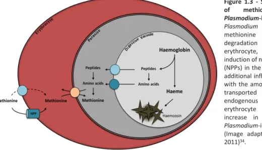
Figure 1.3 - Schematic representation of methionine transport in
Plasmodium-infected erythrocyte.
Plasmodium inability to obtain
methionine from haemoglobin degradation within the host erythrocyte, is overtaken by the induction of new permeability pathways (NPPs) in the host cell membrane. This additional influx of methionine coupled with the amount of methionine that is transported by the L-system - an endogenous transporter of the erythrocyte - results in a 15-fold increase in methionine uptake by
Plasmodium-infected erythrocytes
(Image adapted from Cobbold et al, 2011)34.
Much less is known about Plasmodium dependency on methionine during the other developmental stages of its life cycle. During the liver-stage each sporozoite generates a progeny of thousands of new parasites. Thus, the availability of large amounts of nutrients is mandatory for Plasmodium development and survival. Inside hepatocytes Plasmodium lives and replicates in a haemoglobin-free environment meaning that an alternative source is required38. However, the metabolism of
methionine during Plasmodium liver-stage development remains elusive.
The interactions established between Plasmodium and the host inescapably lead to variations in the equilibrium between pro-oxidant and antioxidant molecules. This imbalance coupled with an excessive production of oxidizing species triggers an oxidative stress that can result in parasite death. The role of oxidative stress during malaria infection is still uncertain and some authors suggest a protective role, whereas others propose a close relation to the physiopathology of the disease39. Despite that,
recent studies suggest that the excessive synthesis of ROS together with an oxidative stress play a pivotal role in the development of systemic complications associated to malaria40. Plasmodium
infection inevitably leads to an increased oxidative stress both in the mammalian host as well as in the mosquito vector. In the mammalian host, oxidative stress occurs mostly due to: 1) the generation of hydroxyl radical (OH·) in the liver which can damage molecules like DNA, proteins and lipids leading
consequently, to apoptosis41; and 2) due to the pro-oxidative environment of the erythrocyte. The
erythrocyte has a high content of oxygen and iron, the key elements of the Fenton reaction - one of the major source of ROS production. Furthermore, the toxic free haeme that is released from digested haemoglobin inescapably leads to oxidation of Fe2+ into Fe3+ and to the release of superoxide anions,
contributing also to an increased oxidative stress26,40. In fact, results obtained by Atamna et al.
demonstrated that during the erythrocytic stage of infection, P. falciparum-infected erythrocytes produce twice more OH• radicals and H
2O2 when compared to normal erythrocytes42.
During the mosquito-stage of development due to the innate immune response of the vector, Plasmodium is also exposed to a high oxidative stress mainly during ookinete and sporozoites development, which results in relevant parasite losses26. In order to survive and succeed in these
oxidative environments, Plasmodium evolved efficient antioxidant systems namely superoxide dismutases and thioredoxin-dependent peroxidases. However, Plasmodium lacks catalase and glutathione peroxidases which is counterbalanced by the presence of a fully functional glutathione redox system43.
AIMS | 7
AIMS
Plasmodium heavily depends on methionine to complete a successful development, as evidenced by a severe impairment in parasite development when perturbations in the methionine cycle, or in any of its branches (transsulfuration and aminopropylation) occur44,45. This dependence on methionine is
more remarkable during the erythrocytic stages of Plasmodium life cycle, resulting in a 15-fold increase in methionine influx-rate into infected erythrocytes. Since there is a close connection between Plasmodium metabolism and the onset of the disease, unravelling the pathways and the mechanisms involved in the metabolism of this essential amino acid may be a rational and promising tool for developing both prophylactic agents and new chemotherapies against malaria16. Given the key role of
the liver in: 1) methionine metabolism; 2) SAMe-dependent transmethylation reactions; and in 3) GSH synthesis and storage, the main aims of the present study focused the characterization of the importance of two main enzymes involved in the methionine metabolic pathway of Plasmodium: 1) GSH synthetase (GS) that is responsible for the de novo synthesis of glutathione and; 2) S-adenosylmethionine synthetase (SAMS) which allows SAMe synthesis, a crucial metabolite in the methionine cycle.
For this purpose, we will use the rodent malaria parasite P. berghei which, despite its phylogenetic distance, is analogous to human and primate malaria parasites sharing essential aspects as biology, physiology and life cycle. Additionally, P. berghei do not pose direct harm to humans and in vivo infected livers are directly accessible for analysis46. The availability of efficient reverse genetics
technologies for P. berghei coupled with an ability to study and analyse its development throughout Plasmodium life cycle, make this species one of the greatest model to analyse parasite gene function47,48. Thus, we will characterize the role of P. berghei GS and SAMS enzyme during the
mosquito-stage and in both the blood- and liver-stage in vivo, using rodent models and in vitro, using cell lines.
8 | MATERIALS AND METHODS
MATERIALS AND METHODS
1. Ethics Statement
All in vivo protocols were approved by the internal animal care committee of Instituto de Medicina Molecular and were performed according to the national and European regulations.
2. Mice and Parasites
Balb/c and C57BL/6J mice (6-8 weeks of age, female or male) were purchased from Charles River® Breeding Laboratories and housed in the Rodent Facility of IMM.
Wild-type Plasmodium berghei (P. berghei) ANKA strain was obtained from the MR4 repository (Manassas, Virginia). Transgenic P. berghei parasite lines, namely GSH synthetase knockout (Pbgs-),
SAMS conditional knockdown (PbSAMS-DD) and SAMS-green fluorescent protein (GFP) tagged (PbSAMS-GFP), were generated in our laboratory. Asexual blood-stage parasitic forms were maintained through passage of infected blood in mice. All recombinant P. berghei parasite lines carry the human dihydrofolate reductase (hdhfr) gene cassete that confers resistance to pyrimethamine, allowing the selection of recombinant parasites. Confirmation of transgenic parasites genotype, construct integration at the desired genomic loci and elimination of WT locus were assessed by performing a polymerase chain reaction (PCR). To that end, blood from the infected mice tail vein was collected in 200µL of 1x PBS and genomic DNA (gDNA) was isolated using the NZY Blood gDNA Isolation Kit (NZYTech), according to manufacturer’s guidelines. The PCR conditions used for genotyping were equivalent for all parasite lines and consisted of an initial denaturation at 95oC for 10 min, followed by
an amplification for 40 cycles of: 10 sec at 95oC, 30 sec at 50 oC and 1 min at 68 oC. Amplification was
followed by an additional extension step at 68 oC for 10 min. Sequences of primers used for genotyping
are provided in Table 1, Annexes section. Stabilisation of PbSAMS-DD fusion protein throughout infection was achieved in vivo, by administration of trimethoprim (TMP) to mice (0.25 mg/ml of TMP in drinking water) and to mosquitoes (0.50 mg/mL of TMP in the mosquito food) or in vitro, to HepG2 hepatoma cells (0.5 or 5mg/mL of TMP in culture medium). The time of the treatment was set according to the experimental protocol.
3. Mosquitoes
Anopheles stephensi (A. stephensi) mosquitoes were obtained from the breeding facility at IMM and both housing and infections were performed at IMM. Mosquitoes were housed at 20oC, 80% relative
humidity and fed ad libitum with 10% glucose supplemented with 2% paraminobenzoic acid (PABA). Female mosquitoes were infected through feeding on mice infected with P. berghei ANKA WT, Pbgs-,
PbSAMS-DD or PbSAMS-GFP parasites.
3.1 Midguts dissection
To assess oocysts number and size, midguts were dissected in 1x PBS at day 12 or 16 (Pbgs-) post
mosquito infection and incubated with mercurochrome 0.1% (in H2O) for 10 minutes. Stained midguts
were washed in 1x PBS, mounted on glass slides and immediately analysed by light microscopy using the Leica DM2500 microscope (10x objective). To compare ROS levels, midguts were incubated with a
MATERIALS AND METHODS | 9 2 µM solution (in DMSO) of the general oxidative stress indicator 6-carboxy-2',7'-dichlorodihydrofluorescein diacetate (Carboxy-H2DCFDA from Molecular Probes) for 20 minutes at room temperature (RT) under dark conditions. After incubation, midguts were rinsed in 1x PBS, mounted on a glass slide and immediately analysed by fluorescence microscopy using the Leica DM5000 B microscope (10x objective). Comparison of fluorescent levels between different conditions wasperformed as the same camera exposure times were used.
3.2 Salivary glands dissection
Sporozoites for infection were obtained by dissection of infected salivary glands 21 and 28 (Pbgs-) days
post mosquito infection. Salivary glands were dissected in Dulbecco’s Modified Eagle’s Medium (DMEM) and sporozoite number was assessed by light microscopy, using an improved Neubaeur cell counting chamber (depth 0.1 mm). Sporozoites were used for in vitro infection of HepG2 hepatoma cells or for in vivo infection of mice.
4. Parasite infections
In vivo infections
For liver-stage experiments mice were infected through intravenous (i.v.) retro-orbital injection of 5x10⁴ sporozoites while for blood-stage experiments mice were infected through i.v. injection of 103
sporozoites. To assess parasite liver load, livers were harvested 48 hours post-infection (hpi). To assess parasitemia levels in the blood throughout infection, thin blood smears fixed in 100% methanol were stained with Giemsa (10% v/v). Prepared slides were allowed to dry overnight before imaging and quantification was preformed using the Leica DM 2500 microscope (100 x oil objective)
4.2 In vitro infection
Liver-stage in vitro infections were performed in HepG2 hepatoma cells cultured in DMEM, supplemented with 10% fetal bovine serum, 2mM glutamine, 100 U/mL penicillin and streptomycin and maintained in a 5% CO2 humidified incubator at 37oC. For P. berghei infection, 6 X 10⁴ HepG2 cells
were platted (per well) in a 24-well culture plate and infection was performed 24 h post-seeding with 3 x 10⁴ salivary gland sporozoites. Two hours after infection, culture medium was supplemented with
0.3% fungizone and infection was proceeded until suitable.
5. RNA isolation and analysis by RT-qPCR
Mosquito tissue: To isolate RNA from midguts and salivary glands, infected mosquitoes were dissected
in 1x PBS supplemented with 1x Ribonuclease Inhibitors (NZY Tech). Tissue lysis was performed by adding 1mL of PureZOL RNA isolation reagent (BioRad®).
Mouse Blood: Blood from mice infected with synchronous parasites was collected at developmentally
relevant time points post infection, by cardiac punction, allowing for isolation of RNA from ring- (6 hpi and 10 hpi), trophozoite- (14 hpi and 18 hpi) and schizont-stage (22 hpi) parasites. Infected blood was lysed by adding 1mL of TRIzol® LS Reagent (ThermoFisher scientific).
10 | MATERIALS AND METHODS
Mouse liver tissue: To extract RNA from mouse liver, whole livers were harvested 48 hpi in 1 mL of
PureZOL RNA isolation reagent. Zirconia/Silica beads (1 mm) were added and tissue was disrupted using the Mini Bead Beater for 90 seconds.
Hepatoma cells: HepG2 cells were lysed at developmentally relevant time points post infection,
through addition of 1mL of PureZOL RNA isolation reagent directly to the well.
For RNA extraction, chloroform was added to the lysates (240 µL chloroform per 1 mL of PureZOL/TRIzol) followed by vigorously vortex for 15 seconds and an incubation step at RT for 5 minutes. Samples were then centrifuged at 12000 x g for 15 minutes at 4oC allowing phase separation.
The upper aqueous phase was collected and 100% ethanol (1.5x volume) was added to precipitate RNA. All subsequent steps were performed using the NZYtech Total RNA Isolation Kit, according to manufacturer’s instructions. cDNA synthesis was performed using 1 µg of RNA and the NZY First-Strand cDNA Synthesis kit (NZYtech) according to manufacturer’s instructions. PCR conditions used for cDNA synthesis were: 25°C for 10 min, 55°C for 30 min and 85°C for 5 min. Isolated cDNA was used to perform a quantitative reverse transcription-PCR (RT-qPCR) in a 10 µL reaction mix, containing 1 μL of sample cDNA. For each experimental condition negative controls were performed, either without RNA or reverse-transcriptase enzyme. For relative quantification of Pbsams expression levels, adenylosuccinate lyase (PBANKA_0304300) and serine-tRNA ligase (PBANKA_0615400) expression levels were used for normalization, whereas for infection load Pb18S rRNA levels were normalized against mouse hypoxanthine guanine phosphoribosyltransferase (hprt) housekeeping gene (ΔCt). The RT-qPCR conditions used consisted of an initial denaturation at 95°C for 5 min followed by amplification for 40 cycles at 95°C for 3 sec and at 60°C for 30 sec, with fluorescence acquisition at the end of each extension step. Gene expression values were then calculated based on the ΔΔCt method using the mean of control group as calibrator to which all other samples were compared. Primer pairs used to detect the target gene transcripts are listed in Table 1, Annexes section.
6. Immunofluorescence assays
6.1 PbSAMS localisation studies
Mouse blood: For blood-stage experiments, blood smears from PbSAMS-GFP-infected mice were fixed
in 4% PFA (in PBS) for 10 min. Fixed cells were washed in 1x PBS and then permeabilized in 0.1 % Triton X-100 (in PBS) for 10 min. Blocking was then performed in 3% BSA (in PBS) for 30 min.
Hepatoma cells: For liver-stage experiments in vitro, HepG2 hepatoma cells cultured in coverslip were
fixed in 4% PFA for 10 min, rinsed in 1x PBS and then permeabilized in 0.1% Triton X-100 for 10 min. After permeabilization, cells were blocked in 3% BSA for 30 minutes.
Mosquito tissue: For mosquito-stage experiments PbSAMS-GFP-infected midguts and salivary glands
were dissected in 1x PBS and DMEM, respectively. Midguts were fixed for 20min and salivary glands for 10min in 4% PFA. Fixed tissue was washed in 1x PBS, permeabilized in 0.1% Triton X-100 for 10 min and then blocked in 3% BSA for 30 min.
After blocking and permeabilization samples were incubated in the following primary antibodies: goat anti-PbUIS4 (1:400, from Sicgen) and mouse anti-HSP70 (1:400, produced in house), for 2 hours at RT. The secondary antibodies used were: rabbit anti-GFP monoclonal antibody conjugated to Alexa Fluor 488 (1:400, from Thermo Fisher scientific), donkey anti-goat conjugated to Alexa Fluor 568 (1:400, from Life Technologies) and donkey anti-mouse conjugated to Alexa Fluor 647 (1:400, from Jackson ImmunoResearch Laboratories). DAPi (1:1000, from Sigma) was used for nuclear staining. Incubation with secondary antibodies was performed for 1 hour at RT. Samples were mounted with
Fluoromount-MATERIALS AND METHODS | 11 GTM (from Southern Biotech) and prepared slides were allowed to dry overnight before imaging. All
images were obtained by fluorescence microscopy using the Zeiss Axio Observer microscope.
6.2 PbSAMS-DD stabilisation by TMP treatment
For PbSAMS-DD stabilisation verification, blood-stage and liver-stage in vitro samples were processed as described above (section 6.1) with exception of the antibodies employed for PbSAMS detection. Additionally, PbSAMS stabilisation during the liver-stage was also assessed in vivo. For this, the median liver lobe was collected and fixed in 4% PFA for 2 hours at RT, washed in 1x PBS and then sliced into 40-µm-thick sections using the Vibratome VT 1000S (Leica). Fixed lobes were blocked in 3% BSA and permeabilized in 0,3% Triton X-100 for 3 hours at RT. Samples were incubated overnight at 4oC in the
following primary antibodies: mouse anti-HA (1:400, from Covance), goat anti-PbUIS4 (1:400, from Sicgen) and rabbit anti-PbBip (1:400, from Thermo Fisher scientific). The secondary antibodies used were: donkey anti-mouse conjugated to Alexa Fluor 488 (1:400, from Jackson Immonoresearch), donkey anti-goat conjugated to Alexa Fluor 568 (1:400, from Life Technologies) and donkey anti-rabbit conjugated to Alexa Fluor 647 (1:400, from Jackson ImmunoResearch Laboratories). For nuclei staining DAPi (1:1000 from Sigma) was used. Incubation with secondary antibodies was performed for 1 hour at RT. Samples were mounted with Fluoromount-GTM and prepared slides were allowed to dry
overnight before imaging. All images of PbSAMS-DD stabilisation were obtained by confocal microscopy using the Zeiss LSM 710 microscope.
7. Parasite pellet extraction for Immuno-blotting
All steps of parasite pellet extraction protocol were performed at 4o C to minimize protein degradation
and all centrifugations were executed at 1000 x g for 10 min. Mice were sacrificed at day 10 post infection and 1mL of blood was collected by cardiac punction. The blood was washed in 10mL of 1x PBS and the pellet of packed erythrocytes was resuspended in 0.15% saponin and centrifuged. The parasite pellet obtained after lysis was washed in PBS containing 1x Proteinase inhibitor cocktail (Roche® cOmplete Protease inhibitor tablets, EDTA free). Parasite pellet was then resuspended in parasite pellet lysis buffer (4 % SDS; 0.5 % Triton X-114 in 1x PBS), incubated on ice for 10 min, centrifuged at 21000 x g for 10 min and the supernatant was collected. Total protein content was determined using the Bio-Rad protein assay kit according to manufacturer’s instructions, as standards different concentrations of bovine serum albumin (BSA) were used. Protein samples diluted in 5x SDS sample buffer (NZYTech) were denatured at 95º for 10 min and resolved in an 8% polyacrylamide gel (SDS-PAGE). Proteins were blotted into a nitrocellulose membrane by wet transfer at 200mA for 2 hours. Primary antibody, mouse anti-HA antibody (1:1000, from Covance) was incubated overnight at 4oC. Secondary antibody, anti-mouse horseradish peroxidase (HRP)-conjugated (1:10000, Santa Cruz
Biotechnology) was incubated at RT for 1 hour. Signal detection was obtained using Luminata Crescendo Western HRP substrate (Merck Milipore®) and the ChemiDoc XRS+ Gel Imaging System (Bio-Rad®). Protein band quantification was performed on Image Lab software (version 5.0).
8. Statistical analysis
Statistically significant differences between two different groups were analysed using the Mann– Whitney test. p < 0.05 were considered statistically significant. Significances are represented in the figures as follows *p < 0.05; **p< 0.01, ***p< 0.001. All statistic tests were performed using GraphPad Prism 5.0 software.
12 | RESULTS
RESULTS
Part I: Importance of Plasmodium berghei glutathione synthetase
(PbGS) during the different stages of the life cycle
Plasmodium is a rapidly multiplying organism that undergoes a complex life cycle between two different hosts – a life style that entails a rapid adaptation to the external milieu. It is well described that during the different stages of the life cycle, Plasmodium is continuously exposed to high levels of ROS and oxidative stress. If not tightly regulated ROS can lead to deleterious effects resulting in parasite death. During the blood-stage of the infection the majority of ROS produced arise from haemoglobin digestion and free-haeme release within the PV of the parasite. Within the anopheline vector, Plasmodium is also exposed to massive amounts of ROS. Inside the midgut ookinetes survival is crucial for the establishment of a new parasite generation, for further production of sporozoites and consequent transmission to the mammalian host. The mosquito midgut is a harsh environment for Plasmodium and during that stage oxidative stress and ROS arise mostly from: 1) the innate immune response of the vector; 2) Plasmodium metabolic switching from glycolysis to oxidative phosphorylation for energy production, resulting in relevant parasite losses49. If produced in excessive
amounts ROS are toxic and harmful to Plasmodium, being ookinete and sporozoite forms the most affected. To sustain life in this noxious environment, Plasmodium depends on its antioxidant defenses. In fact, Plasmodium is adapted to cope and withstand in this toxic environment as evidenced by its ability to fully develop within two completely different hosts40. One of Plasmodium primary lines of
defense against these oxidative species is the tripeptide GSH. Thus, given the major role of GSH in ROS detoxification together with the high amounts of ROS that arise from infection, we decided to study the importance of glutathione synthetase (GS) during Plasmodium development.
1. Generation of a Pbgs knockout parasite line
To study the importance of GS during Plasmodium growth and development, GS-deficient P. berghei lines (Pbgs-) were generated. The GS locus was disrupted by double crossover homologous
recombination and the GS-encoding gene (gs) was deleted and replaced by the human dihydrofolate reductase (hDHFR) – a resistance cassete that allows for selection upon administration of pyrimethamine (Figure 4.1 A). Recombinant clones were obtained by limiting dilution of erythrocytes in a way that each mouse was infected with one single recombinant clone. Infected mice were treated with pyrimethamine in order to eliminate non-transgenic parasites in vivo. Mice parasitemia was assessed by counting GIEMSA-stained thin blood smears.
It has been described that knocking-out γ-GCS-encoding gene had no significant effect in growth and multiplication of blood-stage Plasmodium parasites, in vivo50. As γ-GCS is the enzyme upstream to GS
in the transsulfuration pathway and the rate-limiting enzyme of glutathione synthesis, we predicted that the deletion of gs will not have any effect in Plasmodium development and survival during that stage of development.