1. Rheumatology and Rehabilitation, Faculty of Medicine, Asyut University
2. Biochemistry, Faculty of Medicine, Asyut University 3. Anesthesia, Faculty of Medicine, Asyut University
4. Rheumatology and Rehabilitation, Faculty of Medicine - Suez Canal University
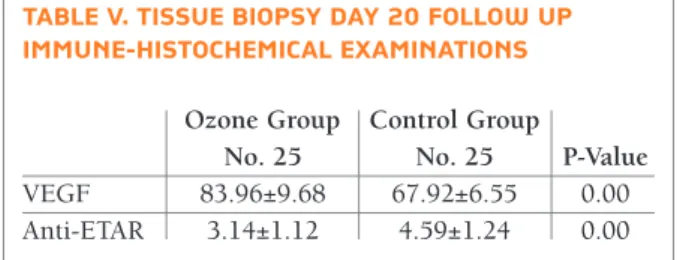
After treatment, the wound sizes in both groups were significantly smaller than before treatment. In group (I), the wound size reduction was significantly more than in group (II) (0.75 ± 0.30 versus 2.44 ± 0.80 mm), (p<0.001). At day 20, VEGF was significantly higher in ozone group (I), than in control group (II), (83.96±9.68) versus (67.92±6.55), (p<0.001) while, ETAR was signi ficantly lower in ozone group (I), than in control group (II), (3.14±1.12 versus 4.59±1.24), (p<0.001).
Conclusion: Ozone therapy may be beneficial tool in
the treatment of DUs in SSc patients, where it promotes the wound healing through a potential induction of VEGF and down-regulation of ETAR at sites of the ulcers.
Trial registration: NCT02733978. Registered April
12, 2016
Keywords: Ozone; Systemic sclerosis; Digital ulcers
IntroductIon
Systemic sclerosis (SSc) is a chronic connective tissue disabling disease associated with fibrosis, microvascu-lar involvement, immunologic dysfunction, increase of extracellular matrix deposition in the skin and variable internal organs involvement1,2.
Vascular disease displays an essential role in the patho-genesis of SSc, including digital ulcers (DUs)3, 4, through involvement of micro-vessels and digital arteries5. DUs are responsible for pain, poor quality of life, impaired function and morbidity associated with SSc6, 7. More-over, they are correlated to disease severity and out-come7.
About one half of patients with SSc report a history of DUs along the course of their disease8. Recurrent DUs occur in approximately 10% of those patients. Three quarters of patients develop their first DUs within 5 years after SSc diag nosis9. Patients with anti-Scl-70 an-tibody develop DUs ∼5 years earlier than those with
Non-invasive Oxygen-Ozone therapy in treating
digital ulcers of patients with systemic sclerosis
Hassanien M1, Rashad S1, Mohamed N2, Elawamy A3, Ghaly MS4
ACTA REUMATOL PORT. 2018;43:210-216
AbstrAct
Background: Digital ulcers (DUs) in systemic sclerosis
(SSc) result from recurrent Raynaud’s phenomenon (RP) and microtrauma with high impact on quality of life. Medical use of ozone (triatomic oxygen) was initi-ated in the 19thcentury. Ozone has multiple therapeu-tic effects in wound healing due to the property of re-leasing nascent oxygen, which has been shown to stimulate antioxidant enzymes. We aimed to assess the effects of ozone therapy on the healing of scleroderma DUs and determine levels of expression of vascular en-dothelial growth factor (VEGF), and endothelin-1 type A receptor (ETAR) autoantibodies in the wounds after treatment.
Subjects and Methods: Fifty SSc female patients with
DUs, were randomized into ozone group (I) (n=25) trea ted with calcium channel blockers plus oxygen-ozone treatment and control group (II) (n=25) treated with calcium channel blockers only. Ozone group recei ved noninvasive oxygenozone treatments for 30 minu tes per day for 20 days using the ozone generator devi -ce. The rapeutic effects were graded into 4 levels according to Zhang and other researchers. The wounds sizes were measured at baseline and day 20, respective -ly. Expressions of VEGF and ETAR autoantibodies proteins were determined by immunehistochemical exa -mination.
Results: Demographics and clinical characteristics of
the 2 groups showed no significant differences at base-line. At day 20, the effective healing rate was signifi-cantly higher in group (I) than in group (II), where it represented 96% (24/25) in ozone group versus 44% (11/25) in control group (p=0.007).
positive anti-centromere10.
Healing process in DUs takes long time, especially if there is an underlying calcinosis11. Amanzi and coworkers (2010) found that the average time to hea -ling was 76.2 days in DUs without calcinosis and 93.6 days in DUs with underlying calcinosis12. DUs are frequently infected and may be complicated by osteomye -litis13. In a retrospective study, 42% infected DUs were associated with osteomyelitis, shown by clinical and radiographic features14.
Management of DUs is a great challenge for clini-cians and often requires the input of a multi--disciplinary team of health professionals6. Medical use of ozone (triatomic oxygen) was initiated in the 19th century and was considered to be an oxidant and a disinfectant15. Currently, ozone is acknowledged as an antiviral and a bactericidal agent16, 17, and has been used for treating coronary artery disease, chronic hepa -titis and chronic low back pain18-20. Also, its effective-ness was proved in healing chronic wounds such as tro phic ulcers, ischemic ulcers and diabetic foot ul-cers17,21- 22.
The mechanism through which ozone achieves healing in chronic wounds is not yet well understood. Its therapeutic effect may be related to ozone growth factors stimulation and antioxidant system activa-tion21,23. In addition, it improves microcirculation in the capillary vessels by improving flexibility and sta-bility of the cell membrane and limiting the aggrega-tion and adhesion of platelets24.
To our knowledge, the effectiveness of ozone thera -py in SSc DUs was not assessed before. Therefore, this study aimed at assessing the effect of ozone therapy on healing scleroderma DUs. Moreover, we measured the levels of expressions of vascular endothelial growth factor (VEGF), and endothelin-1 type A receptor (ETAR) autoantibodies after treatment.
subjects And methods PArtIcIPAnts
Fifty SSc female patients with DUs were recruited from Rheumatology and Rehabilitation department at Asyut University Hospital, Asyut, Egypt. SSc was diagnosed and based on the American College of Rheumatolo-gy/European League Against Rheumatism (ACR/EU-LAR) Criteria for the classification of SSc25. Skin thick-ness was evaluated using the modified Rodnan skin score (MRSS)26. DUs were defined according to Amanzi
et al., (2010) as “a loss of epi thelialization and tissues involving, in different degrees, the epidermis, the der-mis, the subcutaneous tissue and sometimes also in-volving the bone”12.
DUs staging was as follow12:
1. Superficial: partial skin loss where involvement is restricted to the epidermis. Clinically, the lesion ap-pears as an abrasion, blister or small crater. 2. Intermediate: full thickness skin involvement where
damage appears in subcutaneous tissue up to, but not through, underlying fascia. Clinically, there is a deep crater with or without affection of adjacent tissue. 3. Deep: full thickness skin involvement with wide
-spread destruction of the muscle down over the fas-cia, supportive structures (e.g. tendon) and bone. Written informed consents were obtained from all patients before entry into the study, according to the Declaration of Helsinki and guidelines of the local ethics committee. The study was approved by Asyut University ethics committee. Trial registration: NCT02733978. Registered April 12, 2016.
Patients were excluded from the study if they had one or more of the following conditions10: (1) gan-grenous ulcers in whole hand, (2) calcinosis ulcer, (3) traumatic ulcer, (4) vasculitis, (5) active osteomyelitis, (6) other connective tissue disease, (7) hyperthy-roidism, (8) pregnancy or nursing, (9) a known aller-gy to ozone.
bAselIne Assessment
At baseline, both groups were assessed for DUs sta -ging12, number of Raynaud’s attacks /day, duration of Raynaud’s attack, ulcer size in mm, ulcer pain assessed by visual analogue scale (VAS) (where 0 = ‘no pain’ and 10 = ‘severe pain’).
Laboratory work included autoantibodies, antinu-clear antibodies (ANA), anti-centromere antibodies (ACA) and anti-topoisomerase antibody (Scl70). An indirect immunofluorescent assay, for semi-quantita-tive determination of anti-nuclear IgG antibodies (ANA) in patients’ serum NOVA LITE™ IFA HEp-2 ANA Complete Kit was used. Antibodies to extractable nuclear antigens ACA and Scl70 were determined by a commercial clinical enzyme linked immunosorbent assay (ELISA).
Tissue biopsies were obtained from the border area of digital ulcers, including the ulcer edge and part of the surrounding skin at day 0. The tissue was homoge -nized in 3mL PBS followed by centrifugation. The su-pernatants were collected for the determination of
VEGF, ETAR autoantibodies proteins by immune-his-tochemical examinations.
InterventIon
Patients who met the criteria were randomly assigned by computer based selection as ratio of 1:1 to one of 2 groups:
Ozone group
It represented the study group that included 25 SSc pa-tients with DUs who received oxygen-ozone treatment in addition to calcium channel blockers (Epilat retard® 40mg /day). Patients received noninvasive oxygen-ozone treatments for 30 minutes per day for 20 days. The treatment session of oxygen-ozone was 52 mg/mL ozone (total volume: 20-50 mL) in a special bag using the ozone generator device (Humazon Promedic, Ger-man).
Control group
It included SSc patients with DUs who received the same standard medical care only in the form of calci-um channel blockers (Epilat®40mg /day)
outcomes
Primary outcomes
Ulcer healing was our primary outcome, which was as-sessed depending on Zhang and colleagues ulcers grading where they graded ulcers into four levels, Grade 0 (no change), Grade 1 (wound size decreased less than ½); Grade 2 (wound size decreased more than ½) and Grade 3 (wound healing)27. Ozone treatment were con-sidered efficient if patients reached grade 1 to 3.
Secondary outcomes
For our secondary outcomes both group were subject-ed to the following after 20 days of intervention: they were reassessed for number of Raynaud’s attacks/day, duration of Raynaud’s attack, ulcer size in mm, and ul-cer pain was assessed by VAS.
In addition, at day 20, VEGF, ETAR autoantibodies proteins were reassessed by immune-histochemical ex-aminations.
To maintain the study blind: 1) researcher who con-ducted 1stclinical assessments were separated from those carrying out and who had access to record, pa-tients’ efficacy and safety data; 2) results from the pri-mary histopathological assessments at baseline were recorded on paper, sealed in an envelope and not dis-closed to any site staff apart from the physician who
performed it; 3) another physician did the follow up as-sessments clinical and histopathologic.
stAtIstIcAl AnAlysIs
Descriptive analysis was presented as mean and stan-dard deviation for quantitative variables and as fre-quency and percentage for categorical variables. Mann--Whitney U test was used for inter-group comparisons. We used the Chi-square test to compare qualitative variables. Pvalue equal or less than 0.05 was consi dered significant in all statistical tests. Statistical ana -lyses were performed using Microsoft Excel and SPSS (SPSS 23.0, IBM, Armonk, NY, USA).
results
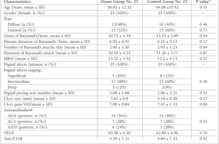
Fifty SSc female patients with DUs were included in the study with a mean age of 40.93 ±11.71 years. Thir-ty-one patients had limited SSc while 19 patients had diffuse type. The mean duration of Raynaud's mani-festation was 8.13 ±5.56 years with 38 patients had pitting scars. There were no statistical significant dif-ferences between two groups in patient demographics, clinical and labo ratory characteristics Table I at base-line.
Results of our primary outcomes are presented in Table II showing the grading of healing of DUs at day 20 in both ozone and control groups. The effective rate was significantly higher in ozone group than in con-trol group, 96% (24/25) versus 44% (11/25), c2 = 7.26, p=0.007. Figure 1 reports photo of the digital ulcers before and after ozone treatments.
At day 20, ozone group showed significant im-provement in the clinical characteristics of secondary outcomes including number of Raynaud’s attacks /day, duration of Raynaud’s attack, ulcer size in mm, ulcer pain (Table III). However, the control group showed statistical significant improvement only for ulcer size (Table IV). Ozone group showed greater improvement in clinical characteristics including number of Ray-naud’s attacks /day, duration of RayRay-naud’s attack, ulcer pain in comparison to control group (P = 0.02, 0.001 and <0.001 respectively).
Concerning wound size, there was no significant dif-ference between two groups at baseline (Table I). At day 20 after treatment, the wound size in both groups was significantly smaller than before (P values were <0.001
and 0.042 respectively). In ozone group the wound size reduction was significantly higher than in control group (0.75 ± 0.30 versus 2.44 ± 0.80 mm), (p<0.001).
At baseline, there was no statistical significant diffe -rence in the expressions of VEGF and ETAR protein
be-tween two groups (Table I). But after treatment at day 20, they were significantly higher in ozone group than in control group (Table V). Also, there were highly signi ficant differences in the expressions of VEGF and ETAR protein in the ozone group after treatment than before treatment (83.96±9.68 versus 63.48±6.20), (p<0.001); (3.14±1.12 versus 4.59 ± 1.31), (p<0.001). In the control group there was only a statistical signi -ficant difference in the expressions of VEGF between baseline and after 20 days (62.80±6.86 versus 67.92±6.55), (P = 0.04).
dIscussIon
DUs are one of the most common complications of SScs at digital level, which has a great impact on patients’ quality of life12. Different researches have shown that tAble I. bAselIne demogrAPhIc, clInIcAl And lAborAtory chArActerIstIcs of the studIed PoPulAtIons
Characteristics Ozone Group No. 25 Control Group No. 25 P value*
Age [Years, mean ± SD] 38.83 ± 12.32 44.08 ±10.42 0.51
Gender [female, n (%)] 25 (100%) 25 (100%) –
Type:
Diffuse [n (%)] 12(48%) 10 (40%) 0.46
Limited [n (%)] 13 (52%) 15 (60%) 0.71
Onset of Raynaud's[Years, mean ± SD] 10.72 ± 4.34 13.33 ± 3.89 0.54 Disease duration of Raynaud’s [Years, mean ± SD] 6.83 ± 6.41 6.21 ± 5.13 0.17 Number of Raynaud’s attacks /day [mean ± SD] 2.90 ± 1.30 2.93 ± 1.23 0.94 Duration of Raynaud’s attack [mean ± SD] 10.10 ± 4.23 11.26 ± 3.77 0.87
MRSS [mean ± SD] 15.32 ± 5.52 13.2 ± 4.13 0.27
Digital ulcers [present, n (%)] 25 (100%) 25 (100%) –
Digital ulcers staging:
Superficial 5 (20%) 8 (32%)
Intermediate 17 (68%) 15 (60%) 0.36
Deep 3 (12%) 2(8%)
Digital pitting scar number [mean ± SD] 3.08 ± 1.68 2.06 ± 2.21 0.31
Ulcer size (mm) [mean ± SD] 3.61 ± 0.8 4.18 ± 0.38 0.27
Ulcer pain VAS[mean ± SD] 7.98 ± 0.84 7.41 ± 1.33 0.06
Autoantibodies# ANA [positive, n (%)] 14 (56%) 12 (48%) ACA [positive, n (%)] 7 (28%) 7 (28%) 0.52 Scl70 [positive, n (%)] 6 (24%) 7 (28%) VEGF 63.48 ± 6.20 62.80 ± 6.86 0.76 Anti-ETAR 4.59 ± 1.31 4.84 ± 1.33 0.42
MRSS: modified Rodnan skin score, VAS: visual analogue scale, ANA: antinuclear antibody; ACA: Anti-centromere antibodies,
Scl70: anti-topoisomerase antibody. # = patient may be positive for more than one autoantibody. VEGF: Vascular endothelial growth factor, Anti-ETAR: endothelin-1 type A receptor autoantibodies,. *p<0.05 adjusted by Bonferroni test
*p<0.05 adjusted by Bonferroni test
tAble II. dIgItAl ulcer grAdIng At dAy 20 In both ozone And control grouPs
Digital Ozone Group Control Group P
grade No. 25 No. 25 value
Grade 0 1 (4%) 14 (56%) 0.001* Grade 1 7 (28%) 3 (12%) 0.032* Grade 2 10 (40%) 5 (20%) 0.018* Grade 3 7 (28%) 3 (12%) 0.032*
the healing process in the wounds is delayed due to hy-poxia, reactive oxygen species, and pro-inflammatory cytokines28.
For more than 100 years, O3 has been used in
dif-ferent ways to treat diseases, infections, and wounds through increasing collagen contents inside the wounds and levels of VEGF, Transforming growth fac-tor beta (TGF-b), and Platelet-derived growth facfac-tor (PDGF) in wound exudates30-32.
Our results revealed the possible effectiveness of a fIgure 1. Systemic sclerosis digital ulcers before (A1) and after ozone treatments (A2) for the same patient and (B, C) for other patients.
A1 A2
b c
VAS: visual analogue scale, *p<0.05 adjusted by Bonferroni test tAble III. clInIcAl chArActerIstIcs follow uP At dAy 20 In ozone grouP
Characteristics Baseline Day 20 P-Value Ulcer size (mm) 3.61±0.8 0.75±0.30 0.00* [mean ± SD] Number of Raynaud’s 2.90±1.30 1.64±1.07 0.00* attacks/day Duration of 10.10±4.23 5.00±2.88 0.00* Raynaud’s attack
Ulcer pain VAS 7.98±0.84 4.04±0.93 0.00*
[mean ± SD] VAS: visual analogue scale, *p<0.05 adjusted by Bonferroni test tAble Iv. clInIcAl chArActerIstIcs follow uP At dAy 20 In control grouP
Characteristics Baseline Day 20 P-Value Number of Raynaud’s 2.93±1.23 2.68±1.02 0.12 attacks /day
Duration of 11.26±3.77 9.80±3.67 0.64 Raynaud’s attack
Ulcer size (mm) 4.18±0.38 2.44±0.80 0.042* Ulcer pain VAS 7.41±1.33 7.02±1.30 0.16
short-term treatment with oxygen-ozone into healing in SSc DUs. In 20 days, we noticed a significant im-provement in the clinical characteristics in comparison to control group. Our study was the first to investigate the effect of oxygen-ozone in treating SSc DUs.
Other studies reported the efficacy of oxygen-ozone on different ulcers. One randomized controlled clini-cal trial in diabetic patients showed better glycemic control, reduced ulcers size, inhibited oxidative stress products and decrease number of amputations in patients treated with ozone for 20 days via rectal in -sufflation than in the control group33. Wainstein et al. (2011) reported that when ozone therapy was added to conventional treatment for 24 weeks, it promoted com-plete wound healing than conventional treatment alone for Diabetic foot ulcers (DFUs)34. Another research showed that oxygenozone treatment for DFUs signi -ficantly promoted early effective rate of the wound healing at day 2027. Degli Agosti and other researchers (2016) suggested in their case report that oxygen-ozone therapy would have a positive role in helping wound healing and reducing pain of complicated wounds35.
We also reported significantly higher expressions of VEGF in the ozone group than in the control group. Multiple growth factors, such as VEGF and TGF-b, have been proved to play an important role in wound heal-ing and lack of their upregulation may explain the poor formation of granulation tissue and chronicity of ulcer epithelialization36. This could explain that the effi cacy of ozone therapy for healing of DUs in SSc may be par-tially to increase endogenous growth factors in local ul-cer. These results were similar to Zhan et al., (2014) who observed a higher expressions of VEGF, TGF-b, and PDGF in ozone group compared to control group34. Moreover, our results revealed downregulation of ETAR autoantibodies at local wound site in ozone group compared to control group. Agonistic
autoanti-bodies against ETAR were recently identified in the sera of SSc patients and are considered to contribute to the pathogenesis of the disease. Anti-ETAR could exacer-bate the inflammatory response in SSc patients through promoting recruitment of immune cells to inflamed tis-sues37. In addition, in vitro studies reported that antiETAR autoantibodies promote the production of colla -gen by skin fibroblasts, the release of reactive oxy-gen species by neutrophils, and affect endothelial repair; these processes lead to progressive fibrosis of the skin and vasculopathies38-39. All these effects indicate that these autoantibodies may have a role in the pathogene -sis of SSc DUs and that downregulation of these au-toantibodies may play a role in promoting DUs healing.
The present study has some potential biases including small sample size, nonblinded patients and outco -me assessors and long-term follow up.
In conclusion, oxygen-ozone treatment significant-ly stimulated earsignificant-ly effective wound healing at day 20 in DUs of SSc patients. We also reported that there were significantly higher expressions of VEGF and down-regulation of anti-ETAR in the ozone group than in the control group. These results show that the efficacy of the ozone treatment for the healing of DUs may be par-tially due to increasing endogenous growth factors and decreasing autoantibodies in the local wounds. How-ever, due to several potential biases oxygen-ozone treat-ment should be tested more carefully in large, blinded and placebo-controlled studies. Also, Further studies are required to assess the long term effects of ozone therapy on DUs.
ethIcs, consent And PermIssIons
Written informed consents were obtained from all patients before entry into the study, according to the Declaration of Helsinki and guidelines of the local ethics committee.
AvAIlAbIlIty of dAtA And mAterIAls
Data sharing is not applicable to this article as no datasets were gene -rated or analysed during the current study.
corresPondence to Mona Sayed Ghaly
Rheumatology and Rehabilitation
Faculty of Medicine - Suez Canal University E- mail: [email protected] references
1. Denton CP, Korn JH. Digital ulceration and critical digital is-chaemia in scleroderma. Scleroderma Care Res. 2003; 1:12-16. 2. Barsotti S, Stagnaro C, and Della Rossa A: Systemic sclerosis: a critical digest of the recent literature. Clin Exp Rheumatol 2015; 33 (Suppl. 91): S3-14.
3. Allanore Y, Simms R, Distler O et al. Systemic sclerosis. Nat VEGF: Vascular endothelial growth factor, Anti-ETAR: endothelin-1
type A receptor autoantibodies. *p<0.05 adjusted by Bonferroni test
tAble v. tIssue bIoPsy dAy 20 follow uP Immune-hIstochemIcAl exAmInAtIons
Ozone Group Control Group
No. 25 No. 25 P-Value VEGF 83.96±9.68 67.92±6.55 0.00 Anti-ETAR 3.14±1.12 4.59±1.24 0.00
Rev Dis Prim 2015. doi.org/10.1038/nrdp.2015.2
4. Matucci-Cerinic M, Kahaleh B, Wigley FM. Review: evidence that systemic sclerosis is a vascular disease. Arthritis Rheum 2013; 65:1953-1962.
5. Rodnan GP, Myerowitz RL, Justh GO. Morphologic changes in the digital arteries of patients with progressive systemic sclero-sis (scleroderma) and Raynaud phenomenon. Medicine 1980; 59:393-408.
6. Michael Hughes & Ariane L. Herrick. Digital ulcers in systemic sclerosis. Rheumatology, Volume 56, Issue 1, 1 January 2017, Pages 14-25, https://doi.org/10.1093/rheumatology/kew047 7. Mihai C, Landewé R, and Van Der Heijde D et al.: Digital
ul-cers predict a worse disease course in patients with systemic sclerosis. Ann Rheum Dis, 2016; 75: 681-686.
8. Ferri C, Valentini G, Cozzi F et al.: Sys¬temic sclerosis: demo-graphic, clinical, and serologic features and survival in 1,012 Ital¬ian patients. Medicine 2002; 81: 139-153.
9. Hachulla E, Hatron P-Y, Carpentier P et al.: Efficacy of sildenafil on ischaemic digital ulcer healing in systemic sclerosis: the placebo-controlled SEDUCE study. Ann Rheum Dis, 2016; 75: 1009-1015.
10. Denton CP, Krieg T, Guillevin L et al. Demographic, clinical and antibody characteristics of patients with digital ulcers in sys-temic sclerosis: data from the DUO Registry. Ann Rheum Dis, 2012; 71:718-721.
11. Zhou AY, Muir L, Harris J, Herrick AL. The impact of magnetic resonance imaging in early diagnosis of hand osteomyelitis in patients with systemic sclerosis. Clin Exp Rheumatol., 2014;32:S232
12. Amanzi LBraschi FFiori G et al. Digital ulcers in scleroderma: staging, characteristics and sub-setting through observation of 1614 digital lesions. Rheumatology 2010; 49:1374-82. 13. Steen V, Denton CP, Pope JE, Matucci-Cerinic M. Digital ulcers:
overt vascular disease in systemic sclerosis. Rheumatology 2009; 48:iii19-24.
14. Giuggioli D, Manfredi A, Colaci M, Lumetti F, Ferri C. Osteo -myelitis complicating scleroderma digital ulcers. Clin Rheuma-tol. 2013; 32:623-627.
15. Bocci V. Ozone as Janus: this controversial gas can be either to -xic or medically useful. Mediators of Inflammation, 2004; 13(1):3-11.
16. Valacchi G, Fortino V, Bocci V. The dual action of ozone on the skin. British Journal of Dermatology, 2005; 153(6): 1096-1100. 17. Kim HS, Noh SU, Han YW, Kim KM, Kang H, Kim HO, et al. Therapeutic effects of topical application of ozone on acute cu-taneous wound healing. Journal of Korean Medical Science 2009; 24(3):368-374.
18. Martínez-Sánchez G, Delgado-Roche L, Díaz-Batista A, Pérez-Davison G, Re L. Effects of ozone therapy on haemostatic and oxidative stress index in coronary artery disease. European Jour-nal of Pharmacology 2012; 691(1-3): 156-162.
19. Gu XB, Yang XJ, Zhu HY, Xu YQ, Liu XY. Effect of medical ozone therapy on renal blood flow and renal function of patients with chronic severe hepatitis. Chinese Medical Journal, 2010; 123(18):2510-2513.
20. Magalhaes FN, Dotta L, Sasse A, Teixera MJ, Fonoff ET. Ozone therapy as a treatment for low back pain secondary to hernia -ted disc: a systematic review and meta-analysis of randomized controlled trials. Pain Physician 2012; 15(2): E115-29. 21. Martinez-Sanchez G, Al-Dalain SM, Menendez S, Re L, Giuliani
A, Candelario-Jalil E, et al. Therapeutic efficacy of ozone in pa-tients with diabetic foot. European Journal of Pharmacology
2005; 523:151-161.
22. De Monte A, van der Zee H, Bocci V. Major ozonated autohe-motherapy in chronic limb ischemia with ulcerations. Journal of Alternative and Complementary Medicine 2005;11:363-367. 23. Re L, Mawsouf MN, Menéndez S, León OS, Sánchez GM, Hernández F. Ozone therapy: clinical and basic evidence of its therapeutic potential. Archives of Medical Research 2008; 39(1):17-26.
24. Danuta Nowicka. Thermography Improves Clinical Assessment in Patients with Systemic Sclerosis Treated with ozone Therapy. BioMed Research International, March 2017. https://doi.org/ 10.1155/2017/5842723.
25. Hoogen F, Khanna D, Johnson SR, et al. Classification criteria for systemic sclerosis: an American College of Rheumatolo-gy/European League against Rheumatism collaborative initia-tive. Arthritis Rheum 2013; 65: 2737-2747.
26. Clements PJ, Lachenbruch PA, Seibold JR, Zee B, Steen VD, Brennan P, et al. Skin thickness score in systemic sclerosis: an assessment of interobserver variability in 3 independent stud-ies. J Rheumatol. 1993; 20(11):1892-1896.
27. Zhang J, Guan M, Xie C, Luo X, Zhang Q, Xue Y. Increased growth factors play a role in wound healing promoted by non-invasive oxygen-ozone therapy in diabetic patients with foot ul-cers. Oxid Med Cell Longev. 2014; 2014:273475.
28. Irene, Degli Agosti, et al. "Effectiveness of a short-term treat-ment of oxygen-ozone therapy into healing in a posttraumatic wound." Case reports in medicine 2016 (2016).
29. A. M. Elvis and J. S. Ekta, “Ozone therapy: a clinical review,” Journal of Natural Science, Biology and Medicine 2011; vol. 2, no. 1,pp. 66-70, 2011
30. J. Zhang, M. Guan, C. Xie, X. Luo, Q. Zhang, and Y. Xue, “In-creased growth factors play a role in wound healing promoted by noninvasive oxygen-ozone therapy in diabetic patients with foot ulcers,” Oxidative Medicine and Cellular Longevity 2014; 8 pages,. 31. H. S. Kim, S. U. Noh, Y. W. Han et al., “Therapeutic effects of top-ical application of ozone on acute cutaneous wound healing,” Jour-nal of Korean Medical Science 2009; vol. 24, no.3, pp.368-374. 32. G. Mart´ınez-S´anchez, S. M. Al-Dalain, S. Men´endez et al.,
“Therapeutic efficacy of ozone in patients with diabetic foot,” European Journal of Pharmacology 2005; vol. 523, pp.151-161. 33. J. Wainstein, Z. Feldbrin, M. Boaz, and I. Harman-Boehm, “Ef-ficacy of ozone-oxygen therapy for the treatment of diabetic foot ulcers,” Diabetes Technology and Therapeutics 2011; vol. 13, no.12, pp. 1255-1260.
34. Degli Agosti I, Ginelli E, Mazzacane B, Peroni G, Bianco S,Guer-riero F, Ricevuti G, Perna S, Rondanelli M. Effectiveness of a Short-Term Treatment of Oxygen-Ozone Therapy into Healing in a Posttraumatic Wound. Case Rep Med 2016; 2016:9528572. 35. H. Galkowska, U. Wojewodzka, and W. L. Olszewski, “Chemokines, cytokines, and growth factors in keratinocytes and dermal endothelial cells in the margin of chronic diabetic foot ulcers,” Wound Repair and Regeneration 2006; vol. 14, no. 5, pp. 558-565.
36. Otavio C. & Gabriela R. Functional autoantibodies targeting G pro tein-coupled receptors in rheumatic diseases. Nature Re-views Rheumatology (2017).doi:10.1038/nrrheum.2017.134. 37. Kill, A. et al. Autoantibodies to angiotensin and endothelin recep-tors in systemic sclerosis induce cellular and systemic events asso-ciated with disease pathogenesis. Arthritis Res. Ther.2014 16,R29. 38. Shi-wen, X. et al. Focal adhesion kinase and reactive oxygen species contribute to the persistent fibrotic phenotype of lesio -nal scleroderma fibroblasts. Rheumatology 2012;51,2146-2154