(Annals of the Brazilian Academy of Sciences)
Printed version ISSN 0001-3765 / Online version ISSN 1678-2690 www.scielo.br/aabc
Magnetic
Parkia pendula seed gum as matrix for Concanavalin A
lectin immobilization and its application in affinity purification
MOACYR J.B.M. RÊGO1, SINARA M. ALMEIDA1,2, SÉRGIO A. BEZERRA1, LUIZ B. CARVALHO JÚNIOR1 and EDUARDO I.C. BELTRÃO1
1Laboratório de Imunopatologia Keizo Asami/LIKA, Universidade Federal de Pernambuco/UFPE, Av. Prof. Moraes Rêgo, s/n, CDU, 50670-901 Recife, PE, Brasil
2Faculdade de Ciências, Educação e Tecnologia de Garanhuns/FACETEG, Universidade de Pernambuco/UPE, Rua Capitão Pedro Rodrigues, 105, São José, 55290-000 Garanhuns, PE, Brasil
Manuscript received on August 2, 2013; accepted for publication on November 26, 2013
ABSTRACT
The present work aimed to magnetize Parkia pendula seeds gum and use it as a matrix for Concanavalin A covalent immobilization. This composite was applied in affinity purification of glycoconjugates. Parkia pendula seeds were hydrated and the gum provenient from the supernatant was precipitated and washed with ethanol and dried. The gum was magnetized in co-precipitation using solutions of Fe+2
and Fe+3 . Matrix activation was accomplished with NaIO4. Magnetized Parkia pendula seeds gumwith covalently immobilized Concanavalin Awas used as an affinity matrix for the recognition of bovine serum fetuin glycoprotein. Fetuin elution was carried out with a solution of glucose (300mM) and evaluated through SDS-PAGE. The efficiency of lectin immobilization and fetuin purification were 63% and 14%, respectively. These results indicate that the composite produced is a promising magnetic polysaccharide matrix for lectins immobilization. Thus, such system can be applied for affinity purification allowing an easy recovery by magnetic field.
Key words: immobilization, magnetization, Parkia pendula seed gum, Concanavalin A.
Correspondence to: Eduardo Isidoro Carneiro Beltrão E-mail: [email protected]
INTRODUCTION
Many biomolecules have been isolated using
separation techniques based on the interaction of
biospecific molecules. Among these molecules of
particular interest there is a class named lectins that
are sugar-specific and cell-agglutinating proteins
of non-immune origin (Sharon 2007). This class
of proteins functions as recognition molecules in
cell-molecule and cell-cell interactions in a variety
of biological systems (Ghazarian et al. 2010).
Because of this, lectin-carbohydrate interactions
are extensively studied, from basic to applied
natural and clinical sciences. Such inter- and
multi-disciplinary approaches corroborate the importance
of developing new methodologies for the study of
lectin-saccharide interactions and their potential in
biotechnology (Gemeiner et al. 2009).
Thus, immobilized lectins have found
applica-tions in the purification and analysis of
poly-saccharides (Fraguas et al. 2003), glycoconjugates
(Yang et al. 2012) and cells (Ribeiro et al. 2012).
For instance, the lectin extracted from
Canavalia
ensiformis
, named Concanavalin A (Con A) has
been extensively used in the isolation, fractioning
and structural characterization of glycoproteins
(Bucur et al. 2004, Uygun et al. 2012) and other
important glycoconjugates bearing glucose and/or
mannose residues (Fraguas et al. 2004).
Among the matrices used in protein
immo-bilization, those which contain carbohydrates have
become the focus of intense interest in biotechnology
(Fraguas et al. 2003, 2004, Mislovicová et al.
2004, Angeli et al. 2009). Furthermore, to increase
their performance, matrices have been magnetized to
decrease the processing time of samples, the utilization
of chemicals as well as to facilitate separation and
process automation (Pan et al. 2005, Angeli et al. 2009).
In our laboratory, several kinds of supports
have been magnetized with Fe
3O
4magnetite
particles prepared by co-precipitating Fe
2+and
Fe
3+. An example is a composite of the levan
carbohydrate from
Zimomonas mobilis
that was
easily ferromagnetized by Angeli et al. (2009)
and subsequently recovered by a magnetic field.
Glycoproteins recognized by lectins attached to the
composite were recovered by washing the composite
with a high ionic strength solution or with the lectin
specific monosaccharide solution. Finally, these
glycoproteins were collected from supernatant and
the composite was reused. The washing procedures
were facilitated by the magnetic field and all the
process can be automated (Angeli et al. 2009).
Natural plant gums are another source of
carbohydrates that can be exploited.
Parkia pendula
(Fabaceae) is a plant with pan tropical distribution
found in the Atlantic Forest in the Northeast of
Brazil and in the Brazilian Rainforest (Anderson
and Pinto 1985). When hydrated with water, its
seeds produce gum. In this way, the aim of this work
was to magnetize the
P. pendula
seeds gum (PpeG)
and use it as a matrix for Con A immobilization for
affinity chromatography of glycoconjugates.
MATERIALS AND METHODS
GUM PURIFICATION
PpeG purification was carried out according to
Rodrigues et al.
(1993). Briefly, seeds of
P. pendula
were hydrated with distilled water for 24 h at 25°C.
The supernatant (75 ml) was diluted in 300 ml of
distilled water, filtered through cheesecloth, and
the gum was precipitated with 750 ml of ethanol for
24 h at 4°C. Afterwards, the precipitate was filtered
through cheesecloth, washed twice with 150 ml of
ethanol at 4°C, and dried at 36°C overnight.
GUM MAGNETIZATION
Dried PpeG (500 mg) was added to 50 mL of 1%
acetic acid solution and stirred for 4 h at 25°C.
Solution (10 mL) containing 1.1M FeCl
3.6H
2O and
0.6M FeCl
2.4H
2O (1:1) was added to the mixture;
the pH was adjusted to 11 with a 28% solution of
NH
4OH and heated in water-bath for 30 min at 80°C.
Finally, the magnetic particles were collected by
centrifugation and washed with distilled water until
the supernatant reached pH 7.0. PpeG magnetized
(
magPpeG) was then dried at 25°C, grounded and
sieved (particle size
≤
250
µ
m). From this time on
the
magPpeG particles were collected by a magnetic
field (6,000 Oe). This procedure was performed
according to Carneiro Leão et al. (1991), except for
incubation time (30 min), temperature (80°C) and
final pH of the mixture (11). Particle sizes produced
by this method were previously determined by
Maciel et al. (2012), and in our work the same sieve
was used in the final step.
MATRIX ACTIVATION AND CON AIMMOBILIZATION
One milliliter of sodium meta-periodate solution
(100 mg/mL in 0.01M sodium phosphate buffer pH
7.4 - from now on called buffer) was added to 50 mg
of
magPpeG. Activation reaction was developed in
the dark, under stirring for 4 h at 25°C. The matrix
was washed 10 times with buffer and incubated
buffer) under stirring for 20 h at 4°C. Afterwards,
mag
PpeG-Con A was washed with buffer (5 times) and
incubated with 1 mL of a solution of 0.03M sodium
borohydride for 2 h at 4°C. The matrix was washed
10 times with the buffer and kept at 4
oC until use.
Efficiency of Con A immobilization onto
magPpeG
was determined by the difference between the protein
content of Con A offered and the one in the supernatant
after immobilization process using Lowry et al. (1951).
All experiments were carried out in quintuplicates.
AFFINITY BINDING WITHMAGPPEG-CON A
One milliliter of a solution of fetuin (400 μg/
mL in buffer) was incubated with
magPpeG-Con A
(50mg) for 1 h under stirring at 25°C. Afterwards,
the magnetic particles were collected and washed
twice with the buffer. Protein determination was
established for the supernatants according to Lowry
et al. (1951). The washed magnetic particles were
incubated with 0.3M glucose (1 mL) for 1 h at 25°C in
order to disrupt the Con A-fetuin complex. Efficiency
of fetuin recognition by Con A immobilized on the
mag
PpeG was determined by the difference between
the fetuin offered and the one in the supernatant. All
experiments were carried out in quintuplicates.
SDS-PAGE
Samples were dialyzed, lyophilized and resuspended
in distilled water prior to the gel running. SDS-PAGE
(12.5%) was carried out according to Laemmli
(1970) and visualized with silver staining according
to Keshoven and Dernick (1985). Protein standards
used were bovine serum albumin (66 kDa), fetuin
(64 kDa) egg-albumin (46 kDa), gliceraldehyde-3-P
enzyme (36 kDa), carbonic anhydrase (29 kDa),
trypsinogen (24 kDa) and Con A (32 kDa), obtained
from Sigma-Aldrich (St. Louis, MO, USA).
PHYSICAL-CHEMICAL PARTIAL CHARACTERIZATION
PpeG and
magPpeG were analyzed for elemental
content and infrared spectrometry in the Analytical
Laboratory of the Chemistry Department at the
Federal University of Pernambuco (UFPE), Brazil.
Fourier transform infrared (FTIR) spectrum from
the KBr pellet method in the range of 4000–400 cm
-1was recorded in a BRUKER instrument model IFS
66. Elemental analysis of samples was determined
by a LECO Elemental Analyzer CHNS O932 and
Unicam 929 AA spectrophotometer (USA).
RESULTS AND DISCUSSION
PHYSICO-CHEMICAL PARTIAL CHARACTERIZATION
The element’s composition of the free and magnetized
PpeG (Table I) showed the presence of carbon and
hydrogen only. Nitrogen and sulfur detections were not
observed in our preparation of PpeG. Furthermore, no
amine and amide characteristic bands were visualized
in the gum infrared analysis (Fig. 1). Anderson and
Pinto (1985)
evaluated the gum exudates from
Parkia
bicolor
and
P. biglobosa,
and the gum extracted from
the seed pods of
P. pendula.
According to their results
all three gum polysaccharides were of high
weight-average molecular weight and intrinsic viscosities.
The major features revealed that there are close
similarity of the exudate gums from
P. bicolor
and
P. biglobosa,
and the extent of their differences from
P. pendula
seed-pod gum. These differences were
observed both in ratios of found carbohydrates and
in nitrogen values. The presence of 0.92 (
P. bicolor
)
and 0.95 % (
P. biglobosa
) N content indicates the
presence of 6% of protein. However, the N content of
P. pendula
was only 0.35% which indicates a lower
N content and revels that the gum polysaccharide
from the seed pods of
P. pendula
must be regarded
as a typical plant gum (Anderson and Pinto 1995).
The lack of N content in PpeG confirms the existence
of differences among gums extracted from different
species of this plant, besides variation between the
same species collected in distinct places. Elemental
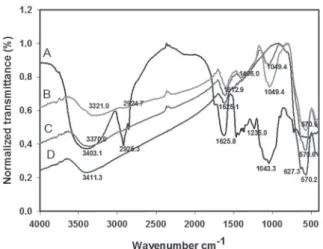
Figure 1 presents the infrared spectra of
the free PpeG (A), magnetic PpeG at different
concentrations 0.500 mg/mL (B) and 0.100 mg/
mL (C) and magnetite (D). Infrared spectroscopy
showed that O–H groups are present in the PpeG
polysaccharide, magnetite and magnetic PpeG near
wavenumber of 3400 cm
-1with higher intensity for
PpeG (3411.3). These O–H groups correspond to
those present in organic compounds. The magnetic
PpeG presented absorption bands in 2924.7 cm
-1due to stretching vibration of C–H bond band in
1049.4 cm
-1and due to stretching vibration of
C–OH bond. These bonds are also present in the
PpeG polysaccharide with bands in 2925.3 cm
-1(stretching vibration of C–H bond), band in 1043.3
cm
-1(stretching vibration of C–OH) indicative of
the presence of polysaccharide in the magnetic
particles. Previous studies (Waldron 1955, Pan et
al. 2005) reported that the characteristic absorption
bands of the Fe–O bond of bulk magnetite were
around 570 cm
-1. However, Ma et al. (2003)
observed that these two bands shift of about 600
and 440 cm
-1respectively, and the band near
600 cm
-1is split into two peaks of 631.4 and
582.9 cm
-1. Here, a band near 600 cm
-1is also
shown split in two peaks of 627.3 and 570.2 cm
-1for magnetite. Magnetic PpeG particles presented
a similar band at 570.6 cm
-1with a lower peak at
627.3. This difference was also observed by Maciel
et al. (2012) for magnetic levan. According to the
authors, a difference in these bands can indicate that
interactions between polysaccharide and magnetite
had inter-molecular origins. The results confirm the
success of the magnetization process of PpeG.
PpeG infrared spectrum is typical for
poly-saccharide (Fig. 1 A) as those reported for cellulose
(Corti et al. 2004) and the cashew gum (Silva
et al. 2004). The C=O axial deformation at the
wavenumber 1736 cm
-1typical of glucuronic acid
is present for the PpeG. Corti et al. (2004) observed
that 1656, 1631 and 1558 cm
-1infrared bands
corresponded to axial deformation of carbonyl in
amide (in our case related to carbonyl of peptide
bonds), N-H angular deformation of amine and
N-H angular deformation of amide, respectively.
Similar bands were observed by Pan et al. (2005).
The absence of these bands in the spectrum of
PpeG and the elemental analysis confirmed that no
proteins were found in this structure.
IMMOBILIZATION AND ACTIVITY OF CON A
The partial oxidation of the gum by NaIO
4aimed
to randomly introduce aldehydes groups in the
vicinal hydroxyls of the carbohydrates
(Martinez-Barragan and Angel 2001, Hong et al. 2004). These
aldehydes groups then react to amine group from
amino acids chains such as lysine, sulfhydryl group
from cysteine and imidazole group from histidine
(Fraguas et al. 2004).
Fig. 1 - Infrared spectra of P. pendula gum (A), magnetized gum using 0.500 mg/mL (B) and 0.100 mg/mL (C) of the gum and magnetite (D).
Sample %
Nitrogen Carbon Hydrogen Sulfur PpeGa
(0.500 mg/mL) 0 35.83 5.29 0
magPpeG a
(0.500 mg/mL) 0 9.24 1.98 0
TABLE I
Element composition of free and magnetized P. pendula seed gum.
a
The immobilization of Con A on the PpeG
retained about 62% of the offered protein (Table
II). Kobayashi and Ichishima (1991) reported 40%
of retention immobilizing bovine serum albumin
on cellulose. Cavalcante et al. (2006)
immobilized
trypsin onto a membrane of a cellulosic
exopoly-saccharide produced by
Zoogloea sp
. in sugarcane
molasses. Carbonyl groups were introduced into
the matrix by sodium meta-periodate oxidation
and the enzyme was immobilized either directly
or through bovine serum albumin (BSA) as a
spacer. The trypsin-membrane and
trypsin–BSA-membrane retained 37.2% and 9.16%, respectively.
It is worthwhile to notice that not all carbohydrate
moieties are oxidized by the NaIO
4(Silva et al.
2004). Furthermore, the PpeG structure is not
completely identified yet and its linear and branched
chains are not established.
not accomplished due to steric hindrance caused
by the immobilization procedure.
Besides that, even with the higher quantity
of oligosaccharides present in fetuin and its
recognition by soluble Con A, Johnson and Heath
(1986b), Green et al. (1988)
observed that among
the 23 N-types of oligosaccharides that could
be found in fetuin (di- or tri-branched), only the
di-branched ones would be recognized, which
corresponds to 17%.
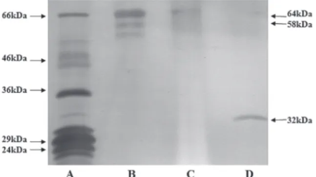
Figure 2 shows three bands (64, 58 e 55
kDa) for the fetuin (Lane B) in the SDS-PAGE.
Nevertheless, only two bands (64 e 58 kDa) appear
for the purified fetuin (Lane C) by affinity binding
to the
magPpeG-Con A. The Con A band (Lane D) is
not present in the Lane C, which demonstrates that
this lectin was covalently linked to the
magPpeG,
and was not detached during the washings.
Fig. 2 - SDS-PAGE of eluted fetuin from the immobilized
magPpeG-Con A. Standard proteins (A), Fetuin (B), Eluted Fetuin (C), Con A (D).
Con A immobilization
Fetuin recognition
Offered (µg/mL) 400 400
Supernatanta (µg/mL) 147 ± 9.6 343.8 ± 11.23 Retaineda (µg) 253 ± 9.7 56.2 ± 11.23 Efficiencya (%) 62.6 ± 2.30 13.8 ± 2.95
TABLE II
Immobilization efficiency of Con A on
magPpeG and efficiency of fetuin recognition
by the immobilized Con A on magPpeG.
a All experiments were carried out in quintuplicates and the results expressed as mean ± Standard deviation.
The fetuin, composed of one polypeptide
chain (Spiro 1963) containing 3 oligosaccharides
N-linked (Spiro and Bhoyroo 1974) and 3 O-linked
(Green et al. 1988), was used as a model. Table
II summarizes the results of this purification and
shows that about 14% of fetuin was complexed to
about 63% of Con A. This relationship accounts
for about 01 mole of fetuin per 08 mole of Con
A considering that the molecular weights are
respectively 64 kDa (Johnson and Heath 1986a)
and 32 kDa (Fontaniella et al. 2004). The ratio of
1:1 mole: mole of fetuin and Con A was probably
Johnson and Heath (1986a) observed that
native fetuin, pre-fetuin and glycosilated fetuin
(found in the rough endoplasmatic reticulum)
presented molecular weight of 64, 49 and 58
kDa, respectively, in SDS-PAGE. Therefore, the
observed 58kDa band in Figure 2 would be a fetuin
with two N-type glycosylation.
According to Green et al. (1988) the
recognition of Con A for di-branched fetuin justifies
the absence of the 55kDa band in the lane C of the
(
Leukoagglutinating Phytohemagglutinin
) and
RCA-I (
Ricinus Communis
Agglutinin) interact
with carbohydrates depending on the type of the
bound between the saccharide residue and its
position in the oligosaccharide chain. L-PHA has
strong interaction if the neuraminic acid, present
in fetuin, has one terminal
α
-2,3 bound, linked to
the mannose’s branching
α
-1-6. Otherwise, if the
bound is
α
-2,6 on the same branching (
α
-1-6)
the recognition of the sugar by L-PHA cannot be
made. Furthermore, no significant interference, in
recognition, was observed if the sugar was located
on another branching (Green et al. 1988). So, the
spatial changes caused by certain saccharides
bearing N-type glycosylation in fetuin, would,
possibly, impose a more or less stable interaction
between Con A and fetuin.
Results from our groups have already
demonstrated the use of other magnetized
poly-saccharides such as levan as affinity matrix for direct
lectin purification (Angeli et al. 2009) or as matrix
for trypsin immobilization (Maciel et al. 2012) and
cellulosic exopolysaccharide produced by
Zoogloea
sp. as a film support for trypsin immobilization
(Cavalcante et al. 2006). Here, we demonstrated
that Ppeg was efficiently magnetized and used
as matrix for Con A immobilization.
magPpeG-Con A was used as an affinity chromatography
matrix for purification of fetuin under a magnetic
field indicating that it is a promising matrix for
biotechnology application.
ACKNOWLEDGMENTS
The Conselho Nacional de Desenvolvimento
Científico e Tecnológico (CNPq) supported this work
The authors have declared no conflict of interest.
RESUMO
O presente trabalho objetivou magnetizar a goma de sementes de Parkia pendula e demonstrar seu uso como matriz para imobilização covalente da lectina Concanavalina A. O composto foi aplicado em
puri-ficação de glicoconjugados por afinidade. Sementes de Parkia pendula foram hidratadas e a goma do sobrenadante foi precipitada, lavada com etanol e seca. A goma foi magnetizada pelo método de coprecipitação usando soluções de Fe+2
e Fe+3. A ativação da matriz foi realizada com NaIO4. Goma das sementes de Parkia pendula magnetizada com Concanavalina A imobilizada covalentemente foi usada como matriz de afinidade para o reconhecimento da glicoproteína fetuína sérica bovina. Eluição de fetuína foi realizada com uma solução de glicose (300mM) e avaliada por meio de SDS-PAGE. A eficiência de imobilização da lectina e purificação de fetuína foram 63% e 14%, respectivamente. Esses resultados indicam que o composto produzido é uma matriz polissacarídica magnética promissora para imobilização de lectinas. Esse sistema pode ser aplicado para purificação por afinidade que permite fácil recuperação por campo magnético.
Palavras-chave: imobilização, magnetização, goma da
semente de Parkia pendula, Concanavalina A.
REFERENCES
ANDERSON DMW AND PINTO GL. 1985. Gum polysaccharides from three Parkia species. Phytochem 24: 77-79.
ANGELI R ET AL. 2009. Ferromagnetic Levan Composite: An Affinity Matrix to Purify Lectin. J Biomed Biotechnol Article ID 179106: 1-6.
BUCUR B, DANET AF AND MARTY JM. 2004. Versatile method of cholinesterase immobilisation via affinity bonds using Concanavalin A applied to the construction of a screen-printed biosensor. Biosens Bioelectron 20: 217-225.
CARNEIRO-LEÃO AMA, OLIVEIRA EA AND CARVALHO JR LB. 1991. Immobilization of protein on ferromagnetic Dacron. Appl Biochem Biotechnol 31: 53-58.
CAVALCANTE AHM, CARVALHO JR LB AND C
ARNEIRO-DA-CUNHA MG. 2006. Cellulosic exopolysaccharide produced by Zoogloea sp. as a film support for trypsin immobilisation. Biochem Eng J 29: 258-261.
CÔELHO RAL, JAQUES GA, BARBOSA AD, VELAZQUEZ G, MONTENEGRO SML, AZEVEDO WM AND CARVALHO JR LB. 2002. Magnetic polysiloxane-polyvinyl alcohol composite as solid-phase in chemiluminescent assays. Biotechnol Lett24: 1705-1708.
CORTI GS, BOTARO VR, GIL LF AND GIL RPF. 2004. Estudo da
FONTANIELLA B, MILLANES AM, VICENTE C AND LEGAZ ME.
2004. Concanavalin A binds to a mannose-containing ligand in the cell wall of some lichen phycobionts. Plant Physiol Biochem 42: 773-779.
FRAGUAS LF, BATISTA-VIEIRA F AND CARLSSON J. 2004. Preparation of high-density Concanavalin A adsorbent and its use for rapid, high-yield purification of peroxidase from horseradish roots. Chromatogr B Biomed Sci Appl 803: 237-241.
FRAGUAS LF, PLÁ A, FERREIRA F, MASSALDI H, SUÁREZ N AND BATISTA-VIEIRA F. 2003. Preparative purification of
soybean agglutinin by affinity chromatography and its immobilization for polysaccharide isolation. Chromatogr B Biomed Sci Appl 790: 365-372.
GEMEINER P, MISLOVIČOVÁ D, TKÁČ J, ŠVITEL J, PÄTOPRSTÝ V, HRABÁROVÁ E, KOGAN G AND KOŽÁR T. 2009. Lectinomics II. A highway to biomedical/clinical diagnostics. Biotechnol Adv 27: 1-15.
GHAZARIAN H, IDONI B AND OPPENHEIMER SB. 2010. A glycobiology review: Carbohydrates, lectins and implications in cancer therapeutics. Acta Histochem 113: 236-247. GREEN ED, ALDELT G AND BAENZIGER JU. 1988. The
aspa-ragine-linked oligosaccharides on bovine fetuin. Structural analysis of N-glycanase-released oligosaccharides by 500-Megahertz 1H-NMR spectroscopy. J Biol Chem 263: 18253-18268.
HONG X, GUO W, YUAN H, LI J, LIU Y, MA L, BAI Y AND LI T. 2004. Periodate oxidation of nanoscaled magnetic dextran composites. J Magn Magn Mater 269: 95-100.
JOHNSON WV AND HEATH E. 1986a. Structural features of bovine fetuin revealed from analysis of the primary translation product: anomalous behavior on sodium dodecyl sulfate-polyacrylamide gel electrophoresis is due largely to peptide and not solely to carbohydrate. Arch Biochem Biophys 251: 732-737.
JOHNSON WV AND HEATH E. 1986b. Evidence for Posttransla-tional 0-glycosylation of fetuin. Biochemistry 25: 5518-5525. KESHOVEN HJ AND DERNICK R. 1985. Simplified method for
silver staining of proteins in polyacrylamide gels and the mechanism of silver staining. Electrophoresis 6: 103-112. KOBAYASHI M AND ICHISHIMA E. 1991. Application of
periodate oxidized glucans to biochemical reactions. J Carbohydr Chem 10: 635-644.
LAEMMLI UK. 1970. Cleavage of structural proteins during the assembly of the head of bacteriophage T4. Nature 227: 680-685. LOWRY OH, ROSEBROUGH NJ, FARR AL AND RANDALL RJ.
1951. Protein measurement with the folin phenol reagent. J Biol Chem 193: 265-275.
MA M, ZHANG Y, YU W, SHEN H, ZHANG H AND GU N. 2003. Preparation and characterization of magnetite nanoparticles coated by amino silane. Colloids Surf A 212: 219-226.
MACIEL JC, ANDRAD PL, NERI DFM, CARVALHO JR LB, CARDOSO CA, CALAZANS GMT, ALBINO AGUIAR J AND SILVA MPC. 2012. Preparation and characterization of magnetic levan particles as matrix for trypsin
immobilization. J Magn Magn Mater 324: 1213-1216. MARTINEZ-BARRAGAN JJ AND ANGEL RM. 2001. Identification
of a Putative Coreceptor on Vero Cells That Participates in Dengue 4 Virus Infection. J Virol 75: 7818-7827. MISLOVICOVÁ D, MASAROVÁ J, VIKARTOVSKÁ A, GEMEINER
P AND MICHALKOVÁ E. 2004. Biospecific immobilization of mannan–penicillin G acylase neoglycoenzyme on Concanavalin A-bead cellulose. J Biotechnol 110: 11-19. NERI DFM, BALCÃO VM, CARDOSO SM, SILVA AMS,
DOMINGUES MRM, TORRES DPM, RODRIGUES LRM, CARVALHO JR LB AND TEIXEIRA JAC. 2011. Charac-terization of galactooligosaccharides produced by b-galactosidase immobilized onto magnetized Dacron. Int Dairy J 21: 172-178.
PAN BF, GAO F AND GU HC. 2005. Dendrimer modified magnetite nanoparticles for protein immobilization. J Colloid Interface Sci 284: 1-6.
RIBEIRO A, CATARINO S AND FERREIRA RB. 2012. Multiple lectin detection by cell membrane affinity binding. Carbohydr Res 352: 206-210.
RODRIGUES JF, PAULA RCM AND COSTA SMO. 1993. Métodos de isolamento de gomas naturais e comparação através de goma de cajueiro. Polim Cienc Tecnol 1: 31-36.
SHARON N. 2007. Lectins: Carbohydrate-specific Reagents and Biological Recognition Molecules. J Biol Chem 282(5): 2753-2764.
SILVA DC, PAULA RCM, FEITOSA JPA, BRITO ACF, MACIEL JS AND PAULA HCB. 2004. Carboxymethylation of cashew tree exudate polysaccharide. Carbohydr Polym 58: 163-171. SORIA F, ELLENRIEDER G, OLIVEIRA GB, CABRERA M AND
CARVALHO JR LB. 2012. α-L-Rhamnosidase of Aspergillus terreus immobilized on ferromagnetic supports. Appl Microbiol Biotechnol 93: 1127-1134.
SPIRO RG. 1963. Demonstration of a single peptide chain in the glycoprotein fetuin: terminal amino acid analyses and studies of the oxidized and reduced alkylated preparations. J Biol Chem 238: 644-649.
SPIRO RS AND BHOYROO V. 1974. Structure of the O-glycosidically linked carbohydrate units of fetuin. J Biol Chem 249: 5704-5717.
UYGUN M, UYGUN DA, ÖZÇALIŞKAN E, AKGÖL S AND DENIZLI
AJ. 2012. Concanavalin A immobilized poly(ethylene glycol dimethacrylate) based affinity cryogel matrix and usability of invertase immobilization. Chromatogr B Biomed Sci Appl 887-888: 73-78.
YANG G, CUI T, CHEN Q, MA T AND LI Z. 2012. Isolation and identification of native membrane glycoproteins from living cell by concanavalin A–magnetic particle conjugates Anal Biochem 421: 339-341.