William Sihler(1), Marlinda Lobo de Souza(1), Fernando Hercos Valicente(2), Rosana Falcão(1) and Marcio Martinello Sanches(1)
(1)Embrapa Recursos Genéticos e Biotecnologia, Parque Estação Biológica, Avenida W5 Norte (Final), CEP 70770-917 Brasília, DF, Brazil. E-mail: [email protected], [email protected], [email protected], [email protected] (2)Embrapa Milho e Sorgo, Rodovia MG-424, Km 45, CEP 35701-970 Sete Lagoas, MG, Brazil. E-mail: [email protected]
Abstract – The objective of this work was to evaluate the response of anin vitro host range to Spodoptera frugiperda multiple nucleopolyhedrovirus (SfMNPV), a pathogenic virus to the fall armyworm (Spodoptera frugiperda, Lepidoptera: Noctuidae), for the further development of a biopesticide based on cell culture systems. The cell lines from Bombyx mori (BM-5), Lymantria dispar (IPLB-LD-625Y), Trichoplusia ni (BTI-Tn-5B1-4), Anticarsia gemmatalis (UFL-AG-286), and S. frugiperda (IPLB-SF-21AE and Sf9) were tested for their susceptibility to a highly-virulent Brazilian isolate of SfMNPV. The cytopathic effects induced by the virus, the production of viral particles, and the synthesis of viral polypeptides were examined and compared. Both S. frugiperda cell lines showed hypertrophy of cell nuclei and production of many polyhedra. The SDS-Page of radiolabed proteins showed that the cell protein synthesis was shutoff, while an intense band of about 30 kDa, recognized as polyhedrin, was synthesized. The other cell lines did not show polyhedra production, although some of them underwent morphological changes and protein synthesis shutdown in response to virus infection. The SF-21 and Sf9 cell lines are recommended for further in vitro production of SfMNPV.
Index terms: Baculovirus, biological control, cell culture, fall armyworm, in vitro production, SfMNPV.
Infectividade in vitro de
Spodoptera frugiperda multiple nucleopolyhedrovirus
a diferentes linhagens celulares de insetos
Resumo – O objetivo deste trabalho foi avaliar uma gama de hospedeiros, in vitro, quanto à resposta a
Spodoptera frugiperda multiple nucleopolyhedrovirus (SfMNPV), um vírus patogênico à lagarta-do-cartucho (Spodoptera frugiperda; Lepidoptera: Noctuidae), para o posterior desenvolvimento de um biopesticida baseado em sistema de cultura de células. As linhagens celulares de Bombyx mori (BM-5), Lymantria dispar
(IPLB-LD-625Y), Trichoplusia ni (BTI-Tn-5B1-4), Anticarsia gemmatalis (UFL-AG-286) e S. frugiperda
(IPLB-SF-21AE e Sf9) foram testadas quanto a sua suscetibilidade a um isolado brasileiro de SfMNPV altamente virulento. Os efeitos citopáticos induzidos pelo vírus, a produção de partículas virais e a síntese de polipeptídeos virais foram examinados e comparados. Ambas as células de S. frugiperda apresentaram
hipertrofia dos núcleos celulares e produção de muitos poliedros. O SDS-Page de proteínas com radiomarcação
mostrou que as proteínas celulares apresentaram inibição de síntese, enquanto uma intensa banda de cerca de 30 kDa, reconhecida como poliedrina, era sintetizada. As outras linhagens celulares não apresentaram produção de poliedros, apesar de algumas terem apresentado alterações morfológicas e inibição de síntese proteica em resposta à infecção viral. As linhagens celulares SF-21 e Sf9 são recomendadas para a posterior produção in vitro de SfMNPV.
Termos para indexação: Baculovirus, controle biológico, cultura de células, lagarta-do-cartucho, produção in vitro, SfMNPV.
Introduction
Spodoptera frugiperda multiple nucleo-polyhedrovirus (SfMNPV), genus Alphabaculovirus, family Baculoviridae, is pathogenic to the fall armyworm (Spodoptera frugiperda; Lepidoptera: Noctuidae) and has a great potential to be used as a
biocontrol agent. The insect is a severe pest in most countries, and it is native to the tropical regions of the Western Hemisphere, from the United States to Argentina (Farias et al., 2014).
named as SfMNPV-19 showed the lowest LC50 and the
highest mortality rate at lower concentrations, leading to intense disruption of the host integument (Barreto et al., 2005). In contrast to other reported SfMNPV isolates, however, an isolate named SfMNPV-6nd did not cause liquefaction to larvae, a very important feature for virus pesticides production. The virulence of both isolates was compared by Vieira et al. (2012), who reported that the LC50 of SfMNPV-6nd is not significantly different
from that of isolate 19, although it took considerable longer periods to kill S. frugiperda larvae. In fact, in preliminary experiments conducted by our team, the isolate 19 also showed better results of infection than the isolate 6nd, in the available cell lines.
Spodoptera frugiperda is a very important pest in South America, causing significant damage to several different crops. It is the most important Lepidoptera pest for maize in Brazil, with damages to yield reaching up to 52% (Figueiredo et al., 2006). The use of chemical pesticides and of genetically modified maize to control the pest have been raising environmental concerns and generating resistant biotypes (Farias et al., 2014).
Large-scale production of the SfMNPV baculovirus is still obtained in vivo by infecting healthy larvae. Therefore, the liquefaction of the larvae integument is a disadvantage to this process. Moreover, the large-scale production of baculovirus is also hindered by the cannibalistic behavior of the fall armyworm. This requires larval individualization, which is labor-intensive, increases the risk of contamination, and increases the costs of biopesticide production (Valicente et al., 2013). Therefore, in vitro production has a great potential to overcome these difficulties (Reid et al., 2014).
Cell cultures of insects are an interesting means to select systems that support viral replication (Castro et al., 2006; Almeida et al., 2010; Huynh et al., 2015). These cell systems are useful in studies of virus-host interactions, but the present technical limitations should be solved for their need in large-scale production of biopesticides, such as the accumulation of “few polyhedra mutants” or “defective interfering particles” in the cell culture (Moscardi et al., 2011; Reid et al., 2014). Other application of similar importance for cell culture is the synthesis of recombinant proteins by genetic engineering (Granados et al., 2007). In this sense, the Sf9 (S. frugiperda) and “High-Five” (Trichoplusia ni) cell lines have been studied
extensively as to their ability to grow in suspension cultures using serum-free media (Reid et al., 2014).
Selecting productive cell lines is an essential initial step for the establishment of SfMNPV in vitro production. Previous work with other SfMNPV isolate 18, which is less virulent than the isolate 19, was done exclusively with S. frugiperda cell lines in suspension culture (Almeida et al., 2010). However, it is well known that baculovirus species can infect cell lines originated from other Lepidoptera species. In the case of
Anticarsia gemmatalismultiple nucleopolyhedrovirus
(AgMNPV), the polyhedra production in some cell lines obtained with different Lepidoptera was similar to that obtained with original host cell line, or even better (Castro et al., 2006). The response of insect cells to baculovirus infection involves apoptosis and protein synthesis shutdown (Du & Thiem, 1997). Several baculoviruses overcome cell defense mechanisms by expressing anti-apoptotic genes, as well as host-range factors that prevent global protein synthesis shutdown. Because the production of baculovirus in cell culture is a system independent of the insect, features such as liquefaction are not pertinent. Most important in this context is the quality of the polyhedra formed as for the maintenance of their ultrastructure and virulence. In addition, it is important to select cell lineages that have desirable trais for in vitro culture. Studies on the subject have shown that baculoviruses establish unique interactions with different cell lines, resulting in various types of productive or nonproductive infections.
The objective of this study was to evaluate the response of six different lepidopteran cell lines to SfMNPV-19.
Materials and Methods
Six lepidopteran cell lines were assayed: Trichoplusia ni BTI-Tn-5B1-4 (Granados et al., 1994), also known as “High Five cells”; Anticarsia gemmatalis UFL-AG-286 (Sieburth & Maruniak, 1988); Spodoptera frugiperda IPLB-SF-21AE (Vaughn et al., 1977) and Sf9 (Granados et al., 2007); Lymantria dispar IPLB-LD- 652Y (Goodwin et al., 1978); and Bombyx mori
lactalbumin hydrolysate and yeastolate), except for the UFL-AG-286 cells, which were cultivated in TC-100 (Gibco-BRL, Grand Island, NY, USA).
The cells were infected with the virus isolate SfMNPV-19 (Barreto et al., 2005). Tissue culture plates (60 mm2) were seeded with 1x10⁶ cells and
allowed to attach overnight (12–14 hours). The cultures were inoculated with SfMNPV-19 at a multiplicity of infection (MOI) of 10. Mock-infected cells did not contain any virus. After viral adsorption for 1 hour, cells were washed and incubated in complete medium at 27°C.
In vitro infectivity was evaluated by phase contrast, electron microscopy analysis, and by the kinetics of protein synthesis. At postinfection (p.i) periods of 0, 24, 48, 72, and 96 hours, cells were observed for cytopathic effects by phase-contrast microscopy. Cells were collected at 96-hour p.i for electron microscopy analysis. Infected and mock-infected cells were centrifuged at 3.000 g for 3 min, washed with PBS, pH 6.2, and centrifuged again. Sedimented cells were resuspended in a buffer solution of 0.1mol L-1 sodium
cacodylate and 2.5% glurataraldehyde, and then transferred to microcentrifuge tubes for incubation at 4ºC. Fixed samples were treated with 0.05 mol L-1
sodium cacodylate, and then fixed with 2% osmium tetroxide. After a new wash using sodium cacodylate buffer, samples were dehydrated in ethanol at 30, 70, 90, and 100%, and subjected to a mixture of ethanol:Spurr resin at the rates of 3:1, 2:1, 1:1, ending with 5 mL of pure Spurr. The blocks were mounted in Spurr and kept at 37ºC, for 72 hours. The specimens were then cut in the ultramicrotome with a diamond knife. Meshes containing the samples were stained with 2% uranyl acetate. Samples were photographed in a transmission electron microscope Jeol 1011 (Jeol, Peabody, MA, USA) (Sample et al., 2007).
The [35S] methionine protein labeling of the infected
cells was done based on the protocol described by Sample et al. (2007). Cells were seeded at a the density of 1x10⁶ per 60 mm2 dish and incubated overnight at
27°C to allow their attachment. Subsequently, the cells were inoculated with the virus (SfMNPV-19) at an MOI of 10. The mock-infected control cells received the same treatment, except for the inoculum without virus. After viral adsorption for 1 hour, cells were washed and incubated in complete culture medium at 27°C. The medium was replaced by phosphate
buffered saline (PBS, pH 6.2) at postinfection periods of 24, 48, and 72 hours, and the incubation continued for 30 min (starvation period). The medium was removed, and 50 µCi of [35S] methionine in 0.6 mL
of PBS was added for 1-hour pulse. Mock-infected cells were labeled at the time 0 , after 1-hour of virus adsorption. After labeling, the cells were transferred to a microfuge tube, pelleted by centrifugation for 60 s, rinsed with PBS, and frozen at -20°C. The samples were disrupted and subjected to SDS–polyacrylamide gel electrophoresis (SDS–Page). To visualize the bands, the gel was treated with a fluorographic reagent Amplify (Amersham Biosciences, Little Chalfont, United Kingdom) for 30 min, dried, and exposed to a Kodak X-Omat AR film (Kodak, Rochester, NY, USA) for 5 days, at -70°C. The autoradiogram was analyzed for the presence of labeled proteins.
In order to verify the presence of apoptosis in some cell lines, a test was conducted to observe DNA fragmentation. The apoptosis tests were performed in cells without evidence of polyhedra presence. The cell lines Tn-5B1-4, AG-286, BM-5, and LD-625Y were seeded at 1x10⁶ density per 60 mm2 dish, and
incubated overnight at 27°C to allow their attachment. Subsequently, the cells were inoculated with the virus SfMNPV-19 at an MOI of 10. The mock-infected control cells received the same treatment, except for the inoculum without virus. At 96-hour p.i., the cells were suspended in 0.1 mL PBS, and total DNA was isolated with the DNeasy blood & tissue kit (Qiagen, Hilden, Germany). The DNA was electrophoretically separated in 1.0% agarose gel stained with ethidium bromide.
Results and Discussion
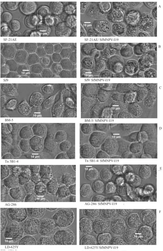
Morphological analysis by phase-contrast microscopy (Figure 1) showed that infected IPLB-SF-21AE and Sf9 cells lead to successful viral replication with many polyhedra formation. Bombyx mori cells (BM-5) seemed to change their morphology from round-refractive to “groundnut-shape”, although without polyhedra production. Anticarsia gemmatalis
A
B
C
D
E
F SF-21AE
Sf9
BM-5BM-5
Tn 5B1-4
AG-286
LD-625Y LD-625Y/SfMNPV-I19
AG-286/ SfMNPV-I19 Tn 5B1-4/ SfMNPV-I19 BM-5/ SfMNPV-I19 Sf9/ SfMNPV-I19 SF-21AE/ SfMNPV-I19
Figure 1. Phase-contrast micrographs of mock-infected cells (left), and cells infected with SfMNPV-19 96-hour p.i. (right) (40X) Spodoptera frugiperda IPLB-SF-21AE (A) and Sf9 (B); Bombyx mori BM-5(C), Trichoplusia ni BTI-Tn 5B1-4 (D);
more polyhedra than Sf9, at 72-hour p.i., the presence of polyhedra became similar in both cells at 96-hour p.i.
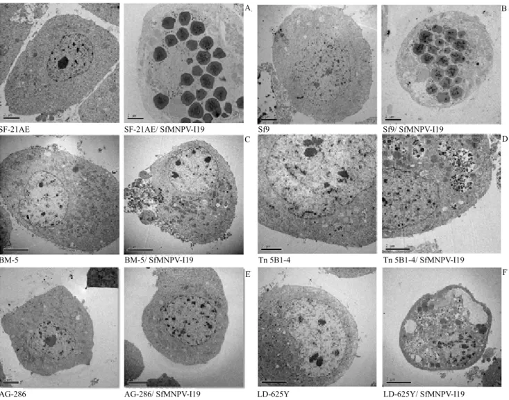
Ultrastructural analysis of the two infected S. frugiperda cell lines showed, as expected, cell nuclei hypertrophy and production of many polyhedra at 96-hour p.i. (Figures 2 A and B). Other typical baculovirus induced effects, such as the formation of virogenic stroma, nucleocapsids, and virions were also observed during the first days postinfection. None of the other cell lines showed virus particles inside the cell nuclei after viral incubation (Figures 2 C, D, E, and F).
However, Lymantria dispar and Trichoplusia ni cells showed several vesicles and vacuoles in the cytoplasm. Both IPLB-SF-21AE and Sf9 cells, infected with SfMNPV and labeled with [35S] methionine, showed
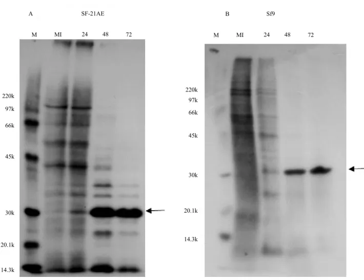
that polypeptide synthesis patterns in the beginning of the infection differ from that observed in the control cells (Figures 3 A and B). In fact, some viral proteins that initially seemed to comigrate with the host proteins were progressively more evident in the cycle of infection, along with the disappearance of host cell protein synthesis. At late times, post-infection (from 48 to72-hour p.i), an intense band of about 30 kDa, was
A B
C D
E F
SF-21AE SF-21AE/ SfMNPV-I19
BM-5/ SfMNPV-I19 BM-5
AG-286/ SfMNPV-I19 AG-286
Sf9/ SfMNPV-I19 Sf9
Tn 5B1-4/ SfMNPV-I19 Tn 5B1-4
LD-625Y/ SfMNPV-I19 LD-625Y
Figure 2. Electron micrographs of mock-infected cells (left) and cells infected with SfMNPV-19 at 96-hour p.i. (right).
Spodoptera frugiperda IPLB-SF-21AE (A) and Sf9 (B); Bombyx mori BM-5 (C), Trichoplusia ni BTI-Tn 5B1-4 (D);
identified as polyhedrin, the main structural protein of NPV. The intensity of the radiolabel incorporation detected in this band is consistent with the high levels of polyhedra production observed in the nuclei of these cells (Figures 1 A and B, and Figures 2 A and B). Polyhedrin synthesis in IPLB-SF-21AE and Sf9 reached its maximum at 72-hour p.i.
The infected BTI-Tn-5B1-4, AG-286, BM-5, and IPLB-LD-625Y cells did not produce polyhedrin. However, they showed low levels of cellular protein synthesis after 48-hour p.i., and a complete shutdown of cellular peptide synthesis at 72-hour p.i. This may have occurred due to interactions between viral proteins and cellular translation machinery, as observed in many virus-host interactions (Walsh & Mohr, 2011).
The infected cell lines showed the same DNA pattern, in comparison to the control without fragmentation.
According to our data, only S. frugiperda cells were susceptible for the in vitro production of SfMNPV. These cells were previously described as permissive to SfMNPV infection (Simón et al., 2008; Almeida et al., 2010; Beperet et al., 2014). Similar observations occurred with another virus from Spodoptera (Yanase et al., 1998). These authors inoculated Spodoptera exigua multiple nucleopolyhedrovirus (SeMNPV) into eight lepidopteran cell lines, and reported that polyhedra were only produced in the cell line derived from its host (Se031 cells). Morphological changes were also induced by SeMNPV in the “High Five” cells, although no productive infection had occurred
Figure 3. Protein synthesis in SfMNPV-infected insect cells at 24, 48, and 72 hours postinfection. Autoradiogram of a 15% SDS–PAGE from insect cells pulse labeled with [35S] methionine. Insect cell lines: Spodoptera frugiperda IPLB-SF-21AE
(A) and Sf9 (B). The arrows indicate the polyhedrin. MI, mock-infected cells; M, molecular mass markers given in kDa. SF-21AE
A
M MI 24 48 72 M MI 24 48 72
B Sf9
220k
97k
66k
45k
30k
20.1k
14.3k
220k
97k
66k
45k
30k
20.1k
(Yanase et al., 1998). Nevertheless, Jain & Das (2004) reported that Spodoptera litura nucleopolyhedrovirus
(SpltNPV), pathogenic to larvae of Spodoptera litura, infects the Sf9 cell line, which is derived from another host. Moreover, Ishikawa et al. (2003) showed that LD-625Y cells underwent apoptosis after infection by SeMNPV and SpltNPV.
Inhibition of protein synthesis has been shown to be a common event in cells infected with viruses (Nguyen et al., 2013). The protein shutoff was observed in the interaction between Autographa californica multiple nucleopolyhedrovirus (AcMNPV) and LD-625Y cells (Du & Thiem, 1997), and both apoptosis and the shutdown were suggested as being separate responses of the insect cells to AcMNPV infection. In the present study, we did not observe the occurrence of apoptosis in “High Five”, BM-5, UFL-AG-286, and LD-625Y cells infected with SfMNPV, but there was a protein synthesis shutdown, suggesting a response to the virus infection.
Host cells apoptosis induced by virus infection plays an important role in baculovirus host range (Wu et al., 2016). The mechanism that triggers apoptosis in baculovirus-infected cells relies on an intricate relationship between cellular and viral functions (Ishikawa et al., 2003). For instance, the AcMNPV, a baculovirus with a wide host range, capable to infect more than 25 cell lines, induces apoptosis in S. litura
and S. littoralis cells (Chejanovsky & Gershburg, 1995; Zhang et al., 2002), but can infect the SF-21 and Sf9 cell lines. The product of anti-apoptotic viral gene p35 was observed as essential for the replication of AcMNPV in the SF-21 cells (Clem et al., 1991). Recently, the anti-apoptotic gene iap3 of SeMNPV was observed as responsible for the expanded host range for a recombinant virus, including nonpermissive cells to the parental viruses (without iap3). This virus was constructed with AcMNPV lacking the p35 gene and a fragment of SeMNPV (Wu et al., 2016).
Accordingly, severe apoptotic response has been shown in a study conducted with LD-625Y infected by
Hyphantria cunea nucleopolyhedrovirus (HycuNPV) with high expression of iap3 (Ishikawa et al., 2003). These authors suggested that the apoptosis induction in NPV-infected LD-652Y cells is largely dependent on inherent cellular properties, rather than on functions of the NPVs, and indicate that defects in progeny virion production are not merely due to the virus-induced
apoptosis. The data of the present study suggest a defense response of the cells to the baculovirus infection, as well as an anti-apoptotic function mediated by the virus. The genome of SfMNPV-19 is completely sequenced, and the presence of iap3 was pointed out (Wolff et al., 2008). However, further analysis on gene function is necessary to confirm this hypothesis.
A broader host range could be important for selecting more productive cell lines to be used in large-scale production. The baculovirus AgMNPV has been largely used as a biopesticide in Brazil for controlling A. gemmatalis in soybean (Moscardi et al., 2011). In comparison to SfMNPV-19, AgMNPV-2D has a broad capacity of in vitro infection, including cells from other hosts (S. frugiperda and T. ni). In addition, polyhedra were produced faster at 48-hour p.i. (Castro et al., 2006).
Our study indicates that IPLB-SF-21 cells in static culture are more productive than Sf9 in the beginning of infection with SfMNPV-19. However, at 96-hour p.i., the production of occlusion bodies (OB) became similar in both cell lines. Nevertheless, in a suspension culture infected with another Brazilian isolate (SfMNPV -18), the Sf9 cells seemed to be more productive than SF-21 cells (Almeida et al., 2010). These authors showed that the Sf9 cell-line resulted in better viral production (5.0 x 10⁸ OB mL-1) than the
SF-21 cell-line (2.5 x 10⁸ OB mL-1).
The distinct performance between the viral isolates 19 and 18 could be due to the different conditions of the studies. In static cultures, cell growth is limited to the size of the available surface. In the suspension culture, however, the cells develop freely, and growth is limited by other factors (Wu et al., 1998). Moreover, it was not possible to compare the MOI in neither of the studies, since a fixed volume of the virus inoculum (BVs) was used in the suspension culture after the initial release of the caterpillar-produced occlusion bodies (Almeida et al., 2010). Another culture medium was used in the study with the isolate 18 (HyQ SFX-Insect supplemented with 5% bovine fetal serum), as well as a different initial cell density (5x10⁵ cell mL-1). Several factors have been
The SfMNPV-6nd has a clear advantage to large-scale biopesticide production in vivo because it does not disrupt the larvae integument. However, this characteristic may be unfavorable to control the pest under crop conditions, since it is advantageous for the virus to escape efficiently from the dead larva. Virus polyhedra that remain trapped within the host are less likely to encounter another susceptible individual than virus released via liquefaction (Hawtin et al., 1997). Moreover, the 6nd isolate showed a longer lethal time compared to SfMNPV-19 in bioassays (Vieira et al., 2012).
One of the requirements for a cell line to be considered for in vitro production of a baculovirus is its capacity to produce a useful virus, yielding at least 300 OB per cell (Reid et al., 2014). The potential of SF-21 and Sf9 for the production of SfMNPV-19 are similar, according to preliminary studies (5.0 x 10⁸ OB mL-1
and 300 OB per cell). Therefore, the utilization of SfMNPV-19 as a biopesticide may be more interesting when a large-scale in vitro production strategy of SfMNPV becomes available.
Conclusions
1. The Spodoptera frugiperda SF-21 and Sf9 cell lines are recommended for in vitro production of
Spodoptera frugiperda multiple nucleopolyhedrovirus
(SfMNPV), both with similar polyhedra production in static culture.
2. The cell lines BTI-Tn-5B1-4, AG-286, BM-5, and IPLB-LD-625Y are nonpermissive to SfMNPV.
3. The response to SfMNPV varies according to the cell line, and consists in protein synthesis shutoff and distinct morphological changes in the cells.
Acknowledgments
To Empresa Brasileira de Pesquisa Agropecuária (Embrapa), to Agricultural Innovation Marketplace, to Fundação de Apoio à Pesquisa do Distrito Federal (FAPDF), and to Conselho Nacional de Desenvolvimento Científico e Tecnológico (CNPq), for grants; to the following persons, for providing cell lines: Dr. James E. Maruniak, Sf9, IPLB-SF-21AE, and UFL-AG-286; Dr. Robert Granados, BTI-Tn-5B1-4; Dr. Shänti L. Bilimoria, BM-5; and to Dr. Gary Blissard, IPLB-LD-625Y.
References
ALMEIDA, A.F. de; MACEDO, G.R. de; CHAN, L.C.L.; PEDRINI, M.R. da S. Kinetic analysis of in vitro production of wild-type Spodoptera frugiperda nucleopolyhedrovirus.
Brazilian Archives of Biology and Technology, v.53, p.285-291,
2010. DOI: 10.1590/S1516-89132010000200006.
BARRETO, M.R.; GUIMARAES, C.T.; TEIXEIRA, F.F.; PAIVA, E.; VALICENTE, F.H. Effect of Baculovirus spodoptera
isolates in Spodoptera frugiperda (J.E. Smith) (Lepidoptera: Noctuidae) larvae and their characterization by RAPD.
Neotropical Entomology, v.34, p.67-75, 2005. DOI: 10.1590/
S1519-566X2005000100010.
BEPERET, I.; IRONS, S.L.; SIMÓN, O.; KING, L.A.; WILLIAMS, T.; POSSEE, R.D.; LÓPEZ-FERBER, M.; CABALLERO, P. Superinfection exclusion in alphabaculovirus infections is concomitant with actin reorganization. Journal of
Virology, v.88, p.3548-3556, 2014. DOI: 10.1128/JVI.02974-13.
CASTRO, M.E.B.; RIBEIRO, Z.M.A.; SOUZA, M.L. Infectivity of Anticarsia gemmatalis nucleopolyhedrovirus to different insect cell lines: morphology, viral production, and protein synthesis.
Biological Control, v.36, p.299-304, 2006. DOI: 10.1016/j.
biocontrol.2005.10.002.
CHEJANOVSKY, N.; GERSHBURG, E. The wild-type
Autographa californica nuclear polyhedrosis virus induces apoptosis of Spodoptera littoralis cells. Virology, v.209, p.519-525, 1995. DOI: 10.1006/viro.1995.1284.
CLEM, R.J.; FECHHEIMER, M.; MILLER, L.K. Prevention of apoptosis by a baculovirus gene during infection of insect cells.
Science, v.254, p.1388-1390, 1991. DOI: 10.1126/science.1962198.
DU, X.; THIEM, S.M. Responses of insect cells to baculovirus infection: protein synthesis shutdown and apoptosis. Journal of
Virology, v.71, p.7866-7872, 1997.
FARIAS, J.R.; ANDOW, D.A.; HORIKOSHI, R.J.; SORGATTO, R.J.; FRESIA, P.; SANTOS, A.C. dos; OMOTO, C. Field-evolved resistance to Cry1F maize by Spodoptera frugiperda
(Lepidoptera: Noctuidae) in Brazil. Crop Protection, v.64, p.150-158, 2014. DOI: 10.1016/j.cropro.2014.06.019.
FIGUEIREDO, M. de L.C.; MARTINS-DIAS, A.M.P.; CRUZ, I. Relação entre a lagarta-do-cartucho e seus agentes de controle biológico natural na produção de milho. Pesquisa Agropecuária
Brasileira, v.41, p.1693-1698, 2006. DOI:
10.1590/S0100-204X2006001200002.
GOODWIN, R.H.; TOMPKINS, G.J.; MCCAWLEY, P. Gypsy moth cell lines divergent in viral susceptibility. In Vitro, v.14, p.485-494, 1978. DOI: 10.1007/BF02616088.
GRACE, T.D.C. Establishment of a line of cells from the silkworm
Bombyx mori. Nature, v.216, p.613, 1967. DOI: 10.1038/216613a0. GRANADOS, R.R.; GUOXUN, L.; DERKSEN, A.C.G.; MCKENNA, K.A. A new insect cell line from Trichoplusia ni
GRANADOS, R.R.; LI, G.; BLISSARD, G.W.Insect cell culture and biotechnology. Virologica Sinica, v.22, p.83-93, 2007. DOI: 10.1007/s12250-007-0010-7.
HAWTIN, R.E.; ZARKOWSKA, T.; ARNOLD, K.; THOMAS, C.J.; GOODAY, G.W.; KING, L.A.; KUZIO, J.A.; POSSEE, R.D. Liquefaction of Autographa californica nucleopolyhedrovirus-infected insects is dependent on the integrity of virus encoded chitinase and cathepsin genes. Virology, v.238, p.243-253, 1997. DOI: 10.1006/viro.1997.8816.
HUYNH, H.T.; TRAN, T.T.B.; CHAN, L.C.L.; NIELSEN, L.K.; REID, S. Decline in Helicoverpa armigera nucleopolyhedrovirus occlusion body yields with increasing infection cell density in vitro is strongly correlated with viral DNA levels. Archives of Virology, v.160, p.2169-2180, 2015. DOI: 10.1007/s00705-015-2478-z.
ISHIKAWA, H.; IKEDA, M.; YANAGIMOTO, K.; FELIPE ALVES, C.A.; KATOU, Y.; LAVIÑA-CAOILI, B.A.; KOBAYASHI, M. Induction of apoptosis in an insect cell line, IPLB Ld652Y, infected with nucleopolyhedroviruses. Journal
of General Virology, v.84, p.705-714, 2003. DOI: 10.1099/
vir.0.18815-0.
JAIN, M.; DAS, R.H. Nucleotide sequence and molecular characterization of the structural glycoprotein gp41 gene homologue of Spodoptera litura nucleopolyhedrosis virus (SpltNPV-I). Molecular Biology Reports, v.31, p.231-239, 2004. DOI: 10.1007/s11033-005-1405-x.
MOSCARDI, F.; SOUZA, M.L. de; CASTRO, M.E.B. de; MOSCARDI, L.M.; SZEWCZYK, B. Baculovirus pesticides: present state and future perspectives. In: AHMAD, I.; AHMAD, F.; PICHTEL, P. (Ed.). Microbes and microbial technology. New York: Springer, 2011. p.415-445. DOI: 10.1007/978-1-4419-7931-5_16. NGUYEN, Q.; NIELSEN, L.K.; REID, S. Genome scale transcriptomics of baculovirus-insect interactions. Viruses,v.5, p.2721-2747, 2013. DOI: 10.3390/v5112721.
REID, S.; CHAN, L.; OERS, M.M. van. Production of entomopathogenic viruses. In: MORALES-RAMOS, J.; ROJAS, M.G.; SHAPIRO-LLAN, D.J. (Ed.). Mass production of beneficial organisms: invertebrates and entomopathogens. London: Elsevier, 2014. p.437-482. DOI: 10.1016/B978-0-12-391453-8.00013-3.
RODAS, V.M.; MARQUES, F.H.; HONDA, M.T.; SOARES, D.M.; JORGE, S.A.C.; ANTONIAZZI, M.M.; MEDUGNO, C.; CASTRO, M.E.B.; RIBEIRO, B.M.; SOUZA, M.L.; TONSO, A.; PEREIRA, C.A. Cell culture derived AgMNPV bioinsecticide: biological constraints and bioprocess issues. Cytotechnology, v.48, p.27-39, 2005. DOI: 10.1007/s10616-005-3175-7.
SAMPLE, R.; BRYAN, L.; LONG, S.; MAJJI, S.; HOSKINS, G.; SINNING, A.; OLIVIER, J.; CHINCHAR , V.G. Inhibition of iridovirus protein synthesis and virus replication by antisense morpholino oligonucleotides targeted to the major capsid protein, the 18 kDa immediate-early protein, and a viral homolog of RNA polymerase II. Virology, v.358, p.311-320, 2007. DOI: 10.1016/j. virol.2006.07.009.
SIEBURTH, P.J.; MARUNIAK, J.E. Growth characteristics of a continuous cell line from the velvetbean caterpillar, Anticarsia gemmatalis Hübner (Lepidoptera: Noctuidae). In Vitro Cellular
& Developmental Biology, v.24, p.195-198, 1988. DOI: 10.1007/
BF02623546.
SIMÓN, O.; WILLIAMS, T.; ASENSIO, A.C.; ROS, S.; GAYA, A.; CABALLERO, P.; POSSEE, R.D. Sf29 gene of Spodoptera frugiperda multiple nucleopolyhedrovirus is a viral factor that determines the number of virions in occlusion bodies. Journal of
Virology, v.82, p.7897-7904, 2008. DOI: 10.1128/JVI.00099-08.
VALICENTE, F.H.; TUELHER, E.S.; PENA, R.C.; ANDREAZZA, R.; GUIMARÃES, M.R.F. Cannibalism and virus production in Spodoptera frugiperda (J.E.Smith) (Lepidoptera: Noctuidae) larvae fed with two leaf substrates inoculated with
Baculovirus spodoptera. Neotropical Entomology, v.42, v.191-199, 2013. DOI: 10.1007/s13744-013-0108-6.
VAUGHN, J.L.; GOODWIN, R.H.; TOMPKINS, G.J.; MCCAWLEY, P. The establishment of two cell lines from the insect Spodoptera frugiperda (Lepidoptera: Noctuidae). In Vitro, v.13, p.213-217, 1977. DOI: 10.1007/BF02615077.
VIEIRA, C.M.; TUELHER, E.S.; VALICENTE, F.H.; WOLFF, J.L.C. Characterization of a Spodoptera frugiperda multiple nucleopolyhedrovirus isolate that does not liquefy the integument of infected larvae. Journal of Invertebrate Pathology, v.111, p.189-192, 2012. DOI: 10.1016/j.jip.2012.07.010.
WALSH, D.; MOHR, I. Viral subversion of the host protein synthesis machinery. Nature Reviews Microbiology, v.9, p.860-875, 2011. DOI: 10.1038/nrmicro2655.
WOLFF, J.L.C.; VALICENTE, F.H.; MARTINS, R.; OLIVEIRA, J.V. de C.; ZANOTTO, P.M. de A. Analysis of the genome of Spodoptera frugiperda nucleopolyhedrovirus (SfMNPV-19) and of the high genomic heterogeneity in group II nucleopolyhedroviruses. Journal of General Virology, v.89, p.1202-1211, 2008. DOI: 10.1099/vir.0.83581-0.
WU, C.; DENG, Z.; LONG, Z.; CAI, Y.; YING, Z.; YIN, H.; YUAN, M.; CLEM, R.J.; YANG, K.; PANG, Y. Generating a host range-expanded recombinant baculovirus. Scientific Reports, v.6, article number 28072, 2016. DOI: 10.1038/srep28072.
WU, J.; RUAN, Q.; PETER LAM, H.Y. Evaluation of spent medium recycle and nutrient feeding strategies for recombinant protein production in the insect cell-baculovirus process. Journal
of Biotechnology, v.66, p.109-116, 1998. DOI:
10.1016/S0168-1656(98)00057-1.
YANASE, T.; YASUNAGA, C.; KAWARABATA, T. Replication of Spodoptera exigua nucleopolyhedrovirus in permissive and non-permissive lepidopteran cell lines. Acta Virologica, v.42, p.293-298, 1998.
ZHANG, P.; YANG, K.; DAI, X.; PANG, Y.; SU, D. Infection of wild-type Autographa californica multicapsid nucleopolyhedrovirus induces in vivo apoptosis of Spodoptera litura larvae. Journal of General Virology, v.83, p.3003-3011, 2002. DOI: 10.1099/0022-1317-83-12-3003.