1
Dextran-Coated Magnetic Supports Modi
fied with a Biomimetic
2
Ligand for IgG Puri
fication
3
Sara D. F. Santana, Vijaykumar L. Dhadge, and Ana C.A. Roque*
4REQUIMTE, Departamento de Química, Faculdade de Ciências e Tecnologia, Universidade Nova de Lisboa, 2829-516 Caparica, 5 Portugal
6 ABSTRACT: Dextran-coated iron oxide magnetic particles modified with ligand 22/8, a protein A mimetic ligand, were 7 prepared and assessed for IgG purification. Dextran was chosen as the agent to modify the surface of magnetic particles by 8 presenting a negligible level of nonspecific adsorption. For the functionalization of the particles with the affinity ligand toward 9 antibodies, three methods have been explored. The optimum coupling method yielded a theoretical maximum capacity for 10 human IgG calculated as 568± 33 mg/g and a binding affinity constant of 7.7 × 10⁴M−1. Regeneration, recycle and reuse of 11 particles was also highly successful forfive cycles with minor loss of capacity. Moreover, this support presented specificity and 12 effectiveness for IgG adsorption and elution at pH 11 directly from crude extracts with a final purity of 95% in the eluted fraction. 13 KEYWORDS: magnetic particles, dextran, immobilization, synthetic affinity ligand, IgG purification
1. INTRODUCTION
14Full antibodies and engineered antibody formats can be
15designed to bind to a diversity of antigens with high specificity, 16and further conjugated with other therapeutics for increased 17efficiency.1 For the in vivo administration of antibodies, 18demanding production and purification processes are required 19in order to avoid contaminations and produce safe, pure, and 20consistent products. Simultaneously, industries have the 21challenge to reduce total manufacturing costs. Downstream 22processing can account for 50−80% of the total production 23costs; therefore, there is the need to design purification 24strategies that will target high purity and product yield as well 25as cost minimization.2,3
26 Affinity-based methodologies are widely employed on
27traditional antibody purification processes, and are based on 28the selective recognition between the antibody molecule and a 29complementary ligand immobilized in a solid matrix, commonly 30agarose or derivatives.3 Nonspecific interactions are reduced 31with increased yield and contaminants can be eliminated in a 32single step. The affinity ligands mostly used to capture
33
antibodies are biospecific ligands which are natural
immuno-34
globulin binding ligands (protein A, protein L).4,56 However,
35
these ligands are costly, labile, and can leach under certain
36
conditions. An alternative and promising choice is the use of
37
synthetic affinity ligands mimicking the biological receptors.7−9
38
Although presenting lower binding constants, the purity
39
obtained with the biomimetic ligands is still high with the
40
advantages of being inexpensive, scalable to produce, durable
41
and extraordinarily stable under harsh conditions.3 A good
42
example of biomimetic ligands toward antibodies is ligand 22/8,
43
a protein A mimetic.10 In addition, the support for ligand
44
attachment is also a key step for binding the target molecule.
45
The immobilization of ligands on agarose beads has been
46
extensively studied on literature.3,7 However, packed bed
47
chromatography and bed expanded systems present some
48
limitations, namely clogging and diffusion limitations.3,11 Received: August 3, 2012
Accepted: October 25, 2012
Research Article
www.acsami.org
© XXXX American Chemical Society A dx.doi.org/10.1021/am301551n| ACS Appl. Mater. Interfaces XXXX, XXX, XXX−XXX
49 Iron oxide magnetic particles (MPs) appear as a challenging 50and a suitable choice for bioseparation applications because this
51support can contribute to cost reduction and process
52integration.2,3 MPs present attractive features such as super-53paramagnetism, which greatly facilitates manipulation, recovery, 54and reutilization, particularly in high-gradient magnetic 55separation devices.12,13 Other advantageous characteristics of 56MPs concern the small size of the particles providing a high 57surface area to volume and minimum diffusion limitations.14,15 58MPs present low colloidal stability because of the highly active 59surface and high surface area to volume ratio, which increases 60the particles agglomeration. Both phenomena have impact on 61the size, shape, and stability of the particles. In solution, the
62impact of these might bring some disadvantages in the
63applicability of these supports.15,16The coating of MPs appears 64as an essential strategy for particle stabilization, and different 65coating agents can be applied. MPs coating with polymers, 66particularly biopolymers such as polysaccharides, attracted 67attention of researchers as these are known to increase 68biocompatibility, chemical functionality, and colloidal stability 69of different materials. In addition, biopolymers are renewable,
70nontoxic and biodegradable which make them an
environ-71mental and sustainable choice.15Some of the polysaccharides 72most used for covering MPs, include agarose,17 chitosan,18
73
starch,19 dextran,20 and gum Arabic.21,22 Dextran, a neutral
74
polysaccharide produced by lactic acid bacteria, is a
conven-75
tional polymer used for coating MPs. MPs coated with dextran
76
(MPs_Dex) are mostly used in biomedical applications for
77
resonance magnetic imaging and there are already preparations
78
available in the market.15These supports were also explored for
79
bioseparation and biosensing applications.23,24 In the
biosepa-80
ration field, dextran-coated MPs have already been applied for
81
the separation of proteins,25,26 cells,27 organelles,28 and for
82
isolation of target bacteria by immunomagnetic particles,29
83
through the exploitation of the natural interactions between
84
sugars and biological receptors.
85
This work focused on the preparation of a new magnetic
86
support, based on iron oxide magnetic particles coated with
87
dextran for bioseparation processes, taking into account the
88
characteristics of iron oxide magnetic particles coated with gum
89
Arabic (MPs_GA) previously studied.21 The novelty of this
90
work relies on the combination of a low cost and inert polymer
91
with a robust synthetic ligand mimicking protein A for the
92
purification of IgG from purified and unpurified mixtures.
2. EXPERIMENTAL PROCEDURE
93
Materials. (Aminopropyl)triethoxysilane (APTES) 98%,
3-94
hydroxyanilin 98%, 4-amino-1-naphtol hydrochloride 90%, cyanuric Figure 1.Schematic representation of the synthetic affinity ligand 22/8 Immobilized onto MPs coated with dextran by three different methods: method A, the ligand 22/8 was used in solution phase with a six carbon spacer; method B, the ligand 22/8 was also used in solution phase but without spacer; and method C, the ligand 22/8 was directly synthesized onto the support (ChemDraw 11).
dx.doi.org/10.1021/am301551n| ACS Appl. Mater. Interfaces XXXX, XXX, XXX−XXX
95chloride 99% were acquired from Aldrich (Sintra, Portugal). Sodium 96hydroxide 99% was purchased from Panreac (Cascais, Portugal). 97Albumin from bovine serum, dextran from Leuconostoc mesenteroides,
98glutaric dialdehyde 50 wt % sol in water, gum arabic from acacia tree,
99iron(III) chloride hexahydrate 98%, iron(II) chloride tetrahydrate
10099%, and N,N-dimethylformamide 99% were acquired from Sigma
101(Sintra, Portugal). Anthrone 97%, sodium bicarbonate 98%, and
102sulfuric acid 98% were from Sigma−Aldrich (Sintra, Portugal). Human
103normal immunoglobulin (Gammanorm) was purchased from
104Octapharma (Lisboa, Portugal). Protein quantification assay used 105was bichinchoninic acid (BCA) kit from Sigma. For SDS-PAGE gels,
106the reagents used were 30% acrylamide/bis solution 37.5:1, sodium
107dodecyl sulfate solution 10% purchased from BIO-RAD. Ammonium
108persulphate 98% (PSA), N,N,N,N-tetramethylethylenediamine 99%
109(TEMED), and bromphenol blue sodium salt were acquired from
110Roth (BetaLab, Queluz, Portugal). Glycerol 99% purchased from
111Sigma−Aldrich (Sintra, Portugal). SDS micropellets 99% (sodium 112dodecyl sulfate), tris base 99.9% ultrapure for molecular biology, and
113glycine 99% ultrapure for molecular biology were purchased from
114NZYTech (Lisboa, Portugal). 2-Mercaptoethanol 99% purchased from
115Aldrich (Sintra, Portugal). Hydrochloric acid 37% (concentrated) was
116acquired from Panreac (Cascais, Portugal). To stain polyacrylamide
117gels , we used the Silver Stain Plus kit from BIO-RAD (Amadora,
118Portugal). LMW-SDS Marker Kit (18.5 kDa −96 KDa) was from 119NZYTech (Lisboa, Portugal).
120 Methods. Synthesis, Amination, Stability Study, and
Character-121ization of Bare and Dextran-Coated MPs. Bare MPs and dextran-122coated MPs were synthesized by the coprecipitation of FeCl3and FeC2
123salts, using a Fe2+/Fe3+molar ratio of 0.5, through the addition of a
124base under an inert atmosphere, following the Massart method.30The
125syntheses were performed at room temperature for the bare MPs and
126at 60°C for the dextran-coated MPs (MPs_Dex). For the MPs_Dex,
1272.0 g of a 50 mg/mL aqueous solution of the biopolymer was added
128dropwise immediately after the addition of the iron solution. The
129synthesized MPs were washed several times with distilled water using a 130magnet for separation. To quantify the yield of biopolymer coating 131MPs, we analyzed the amount of biopolymer in the washes after 132synthesisby the anthrone method.31MPs were then aminated by using 1333-aminopropyltriethoxy silane (APTES),21 yielding amination
den-134sities of 214± 44 μmoL NH2/g MPs. Finally, to evaluate the storage
135stability at 4°C and the stability of the supports on amination, we
136analyzed all the washes performed in the intermediate steps by the
137anthrone method to determine the quantity of biopolymer released.
138All samples were characterized by Fourier transform infrared (FTIR)
139spectroscopy on a Perkin-Elmer Spectrum BX instrument. Samples
140were prepared by grounding and mixing with KBr in a proportion of
1411:100. The magnetization of the magnetic particles in solution were
142characterized by using a vibrating sample magnetometer (VSM)
143(DSM 880 VSM) at INESC-MN facilities (Lisbon, Portugal). The
144samples were prepared in milli-Q water with a concentration of 6.1
145mg/mL and were used 30μL of each sample in a vertical quartz rod.
146Transmission electron microscopy (TEM) was utilized for the
147characterization of particle morphology and estimation of the size of
148the magnetic core. The dried particle samples were prepared by
149evaporating dilute suspensions on a carbon-coated film and TEM
150performed in an Analytical TEM Hitachi 8100 with Rontec standard
151EDS detector and digital image acquisition. For all supports the
152physical properties (hydrodynamic diameters and zeta potential) were 153determined by Dynamic light scattering (DLS), using a Zetasizer Nano 154ZS from Malvern. For these analyses, samples with a final 155concentration of 0.05 mg/mL in milli-Q water were prepared.
156 Immobilization of the Biomimetic Ligand 22/8 onto
Dextran-157Coated MPs. For the immobilization of the biomimetic ligand 22/8 f1 158onto MPs_Dex, three different methods were tested (Figure 1). In 159method A, the ligand 22/8 has a six carbon space arm and was
160previously synthesized in solution phase and purified7 by Dr. Abid
161Hussain from our group. For the immobilization procedure, the
162aminated particles (10 mg/mL) were washedfive times with distilled
163water and resuspended in a solution of glutaraldehyde with a final
164concentration of 5% (v/v). The suspensions were sonicated for 10 min
165
and subsequently incubated for 1 h at room temperature with constant
166
shaking. Afterward, the particles were washedfive times with milli-Q
167
water. The support was then incubated in a 1:1 stoichiometry (taking
168
into account the number of amines available) with the ligand 22/8
169
previously dissolved in DMF:H2O (50:50) and centrifuged for 5 min
170
at 13000 rpm to make sure the insoluble part was discarded. The
171
incubation proceeded for 1 h at room temperature at 300 rpm in an
172
orbital shaker. Finally, to block the remaining functional groups, we
173
washed modified supports five times with distilled water and were
174
incubated 1 h at room temperature with constant shaking in the
175
presence of a solution of 100 mMol/L glycine in distilled water.
176
For method B, the ligand 22/8 was synthesized in solution phase32
177
and kindly provided by Telma Barroso from our group. For this
178
immobilization procedure the aminated MPs were incubated with 5
179
mol equiv (taking into account the number of amines available) of the
180
ligand 22/8 dissolved in DMF:H2O (1:12) and with 1 equivalent of
181
sodium bicarbonate. Incubation occurred for 2 days at 85 °C with
182
constant shaking. In methods A and B,final washes were collected in
183
order to quantify the amount of ligand bound to the particles (by
184
measurement of absorbance at 280 nm). However, it was not possible
185
to quantify the exact amount of ligand bound because of the extremely
186
low solubility of the ligand.
187
Finally, in method C, ligand 22/8 was synthesized directly on the
188
particles. The aminated support was resuspended in 50% (v/v)
189
acetone/water and reacted with 5 mol equiv (according to the amount
190
of amines available) of Cyanuric chloride, dissolved in acetone, during
191
2 h at 0 °C at 300 rpm. In the end of this reaction, the MPs were
192
washed one time with acetone, one time with 50% (v/v) acetone/
193
water and finally five times with water. The first nucleophilic
194
substitution on triazine ring was then performed by adding 2
195
equivalents (relative to the amount of amines) of 3-hydroxyanilin in
196
water. This reaction proceeded for 24 h with stirring at 30°C and after
197
the reaction the particles were washedfive times with water. Finally,
198
for the second nucleophilic substitution, 5 mol equiv of
4-amino-1-199
naphtol- hydrochloride, in the presence of 5 equiv. of sodium
200
hydroxide, dissolved in 50% (v/v) DMF/water, were added to the
201
reaction and left to incubate for 48 h with stirring at 90°C.
202
After every procedure in methods A, B, and C, the particles were
203
washed sequentially with 50% (v/v) DMF/water, water, and finally
204
resuspended in water for storage.21
205
Assessment of Human IgG and Bovine Serum Albumin Binding
206
to Affinity Magnetic Supports. The MPs_Dex modified with affinity
207
ligand 22/8 (250μL at 6.0 mg/mL) were tested with a pure solution
208
of human IgG (hIgG), and with a pure solution of Bovine Serum
209
Albumin (BSA). The particles suspensions were washed with
210
regeneration buffer (0.1 M NaOH in 30% (v/v) isopropanol),
211
followed by deionized water to neutralize the pH. These cycles of
212
washes were repeated two times. Then, particles were equilibrated with
213
binding buffer (50 mM phosphate, pH 8). After preparation of the
214
supports, 250μL of a hIgG or BSA solution in binding buffer (1 mg/
215
mL) was added to the particles and incubated for 15 min at room
216
temperature with constant stirring, after which the supernatants were
217
separated by magnetic separation and removed. Particles were then
218
washed five times using binding buffer (250 μL) following the same
219
methodology. Bound protein was then eluted with a 50 mM Glycine−
220
NaOH, pH 11 buffer. Reuse of the modified supports were repeated
221
five times for the binding of hIgG, where after each cycle of adsorption
222
and elution the supports were regenerated two times using
223
regeneration buffer followed by deionized water to neutralize the
224
pH. All samples were analyzed by BCA assay (microplate reader
225
assay), in order to quantify the amount of protein bound to and eluted
226
from the supports.21 Nonmodified particles (MPs and MPs_Dex)
227
were tested at the same time and in the same conditions. To assess
228
biopolymer and iron leaching, we incubated the magnetic supports
229
separately with binding, elution, and regeneration buffers, and the
230
supernatants recovered by magnetic separation. The amount of
231
biopolymer and iron in the supernatants were quantified by the
232
anthrone31 and magnetite33 methods, respectively. Adsorption
233
isotherms of hIgG on the magnetic supports were estimated by
234
partition equilibrium experiments. Solutions of hIgG (0−18 mg/mL;
ACS Applied Materials & Interfaces Research Article
dx.doi.org/10.1021/am301551n| ACS Appl. Mater. Interfaces XXXX, XXX, XXX−XXX
235250μL) in phosphate buffer (50 mM, pH 8) were incubated with 250
236μL at 6.1 mg/mL of MPs_Dex functionalized with ligand 22/8 by 237method C, as previously described in literature.21
238 Assessment of Monoclonal Antibody Magnetic Purification from 239Crude Extracts. The functionalized (MPs_Dex_22/8 by Method C)
240and nonfunctionalized supports (MPs_Dex) (500μL with 54 mg/mL)
241were washed sequentially with regeneration and binding buffers, as 242described above, and then incubated for 15 min at 4°C with 500 μL of
243a CHO cell culture supernatant. The solution in which the particles
244were suspended was removed by magnetic separation, and then MPs
245were washedfive times with binding buffer (500 μL). After washing,
246MPs were divided in two equal portions and protein recovery was
247tested for two elution buffers: (i) 50 mM glycine−HCl, pH 3 and (ii) 24850 mM glycine−NaOH, pH 11. All collected samples (loading, 249flowthrough, and elutions) were analyzed by SDS-PAGE 12.5% 250Acrylamide/Bisacrylamide in denaturing conditions and stained with
251Silver Staining kit (BioRad). A BCA assay was also performed in order
252to quantify the amount of total protein in each of the samples
253collected.
3. RESULTS AND DISCUSSION
254 Preparation and Characterization of Affinity Magnetic
255Supports. Magnetic supports were prepared by the chemical
256
coprecipitation of iron salts and coated with dextran, a neutral
257
polysaccharide well-known as a coating agent. Upon MPs
258
coating, dextran presented high stability toward storage and
259
modification with amino-silanes, as no biopolymer was released
260
over a period of 160 days and during the amination step. The
261
prepared magnetic particles were then characterized by FTIR,
262 f2
VSM, TEM and DLS. The analysis of FTIR spectra (Figure 2−
263
A) confirmed the presence of dextran on the surface of the
264
particles. The characteristic dextran peaks at 1427 cm−1, due to
265
C−H bond bending, and around 1000 cm−1, due to the
266
stretching vibration of the alcoholic hydroxyl (C−OH), were
267
visible in the spectra of coated MPs. The characterization by
268
TEM revealed the existence of spherical magnetic cores (Figure
269
2C) with an average diameter of 12 nm (Figure 2D) and a size
270
distribution between 8−12 nm, as observed previously by
271
Batalha and co-workers.21The spherical magnetic cores tend to
272
form agglomerates, more pronounced upon dextran coating, as
273
assessed by an increase on the hydrodynamic diameter (Figure
274
2E) of MPs_Dex. This phenomenon has already been observed
275
in other works and might be attributed to the noncovalent
276
interactions between the coating biopolymers and neighbor
Figure 2.(A) Magnetic particle characterization by FTIR spectra for dextran (curve a), bare MPs (curve b), dextran-coated MPs (curve c), and MPs_Dex functionalized with 22/8 (curve d). (B) VSM curves for bare MPs (curve a), dextran-coated MPs (curve b), and MPs_Dex functionalized with 22/8 (curve c). (C) TEM image of dextran-coated MPs. (D) Grain size distribution from TEM. (E) Hydrodynamic diameter. (F) Zeta potential (n = 2).
dx.doi.org/10.1021/am301551n| ACS Appl. Mater. Interfaces XXXX, XXX, XXX−XXX
277particles.21,34 The hydrodynamic diameter for MPs_Dex 278agglomerates decreases slightly upon modification with ligand 27922/8, since this functionalization can create steric restrictions, 280alteration of surface charge and increased hydrophobicity.21 281Through zeta potential analysis (Figure 2F), the presence of the 282dextran was confirmed as well as the modification of the surface 283of the particles with ligand 22/8. When coated with dextran, the 284particles presented a zeta potential of−1.88 mV, because of the 285neutral charge of the biopolymer, which is corroborated with 286the values determine by Xu and co-workers.35 After chemical 287modification of MPs_Dex with ligand 22/8, the zeta potential 288of the supports became more negative. These changes in the 289zeta potential show a surface charge rearrangement due to the 290presence of new functionalization groups.21
291 Finally, through VSM analysis, it was possible to ascertain the 292magnetic properties of the supports. The curves represented in 293Figure 2B show reversibility and symmetry which represents a 294typical no hysteresis curve characteristic of the super-295paramagnetic behavior of the particles synthesized. In terms 296of saturation magnetization, the values obtained were 41.5
297emu/g for bare MPs (0.9955), 52.0 emu/g for MPs_Dex
298(0.9946), and 62.0 emu/g for MPs_Dex modified with ligand 29922/8 (0.9933). The saturation magnetization value obtained for 300the bare MPs is consistent with the values referenced in the 301literature.36
302 Affinity Magnetic Separation of Antibodies. Our group
303has previously shown the suitability of gum Arabic as a coating 304agent to produce magnetic supports modified with the affinity 305ligand 22/8 for antibody separation. However, the charged 306nature of gum Arabic can interfere with the adsorption of
307
biocomponents and increase nonspecific interactions. The
308
inertness of MPs_Dex magnetic supports for binding hIgG
309
has been assessed and compared with bare agarose, the
310
traditional support for chromatography, bare MPs and gum
311
Arabic coated MPs. Agarose presented the lowest nonspecific
312
interactions (0 mg/g hIgG bound to unmodified agarose),
313
followed by MPs_Dex (4 ± 4 mg of hIgG per gram of dried
314
MPs), MPs coated with gum arabic (28± 3 mg of hIgG per
315
gram of dried MP), and bare MPs (60± 2 mg if hIgG per gram
316
of dried MP).21MPs_Dex presented seven times less capacity
317
for binding to hIgG, when compared with gum Arabic coated
318
MPs.21 The differences in the chemical composition of the
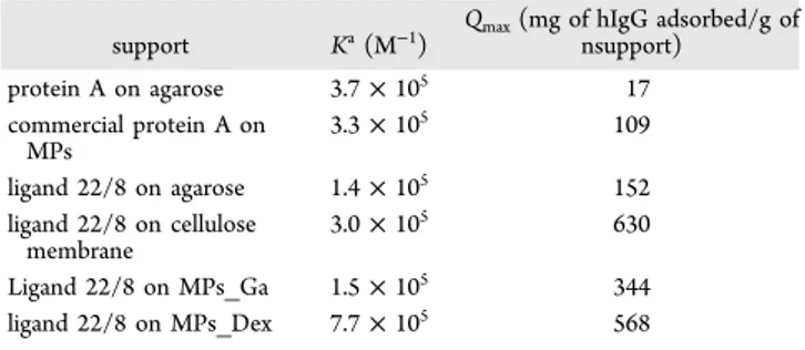
319
biopolymers can explain the different reactivity they impair to
320
the magnetic supports. Nonetheless, coating MPs with
321
biopolymers is likely to create a net of porous structures that
322
leaves reactive iron oxide partly exposed to create interactions
323
and might have some contribution in the nonspecific
324
adsorption of each support.MPs_Dex particles were further
325
on explored for hIgG purification from pure solutions, through
326
the conjugation of a synthetic affinity ligand mimicking protein
327
A, named as ligand 22/8. Three different methods for the
328
covalent attachment of the synthetic ligand onto MPs have
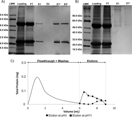
329
been tested (Figure 1). In method A, ligand 22/8 was
330
synthesized in solution-phase with a six carbon spacer. In
331
method B, ligand 22/8 was also synthesized in solution-phase
332
but without a six carbon spacer. Finally, for method C, ligand
333
22/8 was synthesized directly on the solid support. In method
334
A, there is the need to use a strong cross-linker
335
(glutaraldehydem) which can also react with amine groups
336
from neighboring particles, therefore reducing the free aldehyde
Figure 3.(A) Binding and elution of hIgG to MPs_Dex modified with ligand 22/8 (n = 2); (B) binding of BSA and hIgG to MPs_Dex modified with ligand 22/8 through Method C (n = 2); (C) reutilization of MPs_Dex modified with ligand 22/8 through Method C for binding and elution of hIgG (n = 2); and (D) binding of hIgG at the surface of MPs_Dex modified with ligand 22/8 by Method C. Representation of q (the amount of bound hIgG in equilibrium per mass of solid support) as function of Ceq(the concentration of hIgG in equilibrium). Experimental data werefitted
with the expression q = (Qmax× Ceq)/(Kd+ Ceq) for the Langmuir isotherm (OriginLab 6.1 software), where Qmaxcorresponds to the maxium
concentration of the matrix sites available to the partitioning solute (which can also be defined as the binding capacity of the adsorbent), and Kdis
the dissociation constant (n = 2).
ACS Applied Materials & Interfaces Research Article
dx.doi.org/10.1021/am301551n| ACS Appl. Mater. Interfaces XXXX, XXX, XXX−XXX
337groups available to react with the amine groups from the ligand. 338In addition, the solubility of the ligand is very poor. Method B 339is performed at high temperature (80−90 °C), at which the less 340reactive chloride of the ligand is substituted. Consequently, the 341quantity of ligand that is immobilized on the support may be 342compromised. In the case of method C, this is a multistep 343reaction where the coupling of the triazine ring is done at 0°C 344through the most reactive chloride, and therefore less likely to 345result in low reaction yields. Previous works have also shown 346that immobilization of very insoluble triazine ligands through 347direct directly on the solid support yields best results for 348protein adsorption.9
349 By analyzing the quantity of hIgG bound and eluted from the f3 350supports (Figure 3A), method A revealed to be the less suitable 351method followed by method B. Method C seems to be the best 352method to immobilize ligand 22/8 and to produce affinity
353magnetic supports toward IgG To assess the recovery of
354protein, we studied the elution buffer 50 mM glycine−NaOH, 355pH 11, because of iron leaching at acidic pH, previously 356observed.21In Method A it was not possible to quantify eluted 357protein. In method B, it was possible to elute 42 ± 1 mg of 358hIgG eluted/g of MPs which corresponds to 37% of the bound 359protein, whereas for method C, 46% of bound protein was 360eluted. As a result of these studies, MPs_Dex with ligand 22/8
361immobilized by Method C (MPs_Dex_228) appear as the
362most promising magnetic supports with a binding capacity of 363130± 5 mg of hIgG/g of MPs and a elution capacity of 60.1 ± 3640.7 mg of hIgG/g of MPs, and further studies were performed 365with this magnetic support.
366 MPs_Dex_22/8 were tested for binding to a model
367contaminant protein, bovine serum albumin (BSA), for which 368the support should not present affinity. The magnetic support 369bound 12± 2 mg of BSA/g of MP, a 10-fold lower value when 370compared to the quantity of hIgG bound (130± 5 mg of hIgG
371bound/g of MP) (Figure 3B). The regeneration and reuse
372capacity of the particles was also studied. As shown in Figure 3733C particles retain about 70% of the initial protein binding and 374elution capacity until the fifth stage of recycling. The pH 375resistance of the support was evaluated in order to assess the 376release of iron and dextran and therefore infer on eventual 377ligand leaching, which is covalently bound to the polymer. The 378total amount of dextran released after using five times the 379support, was 0.0007% of the total amount of dextran initially 380adsorbed, and during thefirst and second cycle of reutilization 381there was no dextran release. In terms of magnetite release, we 382observed that after five cycles of reutilization the support lost 3830.39% of the initial magnetite which corresponded to 19 ng of 384iron. In the first cycle of reutilization there was a leaching of 3850.09 mg/L Fe (corresponding to 0.0006% of initial iron) during 386the elution step, that is comparable with the results of Batalha 387and co-workers.21 These observations, together with the 388retention of protein attached to the support after elution and 389regeneration, can account for the loss of capacity of the support 390throughout the reutilization cycles.
391 The adsorption isotherm of human IgG on the magnetic
392support MPs_Dex_22/8 (Figure 3D) wasfitted in a Langmuir 393type isotherm and compared with data available in the literature t1 394(Table 1). The experimental adsorption values of human IgG 395on MPs_Dex_22/8 was found to be 130 mg of hIgG adsorbed/ 396g of MPs. The commercially available protein A modified MPs
397show experimental adsorption of 109 mg hIgG adsorbed/g
398MPs.37Through thefitting of the adsorption curve of hIgG, an 399affinity constant of 7.7 × 10⁴ M−1 (Ka) and a theoretical
400
maximum capacity of 568 ± 33 mg hIgG adsorbed/g MPs
401
(Qmax) were obtained with a correlation factor of 0.95.The
402
affinity constant value is in the same order of magnitude to the
403
Protein A and ligand 22/8 immobilized on different supports.
404
The Qmax value for MPs_Dex_22/8 is nearly two times higher 405
than the same ligand immobilized on MPs_GA,21 four times
406
higher than the same ligand immobilized on agarose and thirty
407
times higher than the natural Protein A immobilized on
408
agarose.7 Only the cellulose membrane revealed a higher
409
binding capacity, which was not compensated by the low
410
recovering capacity shown by this support.32
411
The magnetic support MPs_Dex_22/8 wasfinally employed
412
in the small-scale purification of an IgG monoclonal antibody
413 f4
directly from CHO cell culture supernatants (Figure 4 - A)
414
without any initial step to remove impurities. The recovery of
415
pure IgG was visible at pH 3 and pH 11, but in larger yields for
416
the latter. From 56% of total protein bound to the support,
417
there was a recovery of 5 and 16% of total protein at pH 3 and
418
11, respectively (Figure 4C). Through analysis of the 2D gel by
419
densitometry analysis with software Image J, it was estimated
420
that the loading sample contains about 60% of IgG (in terms of
421
total protein present) and that the purified IgG presents 95%
422
purity. The inertness of the MPs_Dex particles was also
423
assessed (Figure 4B) with the crude samples, showing the
424
absence of protein bound to or eluted from the support.
4. CONCLUSION
425
Iron oxide magnetic particles with a dextran coating are a
426
promising support for the magnetic separation of biomolecules,
427
because of the ease of preparation and chemical modification,
428
low cost, reduced nonspecific adsorption, and high stability. In
429
particular, the covalent attachment of a synthetic affinity ligand
430
mimicking protein A turned these particles viable for the
one-431
step recovery of IgG. Our results show that the direct synthesis
432
of the ligand on the magnetic support yielded the best
433
antibody-capturing properties. In addition, this support
434
MPs_Dex_22/8 also showed low nonspecific adsorption in
435
the presence of BSA and no major loss of capacity after five
436
cycles of protein purification. Moreover the estimated values for
437
affinity constant for ligand 22/8 were comparable with those
438
found for protein A and ligand 22/8 immobilized on different
439
adsorbents, but with the advantage of presenting considerable
440
higher maximum capacity for antibody adsorption. When
441
contacting the magnetic adsorbent with mammalian cell culture
442
supernatants rich in IgG, the MPs_Dex_22/8 supports were
443
able to purify IgG when eluting at pH11 with a purity of 95%. Table 1. Comparison of Binding Isotherm of Human IgG to Immobilized Protein A and Ligand 22/8 onto Different Supports and to Ligand 22/8 Immobilized on MPs_Dex through Method C
support Ka(M−1) Qmax(mg of hIgG adsorbed/g ofnsupport) protein A on agarose 3.7× 10⁵ 17 commercial protein A on MPs 3.3× 10⁵ 109 ligand 22/8 on agarose 1.4× 10⁵ 152 ligand 22/8 on cellulose membrane 3.0× 10⁵ 630 Ligand 22/8 on MPs_Ga 1.5× 10⁵ 344 ligand 22/8 on MPs_Dex 7.7× 10⁵ 568
dx.doi.org/10.1021/am301551n| ACS Appl. Mater. Interfaces XXXX, XXX, XXX−XXX
444
■
AUTHOR INFORMATION 445Corresponding Author446*E-mail: cecilia.roque@fct.unl.pt Tel.: +351212948385. Fax: 447+351212948550.
448Notes
449The authors declare no competingfinancial interest. 450
■
ACKNOWLEDGMENTS451The authors thank thefinancial support from Fundação para a 452Ciência e a Tecnologia through Grant PEst-C/EQB/LA0006/
4532011 and contracts no. PTDC/EBB-BIO/102163/2008,
454PTDC/EBB-BIO/098961/2008, PTDC/EBB-BIO/118317/
4552010, SFRH/BD/72650/2010 for V.L.D, and Santander
456Totta Bank− Universidade Nova de Lisboa for the Scientific
457Award 2009/2010. The authors are grateful to Dr. Abid
458Hussain and M. Telma Barroso (REQUIMTE, FCT-UNL,
459Portugal) for the preparation of the synthetic affinity ligands, to 460Lonza Biologics, U.K. (Dr. Richard Alldread), and the Animal 461Cell Technology Unit of ITQB-UNL/IBET (Dr. Paula M Alves 462and Dr. Ana Teixeira) for providing the cells and the culture 463bulks and to Mr. Filipe Cardoso and Prof. Paulo Freitas
464(INESC-MN, Lisbon, Portugal) for the help with the VSM
465measurements.
466
■
ABBREVIATIONS467MPs, oxide magnetic particles; MPs_Dex, iron oxide magnetic 468particles coated with dextran; MPs_GA, iron oxide magnetic 469particles coated with gum Arabic; MPs_Dex_22/8, iron oxide 470magnetic particles coated with dextran modified with ligand 22/ 4718; hIgG, human IgG; BSA, bovine serum albumine
472
■
REFERENCES(1)Wang, W.; Singh, S.; Zeng, D. L.; King, K.; Nema, S. J. Pharm. Sci.473 474
2007, 96, 1.
(2)Birch, J. R.; Racher, A. J. Adv. Drug Delivery Rev. 2006, 58, 671.475
(3)Roque, A. C. A.; Silva, C. S. O.; Taipa, M.Â. J. Chromatogr., A 476 477
2007, 1160, 44.
(4) González, Y.; Ibarra, N.; Gómez, H.; González, M.; Dorta, L.; 478 479
Padilla, S.; Valdés, R. J. Chromatogr., B 2003, 784, 183.
(5)Kriangkum, J.; Xu, B.; Gervais, C.; Paquette, D.; Jacobs, F. A.; 480 481
Martin, L.; Suresh, M. R. Hybridoma 2000, 19, 33.
(6) Powers, D. B.; Amersdorfer, P.; Poul, M.-A.; Nielsen, U. B.; 482 483
Shalaby, M. R.; Adams, G. P.; Weiner, L. M.; Marks, J. D. J. Immunol.
484
Methods 2001, 251, 123.
(7)Teng, S. F.; Sproule, K.; Husain, A.; Lowe, C. R. J. Chromatogr.,485 486
B: Biomed. Sci. Appl. 2000, 740, 1.
(8)Roque, A. C. A.; Taipa, M.Â.; Lowe, C. R. J. Mol. Recognit. 2005a,487 488
18, 213.
(9) Roque, A. C. A.; Taipa, M.Â.; Lowe, C. R. J. Chromatogr., A 489 490
2005b, 1064, 157.
(10) Branco, R. J. F.; Dias, A. M. G. C.; Roque, A. C. A. J. 491 492
Chromatogr., A 2012, 1244, 106.
(11) Rosa, P. A. J.; Azevedo, A. M.; Sommerfeld, S.; Mutter, M.; 493 494
Aires-Barros, M. R.; Bäcker, W. J. Biotechnol. 2009, 139, 306.
(12)Ditsch, A.; Yin, J.; Laibinis, P. E.; Wang, D. I. C.; Hatton, T. A.495 496
Biotechnol. Prog. 2006, 22, 1153.
(13) Laurent, S.; Forge, D.; Port, M.; Roch, A.; Robic, C.; Vander 497 498
Elst, L.; Muller, R. N. Chem. Rev. 2008, 108, 2064.
(14) Boyer, C.; Whittaker, M. R.; Bulmus, V.; Liu, J.; Davis, T. P. 499 500
NPG Asia Mater. 2010, 23.
(15)Dias, A. M. G. C.; Hussain, A.; Marcos, A. S.; Roque, A. C. A. 501 502
Biotechnol. Adv. 2011, 29, 142.
(16)Lu, A.-H.; Salabas, E. L.; Schüth, F. Angew. Chem., Int. Ed. 2007, 503 504
46, 1222.
(17)Dawes, C. C.; Jewess, P. J.; Murray, D. A. Anal. Biochem. 2005, 505 506
338, 186.
Figure 4.Electrophoreses gel 12.5% in denaturation conditions to verify (A) binding capacity of MPs_Dex_22/8 for IgG from a crude extract, (B) inertness of MPs_Dex for IgG. LMW (low molecular weight); loading (sample of the crude extract incubated with the adsorbent); FT (flowthrough); E1 (first elution with 50 mM glycine − HCl, pH 3); E1* (first elution with 50 mM glycine − HCl, pH 11), and (C) washes and elution profiles for IgG onto MPs_Dex_22/8. The squared and circled points represent the elution profiles at pH 11 and 3, respectively.
ACS Applied Materials & Interfaces Research Article
dx.doi.org/10.1021/am301551n| ACS Appl. Mater. Interfaces XXXX, XXX, XXX−XXX
(18)
507 Kievit, F. M.; Veiseh, O.; Bhattarai, N.; Fang, C.; Gunn, J. W.;
508Lee, D.; Ellenbogen, R. G.; Olson, J. M.; Zhang, M. Adv. Funct. Mater. 5092009, 19, 2244.
(19)
510 Dung, T. T.; Danh, T. M.; Hoa, L. T. M.; Chien, D. M.; Duc, N. 511H. J. Exp. Nanosci. 2009, 4, 259.
(20)
512 Hradil, J.; Pisarev, A.; Babič, M.; Horák, D. China Particuol.
5132007, 5.
(21)
514 Batalha, I. L.; Hussain, A.; Roque, A. C. A. J. Mol. Recognit. 2010,
51523, 462.
(22)
516 Roque, A. C. A.; Bicho, A.; Batalha, I. L.; Cardoso, A. S.; 517Hussain, A. J. Biotechnol. 2009, 144, 313.
(23)
518 Kaittanis, C.; Nath, S.; Perez, J. M. PLoS ONE 2008, 3, e3253.
(24)
519 Kaittanis, C.; Santra, S.; Perez, J. M. Adv. Drug Delivery Rev.
5202010, 62, 408. (25)
521 Heebøll-Nielsen, A.; Dalkiær, M.; Hubbuch, J. J.; Thomas, O. R. 522T. Biotechnol. Bioeng. 2004a, 87, 311.
(26)
523 Heebøll-Nielsen, A.; Justesen, S. F. L.; Hobley, T. J.; Thomas, 524O. R. T. Sep. Sci. Technol. 2004b, 39, 2891.
(27)
525 Huang, X.; Zhuang, J.; Chen, D.; Liu, H.; Tang, F.; Yan, X.;
526Meng, X.; Zhang, L.; Ren, J. Langmuir 2009, 25, 11657.
(28)
527 Diettrich, O.; Mills, K.; Johnson, A. W.; Hasilik, A.; Winchester,
528B. G. FEBS Lett. 1998, 441, 369.
(29)
529 Duan, H.; Shen, Z.; Wang, X.; Chao, F.; Li, J. World J. 530Gastroenterol. 2005, 11, 3660.
(30)
531 Massart, R. IEEE Trans. Magn. 1981, 17, 1247.
(31)
532 Leyva, A.; Quintana, A.; Sánchez, M.; Rodríguez, E. N.;
533Cremata, J.; Sánchez, J. C. Biologicals, 2008, 36,
(32)
534 Barroso, T.; Temtem, M.; Hussain, A.; Aguiar-Ricardo, A.; 535Roque, A. C. A. J. Membr. Sci. 2010, 348, 224.
(33)
536 Sabaté, R.; Barnadas-Rodríguez, R.; Callejas-Fernández, J.; 537Hidalgo-Álvarez, R.; Estelrich, J. Int. J. Pharm. 2008, 347, 156.
(34)
538 Williams, D.; Gold, K.; Holoman, T.; Ehrman, S.; Wilson, O. J. 539Nanopart. Res. 2006, 8, 749.
(35)
540 Xu, X. Q.; Shen, H.; Xu, J. R.; Xu, J.; Li, X. J.; Xiong, X. M. Appl.
541Surf. Sci. 2005, 252, 494.
(36)
542 Gupta, A. K.; Wells, S. IEEE Trans. Nanobiosci. 2004, 3, 66.
(37)
543 Borlido, L.; Azevedo, A. M.; Roque, A. C. A.; Aires-Barros, M. R.
544J. Chromatogr., A 2011, 1218, 7821.
dx.doi.org/10.1021/am301551n| ACS Appl. Mater. Interfaces XXXX, XXX, XXX−XXX