Evaluation of Listeria monocytogenes biofilm formation:
Comparison between persistent and sporadic strains
Tomás Albuquerque de Miranda
Dissertation for Master Degree in
Food Engineering – Food Quality and Safety
Supervisor (s): Professor Dr. Maria Luísa Lopes de Castro e Brito
Dr. Paula Cristina Branco Cabrita Cunha
Júri:
Presidente: Doutora Fernanda Maria dos Reis Torroaes Valente, Professora Auxiliar, Instituto Superior de Agronomia da Universidade de Lisboa
Vogais: Doutora Maria Luísa Lopes de Castro e Brito, Professora Auxiliar com agregação, Instituto Superior de Agronomia da Universidade de Lisboa.
Doutora Helena Maria Frazão Rodrigues de Sousa, Professora Auxiliar Convidada, Instituto Superior de Agronomia da Universidade de Lisboa
ii Acknowledgements
It is impossible for any human being to thrive on his own, either by direct or indirect influence; the people surrounding you make you who you are. This work is no exception, being influenced and moulded by several people in every stage of its creation.
I would first like to thank Professor Luísa Brito for the opportunity and availability, letting me return to a lab that had once been my workplace. For her patience and counselling and many enjoyable hours in the workplace making me believe that there still are jobs where you can be happy.
Secondly, to Dr. Paula Cunha for her precious and indispensable aid in comprehending and applying both statistical programs used in this work, as well as solving some “human error” problems during the assays.
In third place, I would like to thank Master Ana Carla Silva for all the practical support and tips given along my thesis as well as availability to show me several new techniques required for its conclusion and to all my lab colleagues including our god given lab technicians, D. Manuela and D. Lena, for their patience showing me around (again); To my two “little ones” – Beatriz and Ana – for their help during each assay, the companionship during those late lab hours and even at holidays.
My biggest acknowledgement goes to my family, in different but nonetheless effective ways of support. To my grandparents for their belief in me and their faith that I would reach as far as I am willing to go, and especially to my grandfather who always said I would become the 2nd “doctor” in our family. To my parents for always pushing me to the limit and demanding Excellency allowing me see that I can always do better and reach further than the last time. To my brother for nagging me into finishing this cycle and stop yammering about lab work all the time. To my girlfriend for her tagging along with many late nights and endless reading hours in writing and translations, as well as giving me a warm cuddling hug whenever I needed it.
Last but not least, a big “I’ve done it” as thanks to all my friends who were responsible for my unwinding and sanity maintenance throughout this process.
iii Abstract
L. monocytogenes has proven to be persistent in several food industries, with different
environmental conditions, including nutritional availability, raising the important question: which is the major factor associated with persistence and can this be related to disinfectant susceptibility. A group of 10 persistent and 10 non persistent strains from different origins were selected and tested for biofilm forming ability using microtiter plate assay crystal violet (CV) and stainless steel coupon (SSC) methods at 25 ºC and 11 ºC for 48 h and 7 days, respectively. Susceptibility of a subset of 12 of these strains was tested by log reduction of cell enumeration from stainless steel coupons exposed to two commercial hydrogen peroxide acidic based disinfectants (P3 OXONIA and MIDIA SAN 315). Results showed no tendency between persistence traits and biofilm forming ability, even though significant differences (p<0.05) were found between both incubation temperatures. Simulation of food industry conditions (cold and nutritional stress) with biofilms on SSC also showed no relation between persistence traits and high biofilm forming ability. Susceptibility results corroborate these results by showing no significant differences in disinfectant susceptibility between persistent and non persistent strains, although P3 OXONIA with acetic and peracetic acid proved to be more effective than MIDIA SAN 315 with citric acid.
iv Resumo alargado
Listeria monocytogenes é uma bactéria gram-positiva com mobilidade a temperaturas
inferiores a 30 °C, podendo sobreviver e crescer entre 1 e 45 °C sendo a temperatura ótima 37 °C. L. monocytogenes tolera valores de pH entre 4,3 e 9,2, com o seu valor de pH ótimo perto da neutralidade.
Este patogéneo tem sido isolado de uma grande variedade de ambientes: solo, plantas e plantas em decomposição e água corrente bem como de diversos produtos transformados ou crus, tais como leite (cru ou pasteurizado), produtos lácteos, carne (bovina, suína, aves) e produtos frescos (repolho, rabanete, etc.). Como tal, o seu efeito na indústria tem uma importância considerável quer pela sua mortalidade, principalmente em grupos específicos (crianças, idosos, gestantes e imunocomprometidos), quer pelos custos associados a indemnizações, retiradas de produtos e despesas hospitalares.
Os biofilmes de bactérias patogénicas, como L. monocytogenes, são de grande relevância em segurança alimentar em várias indústrias, incluindo carne e laticínios. Embora tenha sido sugerido que características como a persistência e a susceptibilidade do agente microbiano em sistemas alimentares podem estar relacionados com a capacidade de formação e as características do biofilme, ainda faltam algumas respostas. Vários estudos têm sido realizados para avaliar a capacidade de formação de biofilme deste patogéneo, bem como a sua susceptibilidade às condições de desinfeção. O objetivo principal deste trabalho foi tentar relacionar a capacidade de formação de biofilme em diferentes condições ambientais e a susceptibilidade a desinfetantes industriais, com o carácter persistente das estirpes. A formação de biofilmes foi avaliada através de dois ensaios: um ensaio de determinação da absorvância do CV retido pelo biofilme, em placas de microtitulação a 25 °C e 11 °C, durante 48 h e 168 h respetivamente; o outro ensaio consistiu na avaliação do crescimento de biofilmes em cupões de aço inoxidável por contagem de UFC/cm2 nas mesmas condições.
Num grupo de 12 estirpes selecionadas, seis persistentes e seis não persistentes foi também avaliada a susceptibilidade a dois desinfetantes comerciais por redução logarítmica após exposição aos dois desinfetantes: P3 OXONIA e MIDIA SAN 315, respectivamente. Os resultados foram analisados estatisticamente recorrendo ao teste de normalidade e de homogeneidade de variância para verificar se os pressupostos de ANOVA eram cumpridos. Quando estes se verificavam o conjunto de dados eram então analisados através de um teste ANOVA a um fator, para determinar diferenças significativas entre médias. Quando os
v pressupostos de ANOVA não eram cumpridos, a análise dos dados era feita através de um teste não paramétrico de dados independentes para determinar também diferenças significativas entre as médias.
A avaliação da capacidade de formação de biofilmes, quer pela técnica de coloração com o cristal violeta quer pela contagem de unidade formadores de colónias SSC, não evidenciou nenhuma relação entre as características de persistência e a capacidade de formação de biofilmes, independentemente das condições de incubação. No entanto, foi possível verificar que, à temperatura de incubação mais baixa, formou-se menos biofilme e que nas condições que mais simulam a indústria as estirpes persistentes mostraram maior capacidade de formação de biofilme.
A avaliação da susceptibilidade do subgrupo de estirpes escolhido não indicou qualquer relação entre as características de persistência e a susceptibilidade aos desinfetantes. No entanto, foi claro que fatores de stresse, tais como temperaturas mais baixas, podem influenciar a susceptibilidade à desinfeção, independentemente dos traços de persistência. Cardoso (2015) combinando stresse frio e stresse nutricional mostrou que essa possibilidade é mais explicativa da menor susceptibilidade à desinfeção e a procedimentos de saneamento e, possivelmente, da persistência das estirpes contaminantes nas indústrias de alimentos.
Estas conclusões abrem a possibilidade de trabalhos futuros com vista a estudar a combinação de outros fatores de stresse com a respetiva susceptibilidade a desinfetantes comerciais de maneira a poder encontrar um ou mais fatores com influência na persistência das estirpes em ambientes industriais.
vi General index Acknowledgements ... ii Abstract ... iii Resumo alargado ... iv General index ... vi
Table and Figure indexes ... i
1. Introduction ... 2
1.1. The genus Listeria: Taxonomy and bacterium characterization ... 2
1.2. Listeriosis and epidemiology ... 3
1.3. Food associated listeriosis ... 6
1.4. Bacterial biofilms ... 8
1.5. Persistence ...11
1.6. Susceptibility and adaptation ...12
1.7. Disinfectants ...13
2. Objectives of this work ...16
3. Materials and Methods ...17
3.1. Work strain collection ...17
3.2. Disinfectant solutions ...18
3.3. Stainless steel coupons (SSC) ...18
3.4. Evaluation of biofilm-forming ability ...18
3.4.1. Crystal violet staining method ...18
3.4.2. Enumeration of viable cells on SSC ...19
3.5. Evaluation of the listericidal activity of the two disinfectants ...20
3.6. Data analysis...20
4. Results and Discussion ...21
4.1. Crystal Violet optimization assay ...21
4.2. Biofilm evaluation by the crystal violet method ...24
4.3. Biofilm evaluation on SSC ...26
4.4. Biofilm disinfectant susceptibility ...29
5. Conclusions ...31
Table and Figure indexes
Table 1 - Clinical syndromes associated with infection by Listeria monocytogenes ... 4
Table 2 – List of several listeriosis outbreaks worldwide with identified serovars ... 5
Table 3 – Food associated with listeriosis... 7
Table 4 - Listeria monocytogenes strains ...17
Figure 1 – Diagram showing the five phases of biofilm development. ...10
Figure 2 - Illustrative diagram representing different ways of dispersion ...11
Figure 3 - Biofilm-forming ability evaluated by CV in 10 non-persistent strains. ...21
Figure 4 - Biofilm-forming ability evaluated by CV in 10 persistent strains. ...23
Figure 5 - Biofilm-forming ability tested by CV at 25º C and 11º C. ...24
Figure 6– Comparison of biofilms CV results between 11 ºC and 25 ºC. ...25
Figure 7 – Biofilm-forming ability tested by SSC for 48 h at 25 ºC ...26
Figure 8 -– Biofilm-forming ability tested by SSC for 168 h (7 days) at 11 ºC ...27
Figure 9 – Comparison of SSC results between 11 º C and 25 ºC ...28
Figure 10 – Comparison of SSC results for 168 h at 11º C. ...28
Figure 11 - Log reduction results after t exposure for 5 min to P3 OXONIA0.5% (v/v) and 0.2% (v/v) on biofilms previously grown at 25 ºC in a 48 h period. ...29
Figure 12 - Log reduction results after exposure for 5 min to MIDIA SAN 315 1% (v/v) and 0.5% (v/v) on biofilms previously grown at 25 ºC in a 48 h period. ...30
2 1. Introduction
1.1. The genus Listeria: Taxonomy and bacterium characterization
The genus Listeria is placed within the Lactobacillaceae family (Ryser & Marth, 2007) and is presently composed of 17 different species: L. monocytogenes, L. seeligeri, L. ivanovii, L. welshimeri, L. marthii, L. innocua, L. grayi, L. fleischmannii, L. floridensis, L. aquatica, L. newyorkensis, L. cornellensis, L. rocourtiae, L. weihenstephanensis, L. grandensis, L. riparia, and L. booriae (Jones et al., 1979; Jacquet et al., 1992; Hartford & Sneath, 1993; Jersek et
al., 1996; Ryser & Marth, 2007; Orsi & Wiedmann, 2016).
The name Listeria has been adopted after its proposition by Pirie in 1940, since the Judicial Commission of the International Committee on Systematic Bacteriology had rejected his first denomination - Listerella (Pirie, 1940).
Morphologically, L. monocytogenes is a gram-positive small, ranging from 1 to 2 μm in length and 0.5 μm in diameter, rod-like shape bacteria with rounded ends. Cells are usually in single units but can be clustered in short chains (3-5 or more) arranged in a V or Y disposition as well as in palisades. It is considered motile showing tumbling motility due to the production of peritrichous flagella when grown below 30 ºC, due to flagelin being produced and assembled in flagella at cell surface. If grown at 37 ºC, flagelin production is reduced to residual (Gray & Killinger, 1966; Ryser & Marth, 2007).
L. monocytogenes can survive and grow at temperatures between 1 and 45 ºC, but
optimum temperature is within the range of 30 to 37 ºC (Walker & Stringer, 1987; Junttila et
al., 1988). L. monocytogenes can grow between a minimum value pH of 4.3 up to a
maximum of 9.2, with its optimum pH value at 7 (Parish & Higgins, 1989; Petran & Zottola, 1989; Buchanan & Klawitter, 1990; George & Lund, 1992) and has the ability to grow at aw values below 0.93 (Parish & Higgins, 1989; Farber et al., 1992).
L. monocytogenes is very demanding in nutritional requirements (Gray & Killinger, 1966;
Seeliger & Jones, 1986) microaerophilic and anaerobic facultative, catalase positive and oxidase negative (Ryser & Marth, 2007). The glucose degradation is done through the Embden-Meyerhof pathway either aerobic or anaerobically (Pine et al., 1989) and it possesses glucose oxidase and NADH oxidase activities (Patchett et al., 1991).
3 1.2. Listeriosis and epidemiology
The first L. monocytogenes to be isolated was from farm animals in Wales, when sheep developed symptoms of “circling disease”, the listeric encephalitis of ruminants. Listeriosis has gained economic and biological importance due to its widespread to humans and animals (Gray & Killinger, 1966; Ryser & Marth, 2007).
The characteristic clinical features of listeriosis manifested by susceptible animals include: localized encephalitis or meningo-encephalitis, in ruminants; septicaemia with more or less focal hepatic necrosis but not involving the brain, in monogastric animals and/or in young ruminants, with undeveloped and non-functional rumen; septicaemia with associated meningitis or meningo-encephalitis, in humans and septicaemia with myocardial degeneration in fouls.
L. monocytogenes has been categorized as a human pathogen since 1929, and its ubiquity
has triggered many studies conferring human listeriosis a general food borne pathology, sporadic or epidemical. In sporadic outbreaks usually can only be determined the organism responsible and not the type of food associated, since many food vehicles can be associated (Garrido et al., 2010; Jordan et al., 2015).
Although most of the infections either in animals or humans are related to L. monocytogenes, some cases of infection were also associated to L. ivanovii and L. seeligeri. The majority of
Listeria infections in humans have occurred in a specific demographic group of hosts named
YOPI – Young, Old, Pregnant and Immunocompromised - liable of being successfully treated with antibiotics, however responsible for a mortality of 20 to 30% (Ryser & Marth, 2007; Garrido et al., 2010).
Each host may develop a variety of symptoms regardless of underlying health predisposition (Table 1) even though, in a not so small number of cases, the host can simply be a carrier. Listeriosis can occur during all the gestational period but tends to have more incidences in the third trimester and symptoms tend to be non-specific as a mild flu-like illness like fever, headaches, myalgias or gastrointestinal manifestations.
4 Table 1 - Clinical syndromes associated with infection by Listeria monocytogenes
Population Clinical presentation Diagnosis
Predisposing
conditions or
circumstances
Pregnant women Fever, ±myalgias, ±diarrhea; preterm delivery; abortion; stillbirth
Blood culture±; amniotic fluid culture
Newborns
< 7 days old Sepsis, pneumonia Blood culture
Prematurity ≥ 7 days old Meningitis, sepsis Cerebrospinal fluid
culture
Non pregnant adults Sepsis, meningitis, focal infections
Culture of blood, cerebrospinal fluid or other normally sterile site
Immunosuppression, advanced age
Healthy adults Diarrhea and fever Stool culture in
enrichment broth Possibly large inoculum From Slutsker and Schuchat in Listeria, Listeriosis and Food Safety (2007)
Listeria is a facultative intracellular pathogen, able to survive macrophage absorption and
highly capable of invading non-phagocytic cells such as epithelial cells, hepatocytes and endothelial cells, whose dissemination after ingestion and phagossome formation can be done through haemolysin membrane disruption (Garrido et al., 2010).
The minimal dose required to cause clinical infection in humans is not known, regardless several authors consider it to be relatively high – at least 106 CFU/g – based on the amount of bacteria detected in food responsible for epidemic outbreaks (Low & Donachie, 1997; Garrido et al., 2010).This does not mean that low dosages are to be discarded of concern, especially if considering the high risk group – YOPI – since the infection is also dependent on the virulence of each strain.
The terms strain, type/subtype and clonal group are often confused and misused when characterizing L. monocytogenes. The definition of isolate of any bacteria is given to a pure culture obtained from a single organism (presumably), whilst strain refers to an isolate/group of isolates of the same species with phenotypic and/or genotypic characteristics. The term subtype is often used in place of the term strain but this terminology is correctly defined as a set of only common genetic or molecular characteristics (Wiedmann, 2002). Evidence has shown in several authors that only three out of the 13 serovars of L. monocytogenes are associated with the majority of human and animal infections worldwide and most of them coincidently are also often present in food as shown in table 2 (Gombas et al., 2003; Garrido
5 Table 2 – List of several listeriosis outbreaks worldwide with identified serovars
Country Year No. of
cases Serovar New Zealand 2000 31 1/2 USA 2000 30 1/2a USA 2000/01 13 4b Sweden 2001 42 1/2a Japan 2001 38 1/2b USA 2002 16 4b Canada 2002 17 4b USA 2003 12 4b United Kingdom 2003 4 1/2 Switzerland 2005 10 1/2a Czech Republic 2006 75 1/2b USA 2007 5 NA Germany 2006/07 16 4b Canada 2008 57 1/2a Chile 2008 119 NA Denmark 2009 8 NA Austria 2008 19 4b
Germany, Austria and Czech Republic 2009/10 34 1/2a NA – Non-available.
Reviewed by Garrido et al. (2010).
It is considered an outbreak if over two listeriosis cases/patients is related to the same strain of L. monocytogenes. Most of listeriosis outbreaks tend to be sporadic, making the identification of its source very difficult. Nevertheless the investigation must determine the contaminated food vehicle in question, in order to remove it from circulation and warn the consumers (Allerberger & Wagner, 2010). In order to conduct a reliable outbreak source investigation it is necessary to collect information about the etiology, food vehicle, outbreak size, duration, geographic location, setting and outcomes , account of illness, hospitalizations and deaths (Cartwright et al., 2013).
In Portugal, there is very few information of human associated listeriosis, due to the lack of monitoring system, although it is obligated to report every case to the EU. The European
Union Summary Report on Trends and Sources of Zoonoses (EFSA, 2015) include such
data along with food associated. Listeriosis in Portugal is only recently considered a mandatory report disease in the national healthcare system and a new platform was created – SINAVE – to facilitate the data registration and transmission of information (Despacho 609-A/2014, 2014).
6 According to EFSA (2008) and EFSA (2015) the last data of human associated listeriosis in Portugal is from 2004 where 38 cases where identified and, since then, no data has been officially available. Nevertheless, there is evidence of a listeriosis outbreak in Portugal between 2009 and 2012, infecting at least 30 persons and resulting in 11 deaths (Magalhães
et al., 2015).
1.3. Food associated listeriosis
Listeria has been isolated from a large variety of environments, animals and transmission
vehicles: soil, plants and decaying plants and running water (Ryser & Marth, 2007; Garrido et
al., 2010; Belessi et al., 2011; Jordan et al., 2015; Orsi & Wiedmann, 2016). Several authors
also isolated Listeria from several – processed or raw- food products such as milk (raw or pasteurized), dairy products, meat (beef, pork, poultry) and fresh produce (cabbage, radish, etc.) (Gandhi & Chikindas, 2007)
Regarding food associated with listeriosis, sampling is made in several products but it is not taken with strict control in all EU member states. Some countries have more tight control over possible risk products or industries (Table 3).
7 Table 3 – Food associated with listeriosis
Year Food (processing) Sampling No. of
cases
2008 RTE (pork) Industry 1
Soft cheese (goat and sheep; low heat treated) Industry 6
2009 RTE (pork) Industry 16
RTE (pork) Retail 61
2010
RTE (pork)t Industry 13
RTE (pork) Retail 148
Soft cheese ( goat and ewe’s milk; low heat treated) Industry 8 Hard cheese( cow’s milk; pasteurized) Industry 29
Poultry Industry 2
Poultry Retail 8
2011 Soft cheese (goat and ewe’s milk; low heat treated) Retail 17 Soft cheese (goat and ewe’s milk; low heat treated) Industry 1
2012
Soft cheese (goat and ewe’s milk; low heat treated) Industry 18 Hard cheese(ewe’s milk; low heat treated) Industry 4 Hard cheese(ewe’s milk; low heat treated) Retail 12
2013
Hard cheese(ewe’s milk; low heat treated) Industry 4
RTE (Sushi) Retail 4
RTE (pork) Industry 15
Soft cheese(cow; low heat treated) Industry 75 Soft cheese (sheep; low heat treated) Industry 8 Soft cheese (sheep; low heat treated) Retail 5
2014
RTE (fishery products) Retail 1 RTE (meat sandwich) Retail 6 Soft cheese (ewe’s milk; low heat treated) Retail 1 Soft cheese (mixed milk; low heat treated) Retail 1 Withdrawn from the European Union Summary Reports on Trends and Sources of Zoonoses (2008 -2014)
Contamination of food products with L. monocytogenes has been one of the major concerns of the food industry, costing up to several billions of dollars, due to fatality cases, patient treatment costs and loss of products (Gahan & Collins, 1991; Costerton et al., 1999; Brooks & Flint, 2008)
8 1.4. Bacterial biofilms
It has been extensively shown that, either in nature or in food systems, microorganisms tend to become attached to solid surfaces, as a preferential growth and survival state having at their disposal a range of nutrients, ions and other organic material that make this proliferation possible (Wong 1998; Poulsen 1999; Donlan 2002).
An initial mass of cells eventually becomes large enough and starts secreting structural organic polymers, allowing further organic and inorganic molecules, nutrients and even other microorganisms’ entrapment, conferring a perfect mechanism to thrive in niches like soil sediments, water and ice systems, plant and/or animal surfaces (Costerton et al., 1999; de Carvalho, 2007; Brooks & Flint, 2008).This structure is named biofilm and is defined by an assemble of microbial cells that is irreversibly associated with a surface and enclosed in a matrix of primarily polysaccharide material (Donlan, 2002).
The definition of biofilm has been subject to some controversy and disagreement between authors. Some refer it as a biologically active matrix of cells and extracellular substances, namely polysaccharides, designated as glycocalyx and associated with a solid surface (Kumar & Anand, 1998; Poulsen, 1999; Harvey et al., 2007; Brooks & Flint, 2008),others as a complex functional consortium of microorganisms attached to a surface and embedded in extracellular polymeric substances (EPS) secreted by them (Simões et al., 2010; Belessi et
al., 2011; Bridier et al., 2011; da Silva & De Martinis, 2013). There is a growing complexity of
definitions when such involve the type of structure developed, the number of species associated and phenotypic and gene expression alterations (O'Toole et al., 2000; Moltz & Matin, 2005; Srey et al., 2013) as well as a vast number of influencing factors that affect the ability and the speed of biofilm formation, among which are surface physicochemical properties, bacterial strain and serotype, flagella expression, EPS composition, pH, temperature and other culture conditions (Costerton et al., 1994; Chae & Schraft, 2000; Djordevic et al., 2002; Borucki et al., 2003; Bonsaglia et al., 2014; Mai & Conner, 2007) It is very common to confuse biofilms with a similar bio structure – the incrustation – which consists of depositions of layers of microorganisms (living and dead) along with decomposing organic debris, without no proliferation or attachment to the solid surface (Poulsen, 1999; Donlan, 2002; Shi & Zhu, 2009). This bio-fouling can create what is known as a conditioning film, characterized by the accumulation of molecules at the solid-liquid interface on food contact surfaces which affect the biofilm formation directly but isn’t the biofilm per se (Kumar & Anand, 1998; Palmer et al., 2007; Abee et al., 2011; Pilchová et al., 2014).
9 There are a vast number of microorganisms that are able to produce biofilms although some are more prone to do it showing different formation rates and transitions from planktonic to sessile forms, such as for example the genera Pseudomononas, Enterobacter,
Flavobacterium, Alcaligenes, Staphylococcus and Bacillus (Kumar & Anand, 1998; Donlan,
2002; Chmielewski & Frank, 2003; Sauer et al., 2007). In nature, single species biofilms are rare, since a synergistic association of species is preferable, and there are no significant differences in the final biomass of single species biofilms when, compared with mixed species (Chmielewski & Frank, 2003; Sauer et al., 2007). Biofilms of contaminant microorganisms, especially if involving pathogenic bacteria such as L. monocytogenes,
Salmonella Entiritidis, Cronobacter sakasakii, Campylobacter jejuni, Bacillus cereus and Staphylococcus aureus, are top priority concerns in the food industry as evidenced ahead
(Poulsen 1999; O'Toole et al., 2000; Moltz & Matin, 2005; Shi & Zhu, 2009; da Silva & De Martinis, 2013).
In general, all authors agree that biofilm formation is a stepwise process with distinct phases and metabolic processes associated to each phase, as well as defined structures and affecting variables. One of the most exploited structural characteristic of biofilms is the EPS (extracellular polymeric substances) matrix that can comprise homo and hetero polysaccharides based on glucose, fructose, mannose, galactose and pyruvate among others, whose quantity within the matrix varies according to Gram+ or Gram – bacteria, proteins, phospholipids, teichoic and nucleic acids and, with minor incidence, mineral crystals and silt particles (Kumar & Anand, 1998; Poulsen, 1999; Shi & Zhu, 2009; Abee et al., 2011). This particular structure can perform functions that go from the facilitation of the initial attachment by mediating the adhesion of cells to the surface (if already present), to the formation and maintenance of micro colony and biofilm architecture stabilizing its structure, conferring resistance to environmental adversities such disinfectants and other biocides, desiccation, UV light, toxic metals, salinity and even turbulent flow, and entrapping free flowing nutrients, in the surrounding media and other molecules that provide extra nutrient source (Poulsen, 1999; O'Toole et al., 2000; de Carvalho, 2007; Barbosa et al., 2013; da Silva & De Martinis, 2013;)
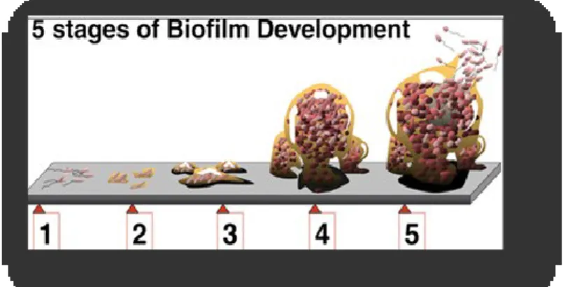
Generally biofilm formation is composed of five steps or phases: I) Initial attachment, II) Cell adhesion, III) Microcolony formation, IV) Maturation and V) Dispersion.
10 The first phase, designated by initial attachment or reversible attachment (Fig.1), consists in a passive or active adhesion to a surface, where motility seems to sets the tone (Kumar & Anand, 1998; Chmielewski & Frank, 2003).
The pre-existence of EPS, fibrils, flagella, fimbriae or pili may account to a more successful attachment in an active adhesion, as well as surface roughness and texture lead to a faster start of the second attachment phase (Poulsen, 1999; Srey et al., 2013). Some authors consider that this initial phase can only begin under certain nutrient conditions – generally a local limitation – and by a molecular conditioning film pre-attachment event that boosts adhesion while providing a source of nutrients for consequent development of the biofilm (Donlan, 2002; Brooks & Flint, 2008).
The transition to the second phase, denominated cell adhesion or irreversible attachment (Fig.1) is characterized by EPS secretion and bonding to the surface, aided or not by flagella and pili. This phase turns the cell biomass unable to be removed from the surface unless by strong shear force, enzyme, detergent, surfactant, sanitizer and/or heat application (Kumar & Anand, 1998; Shi & Zhu, 2009; da Silva & De Martinis, 2013).
Given the initial EPS production for irreversible attachment, other cellular tasks take place suchlike cell enlargement and coalescence and simultaneous growth, with continuous production of more EPS in order to stabilize and define early biofilm architecture (Shi & Zhu, 2009). This third phase in biofilm development – designated by early architecture or microcolony formation (Fig.1) – is usually defined by the hypothesis of quorum sensing events and specialized task initiation, like planktonic cell recruitment, secondary metabolite production, end product removal and even gene expression alterations (O'Toole et al., 2000; Chmielewski & Frank, 2003).
Biofilm maturation, the fourth phase (Fig.1), consists in continuous growth of cells and EPS accumulation in order to form a multilayer structure that can have different shapes specie,
Figure 1 – Diagram showing the five phases of biofilm development - 1) reversible attachment; 2) irreversible attachment; 3) early biofilm architecture; 4) maturation ; 5) dispersion.
11 strain and/or specific growth condition (Møretrø & Langsrud, 2004; Schaudinn et al., 2007; Kadam et al., 2013; Pilchová et al., 2014). In either case a communicating system of highly permeable water channels is present within the biofilm structure, whose sole purpose is to limit external diffusion to the inside of the biofilm, conferring resistance to external factors like disinfectants and other biocides and sanitizers, as well as to allow the transport of secondary metabolites within the community, to remove end products to the outer layers of the biofilm and to retrieve simple digested molecules by entrapped enzymes within the EPS matrix (Costerton et al., 1994; Kumar & Anand, 1998; O'Toole et al., 2000; da Silva & De Martinis, 2013). An oxygen gradient based cell specialization develops as well given the low permeability defined by the bulking EPS matrix.
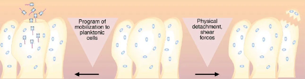
The final phase of biofilm development or, in this case, its termination is the dispersion or detachment phase (Fig.1). There are many factors that can trigger dispersion events such as starvation, the main and most common reason, the internal enzyme degradation leading to EPS or binding proteins destruction and the increase in fluid shear force that may cause structure disruption (Fig.2) (Kumar & Anand, 1998; Costerton et al., 1999; Harvey et al., 2007).
Figure 2 - Illustrative diagram representing different ways of dispersion (Costerton et al., 1999)
1.5. Persistence
The term persistence is a frequent cause of misunderstanding. Persistence has been suspected to be linked to the resistance of the strains to disinfectants. In this context, some authors state that in a situation where the extent of killing by a disinfectant applied at a bactericidal concentration is less than what is expected, persistent strains may arise (Cerf et
al., 2010); others describe it as the long-term survival capacity of pathogens in specific
environments (Carpentier & Cerf, 2011; Ferreira et al., 2014); or as the ability to survive and multiply at low temperatures. The most accepted definition states that a persistent strain is one recurrently recovered in a processing plant over a minimum of a one-year period in several samples after cleaning and disinfection (Møretrø & Langsrud, 2004; Carpentier & Cerf, 2011).
12 There has been a continuous discussion regarding biofilm formation and persistence: some researchers suggest that persistent strains show enhanced adherence or enhanced biofilm formation (Norwood and Gilmour, 2000; Lundén et al., 2003; Borucki et al., 2003), while others found no relation between biofilm formation and environmental persistence (Djordjevic
et al., 2002; Borucki et al., 2003; Chae et al., 2006). The small magnitude of differences
between persistent and sporadic strains observed values and some sporadic isolates that are able to form thicker biofilms when compared with persistent isolates, supports the hypothesis suggesting the existence of other characteristics that confer the persistent trait (Møretrø & Langsrud, 2004; Carpentier & Cerf, 2011).
The usual association between persistency and biofilm-forming ability has a number of factors that support it, like the type of strain, the culture medium and growth time used in the evaluating assay which, can also affect the response to disinfectants and cause further confusion (Lewis 2001; Møretrø & Langsrud, 2004; Carpentier & Cerf, 2011).
1.6. Susceptibility and adaptation
The term resistance is also prone to some definition problems and generally confused with tolerance, two very different concepts but easy to entwine. Resistance is defined as a strain not inactivated by a specific concentration or period of exposure to an antimicrobial agent that inactivates all other strains, or more simply, the killing effect of determined concentration was less than what expected (Bridier et al., 2011; Carpentier & Cerf, 2011). Tolerance is defined by a bacterial strain able to grow in the presence of increasing concentration of disinfectant in sub lethal doses (Lewis, 2005; Carpentier & Cerf, 2011). The most accepted term when comparing biocide efficiency is susceptibility, defined as isolates that are inhibited by the usually achievable concentrations of antimicrobial agent when the recommended dosage is used (Rodloff, et al., 2008).
Susceptibility and adaptation are usually associated with persistence; nevertheless this seems not always to be the case. Costa et al. (2016) tested a set of persistent and non persistent L. monocytogenes strains, trying to attest this relationship and concluded that it was not the case, even though only one type of disinfectants was tested, hydrogen peroxide based commercial sanitizers. These terms can also be associated with high biofilm-forming ability of the strains, assuming that better biofilm producers tend to be persistent and therefore less susceptible to disinfectants (Carpentier & Cerf, 2011).
Regarding biofilm formation and susceptibility and their conjugation, it is true that biofilms tend to be more tolerant to antimicrobials (Simões et al., 2010; Bridier et al., 2011). Some authors attest that the biofilm itself confers a range of resistance to different external
13 aggressions and conditions, including to antimicrobial compounds and show better ability to survive, being up to 1000 times less susceptible than their planktonic counterparts (Lewis 2001; Robbins et al., 2005; Pan et al., 2006; Królasik et al., 2010; Bae et al., 2012).
Biofilms do not have necessarily less susceptibility, they have a set of abilities and traits that help them avoid easy eradication by antimicrobials. These characteristics include restricted penetration of biocides within the EPS matrix, that can cause limited diffusion of molecules within the biofilm (Lewis 2001; Bridier et al., 2011), decreased growth rate within the biofilm associated with population specialization and apearance of genetic variants with resistant phenotypes (Mah & O´Toole, 2001; Królasik et al., 2010),alteration of the cell membrane composition, neutralization of toxic compounds by enzymes entraped in the EPS matrix (Mah & O´Toole, 2001; Bridier et al., 2011) and natural expression or transference of resistant genes within biofilm matrix by plasmid, transposon or integron (Chmielewski & Frank, 2003; Bridier et al., 2011).
1.7. Disinfectants
Processing plant sanitation referes to the combined effect of two processes: cleaning and disinfection, and aims to reduce microorganisms of public health importance to levels considered safe, based on established parameters, without adversely affecting either the quality of the product or its safety (Pfuntner, 2011). The cleaning process sets to remove microorganisms or the required nutrients for microbial growth, having key elements required to achieve effectiveness such as: an understanding of the type and nature of the soil to be removed, the accessibility and type of equipment, an appreciation of what the cleaning process is expected to accomplish, knowledge of the types and capacities of available cleaning agents, the establishment of an effective program carried out by knowledgeable persons. Disinfection is the destruction or irreversible inactivation of microorganism but not necesseraly their spores. In wet surfaces that provide favorable conditions for their growth this cannot be achieved only by thorough cleaning,leading to the application of sanitizers that are costly and their application can be time consuming, hence tend to be used carefully and only when absolutely necessary (Troller & Taylor, 1993).
One of the strategies used for sanitation in food processing plants are the CIP (Cleaning in Place) programs. Listeria is usually associated with several niches within a processing plant such as gaskets, conveyor belts, slicing, dicing and packing machines, containers, knives, tables, drains, floors and walls (Møretrø & Langsrud, 2004).One of the factors that affects the efficiency of these programs is the design of the equipments that must avoid having corners, junctions, crevices and every structure able to accumulate organic debris (Kumar & Anand, 1998; Simões et al., 2010; Srey et al., 2013). CIP programs are not designed to eliminate
14 biofilm formation per se but they can prevent the appearance of conditioning films (Poulsen 1999; Chmielewski & Frank, 2003; Brooks & Flint, 2008).
The choice of the sanitizer to be used must be done considering active compounds adapted to the target microorganism to eliminate, safety to food product and users, easy to use and rinsing off from surfaces (de Carvalho, 2007; Simões et al., 2010). Within the efficiency of the disinfectants some factors are important, like absence of organic material, pH, temperature of application, water hardness, chemical inhibitors, concentration and contact time (Kumar & Anand, 1998; Chmielewski & Frank, 2003; da Silva & De Martinis, 2013).
Disinfectants are different from antibiotics since they do not possess specific targets, conferring to each one a different type of action. Their usual targets in Gram+ or Gram- bacteria are the cell wall, the outer membrane, the cytosolic membrane, functional or structural proteins, DNA, RNA and other cytosolic components (Bridier et al., 2011).
The chemicals used in the food industry are of the following type: acidic compounds, aldehyde-based biocides and caustic products which include active components such as chlorine, lactic acid, peracetic acid (PAA), sodium hypochlorite, quaternary ammonium (QAC), iodine, ozone and hydrogen peroxide (Kumar & Anand, 1998; Chmielewski & Frank, 2003; Simões et al., 2010; da Silva & De Martinis, 2013).
Hypochlorites cause broad microbial mortality by damaging the outer membrane, likely producing a loss of permeability control and eventual lysis of the cell. In addition, these compounds inhibit cellular enzymes and destroy DNA. Spores, however, are resistant to hypochlorites, as the spore coat is not susceptible to oxidation except at high concentrations, coupled with long contact times at elevated temperatures (Pfuntner, 2011).
Quaternary ammonium compounds (QAC’s) are a fairly complex chemical whose action blocks the uptake of nutrients into the microbial cell and prevents the discharge of waste. QAC’s are usually odourless, nonstaining, noncorrosive and relatively nontoxic to users. They function well over a broad temperature range and a wide pH range, although activity is greater at warmer temperatures and in alkaline environments. While QAC’s tolerate small organic loads, heavy soil will decrease QAC activity significantly. Some QAC’s may not function adequately in hard water, but others are formulated with added chelating agents that allow such use (Pfuntner, 2011).
In general, QAC’s are effective against a wide range of microbes, although the spore phase is unaffected. At lower concentrations, Gram + bacteria are more sensitive to QACs than Gram - bacteria. QAC’s cannot be used in food contact surfaces without necessary rinsing after use (Chmielewski & Frank, 2003).
15 Peroxygen sanitizers are disinfectants with stabilized hydrogen peroxide and PAA. Both compounds have a broad spectrum action with bactericidal and endosporicidal elimination activity and working well at low temperatures. Hydrogen peroxide is commonly used in the food industry because it is a strong oxidant able to damage bacterial proteins, DNA and cellular membranes (Yun et al., 2012).PAA solutions can be attenuated by organic load and will begin to lose activity as the pH approaches neutrality. PAA-based sanitizers are environmentally friendly as the compounds therein break down into acetic acid, oxygen, water and are also less corrosive to equipment than hypochlorites (Pfuntner, 2011).
16 2. Objectives of this work
The work developed previously within our research group, by examining different traits of L.
monocytogenes persistent and non-persistent isolates, such as mobility, genetic expression
and biofilm forming ability, was searching for the possible relations between persistence and biofilm-forming ability. In this work a relatively large set of 10 sporadic and 10 persistent strains with high genetic diversity, enclosing seven different serovars, including serovars 4b and 1/2a, and from different geographical origins (Ireland, Norway and Portugal) as well as from different sources (salmon, broiler, cheese and milk) was used. The aim of the work was to investigate characteristics of the strains able to justify the persistence in the food industry environment. The biofilm forming ability of the strains at two different temperatures (11 ºC and 25 ºC), simulating environmental temperature and refrigeration temperature, respectively, was investigated. Similarly, clean and dirty conditions of equipment sanitization were simulated by using, for biofilm growth, culture medium with different nutrient concentration, respectively 1/10 diluted TSB and undiluted TSB. The biofilms produced in those different environmental conditions were tested for susceptibility to two hydrogen peroxide/acidic based commercial disinfectants.
These results will complement other ongoing research aiming to identify targets for persistence in L. monocytogenes.
17 3. Materials and Methods
3.1. Work strain collection
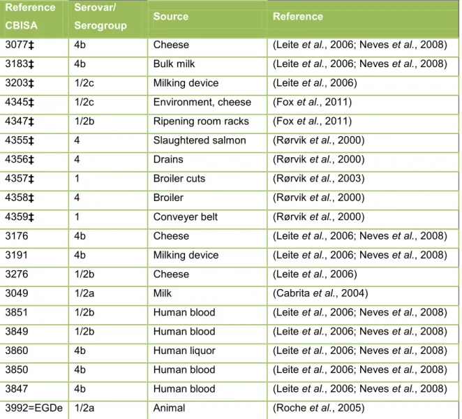
The 20 L. monocytogenes strains used in this work were from the Coleção de Bactérias do Instituto Superior de Agronomia (CBISA) and are described in table 4. Strains withdrawn from – 80 ºC were streaked onto TSA-YE (Tryptone Soy Agar with Yeast Extract) and grown overnight at 37 ºC. A work collection was maintained in semi-solid TSA-YE in cryogenic microtubes at 4 ºC until use.
Table 4 - Listeria monocytogenes strains
Reference CBISA
Serovar/
Serogroup Source Reference
3077‡ 4b Cheese (Leite et al., 2006; Neves et al., 2008)
3183‡ 4b Bulk milk (Leite et al., 2006; Neves et al., 2008)
3203‡ 1/2c Milking device (Leite et al., 2006)
4345‡ 1/2c Environment, cheese (Fox et al., 2011)
4347‡ 1/2b Ripening room racks (Fox et al., 2011)
4355‡ 4 Slaughtered salmon (Rørvik et al., 2000)
4356‡ 4 Drains (Rørvik et al., 2000)
4357‡ 1 Broiler cuts (Rørvik et al., 2003)
4358‡ 4 Broiler (Rørvik et al., 2000)
4359‡ 1 Conveyer belt (Rørvik et al., 2000)
3176 4b Cheese (Leite et al., 2006; Neves et al., 2008)
3191 4b Milking device (Leite et al., 2006; Neves et al., 2008)
3276 1/2b Cheese (Leite et al., 2006)
3049 1/2a Milk (Cabrita et al., 2004)
3851 1/2b Human blood (Leite et al., 2006; Neves et al., 2008)
3849 1/2b Human blood (Leite et al., 2006; Neves et al., 2008)
3860 4b Human liquor (Leite et al., 2006; Neves et al., 2008)
3850 4b Human blood (Leite et al., 2006; Neves et al., 2008)
3847 4b Human blood (Leite et al., 2006; Neves et al., 2008)
3992=EGDe 1/2a Animal (Roche et al., 2005)
18 3.2. Disinfectant solutions
Two commercial disinfectants commonly used in dairies were used in the susceptibility assays. The active ingredients of the first compound - P3-Oxonia active (ECOLAB S.r.l.) - are hydrogen peroxide, acetic and peracetic acid, while the second contains hydrogen peroxide and citric acid - MIDIA SAN 315(Christeyns Food Hygiene S.r.l.).
All disinfectants were diluted in hard water (magnesium chloride, calcium chloride and sodium bicarbonate, pH 7 ± 0.2, prepared according to EN 13697 (European Standard 2001), to achieve the concentrations indicated by the manufacturer. Disinfectant concentrations used were 1% (v/v) and 0.5% (v/v) for MIDIA SAN 315 (MS) and for 0.5% (v/v) and 0.2% (v/v) for P3 Oxonia active (P3).
Engley Neutralizing Broth (D/E) was used in disinfectant testing assays. The Dey-Engley Neutralizing Broth neutralizes a broad spectrum of antiseptics and disinfectants including, quaternary ammonium compounds, phenolics, iodine, chlorine, mercurials, formaldehyde, glutaraldehyde, alcohol, peroxides and acetic and lactic acids.
3.3. Stainless steel coupons (SSC)
Stainless steel coupons (1 x 1 cm) type 316 finish 4b (University of Georgia instrument shop, Athens) were cleaned in acetone to remove grease, rinsed in distilled water and immersed in a phosphoric-acid-based cleaner (CIP 200, Steris Corp., Mississagua, Ontario, Canada) at room temperature for 20 min. The coupons were rinsed again and sterilized individually by autoclaving in test tubes.
3.4. Evaluation of biofilm-forming ability 3.4.1. Crystal violet staining method
For the evaluation of the biofilm-forming ability of the tested strains the crystal violet (CV) method used was based in Djordjevic et al. (2002) with modifications. This methodology is based on the ability that the crystal violet stain has to enter the biofilm. The subsequent extraction of the dye with ethanol, allows its measurement by spectrophotometry.
Five isolated colonies, from 37 ºC overnight plates, were suspended in 5 ml of liquid Tryptone Soy Broth with Yeast Extract (TSB-YE) and incubated at 37 ºC overnight. From this cell suspension 1 ml was added to a 4 ml of fresh TSB-YE media and spun in a vortex for 15 seconds.
A volume of 150 μL from this cell suspension was inoculated in each well of a P96 microtiter plate completing a whole column and one column was the negative control where only 150
19 μL of fresh TSB-YE medium was inoculated. This micro plate was sealed with Parafilm to prevent evaporation and incubated at 25 ºC for 24, 48, 72, 96, 144 and 192 h and at 11 ºC for 7 days (MIR 154-PE cooled incubator, Panasonic, Japan) with a 150 rpm agitation speed (MIS100 Shaker, Panasonic, Japan) to simulate the turbulent flow. For the assay regarding 96, 144 and 192 h the culture medium was refreshed every 48 h, from the 72h incubation period.
After the incubation period the growth medium was drained and the plate rinsed with Ringer’s Solution (RS). The microplate was dried in an inverted position for 30 minutes. Staining was performed with 50 μL of CV solution at 0.1% (v/v) and left to stain for 45 min. The plate was again rinsed with RS and left to air dry in inverted position for another 30 min. Finally 200 μL of 95% ethanol (v/v) was added to each well and left to solubilise for 30 min at 4 º C, in order to avoid alcohol evaporation. From each well, 100 μL was transferred to a new P96 microtiter plate and then the absorbance was read at 600 nm (680 Microplate Reader, BIORAD, Germany). At least three biological replicates were performed, with eight technical replicates each.
3.4.2. Enumeration of viable cells on SSC
Biofilms were formed on 1x1 cm stainless steel coupons according to Costa et al.(2016), as follows: biofilms were grown on stainless steel coupons in P24 microplates (Orange Scientific, Braine-l’Alleud, Belgium). To each well 1.5 ml of inocula was added. Controls with non inoculated coupons were always present. The P24 microplate was sealed with Parafilm (Bemis, USA) and incubated at 25 ºC for 48 h without agitation.
Once the incubation period was over, each coupon was rinsed by pipetting 1 ml of RS on both surfaces, in order to remove all planktonic cells and the coupon was placed inside the respective well in a new P24 plate.
The new microplate had a 20 sterile glass bead (ø= 3 mm) layer per well. The coupon was placed on the top of this layer and 30 sterile glass beads and 1 ml of RS were added in this order. The plate was then sealed with Parafilm and stirred in a Microplate vortex (Tittertek DSG, Flowlabs, Germany) for 1 min, in order to remove the biofilm from the coupon surfaces. From each well, a serial decimal dilution was performed, in order to inoculate TSA-YE plates. Incubation proceeded overnight at 37 ºC, for CFU count assessment. At least three biological replicates were performed with two technical replicates each
20 3.5. Evaluation of the listericidal activity of the two disinfectants
The biofilms for this assay were grown as described previously and P24 plates prepared, stepwise, until RS coupon rinsing was done. Disinfectants were used in the two different concentrations, 1% (v/v) and 0.5% (v/v) for MS 315 and 0.5% (v/v) and 0.2% (v/v) for P3, the SSC were immerged for 5 min.
After this exposure period, each coupon was rinsed with RS, and transferred to another P24 plate where each well had a 20 glass bead layer in the bottom. A set of 30 glass beads was put on the top of each coupon and 1 ml of D/E (Difco, Becton Dickinson; Heidelberg, Germany) was added for a period of 5 min of contact.
After the neutralization step, the plate was sealed with Parafilm and agitated in a Microplate vortex for one minute, to remove biofilm cells from the coupon surfaces. Afterwards, from each well (sanitized or control) a decimal dilution series was performed. One hundred μL of each dilution was spread onto TSA-YE and left overnight at 37 ºC for CFU count assessment.
The treatment was considered effective if a 4 log reduction (difference between log of CFU/cm2 of SSC not exposed and exposed to disinfectant exposed) was observed. At least two biological replicates were performed with two technical replicates each
3.6. Data analysis
For every assay, statistical analysis was conducted based on the principle that each value was part of a continuous distribution of the data and, if so, variance should follow a homogeneous behaviour, being the necessary conditions to perform a one-way factor ANOVA with the Tukey test revealing statistical differences between strains.
When the data did not comply with the normality or the homogeneous distribution of the variance, then the non parametric Kruskal–Wallis median test was used to evaluate statistical differences between strains.
Normality and variance equality tests were performed in MiniTab17 (Minitab, Inc., Pennsylvania, USA) and ANOVA and Non Parametric Kruskal-Wallis test were performed using STATISTICA 8 (Statsoft Inc., Tulsa, USA).
21 4. Results and Discussion
4.1. Crystal Violet optimization assay
In order to optimize the incubation period for the crystal violet (CV) method, biofilm formation was evaluated at 25 °C for 24, 48, 72, 96, 144 and 192 hours in sporadic and persistent strains.
Figure 3 - Biofilm-forming ability evaluated by the CV method in a group of 10 non-persistent strains incubated for different time periods at 25 ºC.
0.000 0.200 0.400 A b s 60 0 n m Time (h)
3049
0.000 0.200 0.400 A b s 60 0 n m Time (h)3176
0.000 0.200 0.400 A b s 60 0 n m Time (h)3191
0.000 0.200 0.400 A bs 60 0 n m Time (h)3276
0.000 0.200 0.400 A b s 60 0 n m Time (h)3847
0.000 0.200 0.400 A b s 60 0 n m Time (h)3849
0.000 0.200 0.400 A bs 60 0 n m Time (h)3850
0.000 0.200 0.400 A b s 60 0 n m Time (h)3851
0.000 0.200 0.400 A b s 60 0 n m Time (h)3860
0.000 0.200 0.400 A b s 60 0 n m Time (h)3992
22
Error bars represent standard deviation. Different letters in the columns indicate significant differences (P<0.05) in average values. Equal letters in the columns indicate no significant differences (P>0.05) in average values.
The results obtained for the non-persistent strains (Fig. 3) showed that except for strain 3992, that showed significant increases (P<0.05) in biofilm formation till the 72 h, for the majority of the non-persistent strains the incubation time did not affect significantly (P>0.05) the biofilm formation.
Even though some strains such as 3176, 3191 3276 and 3850 reveal higher absorbance values at 96 h than at 48 h this may be due to medium renewal after 72 h (see 3.4.1), causing biomass augment which stabilizes from there on.
The results obtained for the persistent strains (Fig. 4) were similar to the ones obtained for the majority of the non-persistent strains. Except for strains 3203 and 4356 that show significant more biofilm formation at 96 h but as before can be associated to medium renewal once their ability stabilizes until 192 h of incubation. Overall, the results obtained after 48 hours of incubation suggest that, in the conditions of this assay, this incubation period is adequate to obtain a mature biofilm as described by Harvey et al. (2007) that in a 14 day assay of biofilm –forming evaluation concluded biofilm production to stabilize after 40 h.
23
Figure 4 - Biofilm-forming ability evaluated by CV method in a group of 10 persistent strains incubated for different time periods at 25 ºC.
Error bars represent standard deviation. Different letters in the columns indicate significant differences (P<0.05) in average values. Equal letters in the columns indicate no significant differences (P>0.05) in average values.
0.000 0.200 0.400 A bs 60 0 n m Time (h)
3077
0.000 0.200 0.400 A b s 60 0 n m Time (h)3183
0.000 0.200 0.400 A b s 60 0 n m Time (h)3203
0.000 0.200 0.400 A b s 60 0 n m Time (h)4345
0.000 0.200 0.400 A b s 60 0 n m Time (h)4347
0.000 0.200 0.400 A b s 60 0 n m Time (h)4355
0.000 0.200 0.400 A b s 60 0 n m Time (h)4356
0.000 0.200 0.400 A b s 60 0 n m Time (h)4357
0.000 0.200 0.400 A b s 60 0 n m Time (h)4358
0.000 0.200 0.400 A b s 60 0 n m Time (h)4359
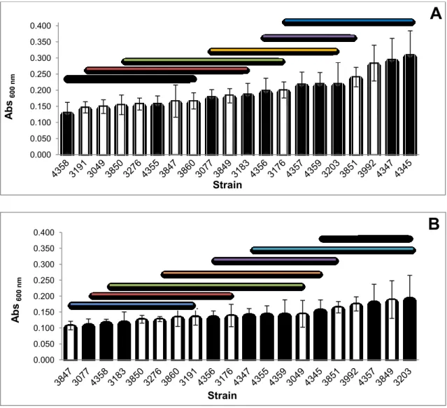
24 4.2. Biofilm evaluation by the crystal violet method
After establishing that an incubation period of 48 h was adequate for this methodology, the tested strains showed differences in biofilm-forming ability after this period of incubation at 25 ºC (Fig. 5A). Nevertheless, though several groups were formed, there was no strict tendency associating the persistent character of the strains with a greater biofilm-forming ability, as was previously suggested by Djordjevic et al. (2002), Borucki et al. (2003) and Chae et al. (2006).
The results obtained at 11 ºC (Fig. 5B), showed similar behaviour in terms of good and bad biofilm-forming ability, independently of the persistent trait of the strains. Therefore setting the incubation temperature to 11 ºC or to 25 ºC, results in no tendency relating biofilm-forming ability and persistence (Fig.5).
Figure 5 - Biofilm-forming ability tested by CV staining. Biofilms were grown for 48 h at 25 ºC (A) and for 168 h at 11 ºC (B).
Dark bars represent persistent strains, white bars represent non-persistent strains. Error bars represent standard deviation. The lines over the bars group strains with no significant differences among them (p>0.05).
0.000 0.050 0.100 0.150 0.200 0.250 0.300 0.350 0.400 A b s 60 0 n m Strain
A
0.000 0.050 0.100 0.150 0.200 0.250 0.300 0.350 0.400 A b s 60 0 n m StrainB
25 Under cold and environment temperatures, persistent strain 4358 and non persistent strains 3191, 3847 and 3860 were always among the low biofilm producer cluster. In opposition, persistent strains 3203, 4345 and 4357 as well as non persistent strains 3851 and 3992 were always in the high biofilm producer’s cluster in both conditions. Even though these results help to confirm the lack of a possible association of biofilm-forming ability and persistence, they also reveal that a higher number of persistent strains were found in both conditions within the high forming ability cluster (Fig.5).
After incubation at 25 ºC the absorbance values registered were between 0.09 and 0.38, similar to results found in Lourenço et al. (2012), whereas for incubation at 11 ºC absorbance values were in the range of 0.08 to 0.32 (Fig.5) also similar to the results published by Nilsson et al. (2011) revealing high plasticity in the biofilm-forming ability among non persistent and persistent strains.
Figure 6– Comparison CV results between biofilms grown at 11 ºC ( ) and at 25 ºC ( ). Statistical significant differences (P<0.05) are marked with [*].
The comparison of biofilms produced in both conditions indicated that 14 (seven persistent and seven non persistent) out of the 20 strains produced significantly more (p<0.05) biofilm at 25 ºC (Fig. 6).This may suggest that some strains may be persistent in particular industries due to other traits triggered by specific temperature conditions of the production facilities. In fact, the number of persistent and non persistent strains that showed statistical differences between the two assays was the same. The same was supported by the results of Kadam et
al. (2013). * * * * * * * * * * * * * * 0.000 0.100 0.200 0.300 0.400 A bs 60 0 n m Strains
26 4.3. Biofilm evaluation on SSC
The SSC assay was performed in order to simulate the food industry systems, where cell deposition is made on stainless steel surfaces and microorganism have different nutritional availabilities as well as growth temperatures. When comparing the biofilm-forming ability of the strains at the two growth temperatures, no tendency was found between biofilm-forming ability and the persistent character of the strains.
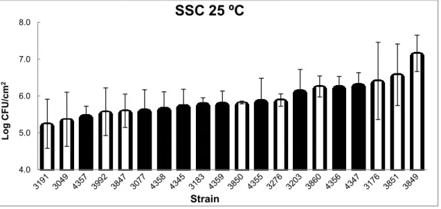
Figure 7 –Biofilm-forming ability tested with SSC with biofilms grown for a period of 48 h at 25 ºC.
Black bars represent persistent strains, white bars represent non-persistent strains. Error bars represent standard deviation.
At 25 ºC in TSB-YE medium, no significant differences were observed between persistent and non-persistent strains as previously reported by Nilsson et al. (2011). At this temperature, the three strains that showed higher values of log CFU/cm2 were all non persistent strains, as previously reported by Costa et al. (2016) where non persistent strains also showed higher CFU counts. In opposition, the three strains that showed lower values of log CFU/cm2 were comprised of persistent and non persistent strains.
4.0 5.0 6.0 7.0 8.0 Lo g CFU/c m 2 Strain
SSC 25 ºC
27
Figure 8 -– Biofilm-forming ability tested with SSC with biofilms grown for a period of 168 h (7 days) at 11 ºC.
Black bars represent persistent strains, white bars represent non-persistent strains. Error bars represent standard deviation. The lines over the bars group strains with no significant differences among them (p>0.05).
Since in the food industries, environmental temperatures are low temperatures (between 4 and 12º C) the SSC assay was also performed at 11 ºC. The incubation time used was, seven days, based on previous results from Moltz et al. (2004), Robbins et al. (2004) Nilsson
et al. (2011) and Lourenço et al. (2014). At 11 ºC two significantly different clusters were
formed, none of them being solely composed of persistent or of non persistent strains. Nevertheless, one of the clusters gathered all the non persistent strains.
In this condition three of the strains of the better biofilm former cluster were persistent as, in opposition; three of the strains of the lower biofilm former cluster were non persistent strains.
4.0 5.0 6.0 7.0 8.0 Lo g CFU/c m 2 Strain
SSC 11 ºC
28
Figure 9 – Comparison of biofilm-forming ability SSC results between biofilms grown at 11 º C ( ) and 25 ºC ( ).
‡ indicates persistent strains. Error bars represent standard deviation. Statistical significant differences (p<0.05) are marked with [*].
When comparing the two growing temperatures (Fig. 9), five (strains 3203, 4345, 4357, 4358 and 4359) out of 10 persistent strains showed significant differences in their biofilm-forming ability. Of the total six strains showing differences, only one is from human origin and all the others are from food (foodstuff or premises). All of them showed higher biofilm-forming ability at the lower temperature (11 ºC) giving strength to the hypothesis of Nilsson et al. (2011) that food isolates form more biofilm at lower temperatures.
Figure 10 – Comparison of biofilm-forming ability results tested by SSC with biofilm grown for a period of 168 h at 11º C.
Dark bars represent TSB-YE medium and light bars represent 1:10 TSB-YE diluted medium.
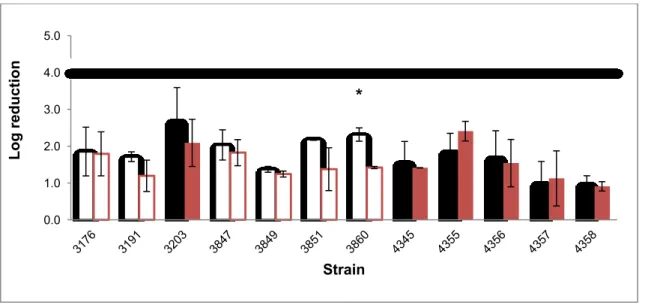
‡ indicates persistent strains. Error bars represent standard deviation. Statistical significant differences (p<0.05) are marked with [*]Results of diluted medium are from Cardoso (2015).
* * * * * * 4.0 4.5 5.0 5.5 6.0 6.5 7.0 7.5 8.0 L o g CF U/cm 2 Strain
SSC - Temperature comparison
* * * * * * * 0.0 1.0 2.0 3.0 4.0 5.0 6.0 7.0 8.0 L o g CF U/cm 2 Strain29 In order to test biofilm performance, besides low temperature, a second stress factor was added to the assay: strains were grown in 1:10 diluted TSB-YE medium, simulating a nutritional deficient environment associated with good cleaning procedures. When comparing at 11 ºC the two growing medium (TSB-YE and 1:10 TSB-YE) seven out of 20 strains showed significantly more biofilm production in richer medium. From this significantly different group three strains were non persistent and four persistent. Once more the results suggest a lack of trend between persistence and response to cold and nutritional stress. This was also observed in Costa et al. (2016).
4.4. Biofilm disinfectant susceptibility
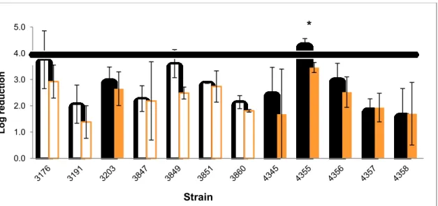
The persistent character of the strains is, as referred previously, usually associated with the ability to become less susceptible to disinfectants eventually due to their higher biofilm-forming ability. Other authors have tested different disinfectants to control L. monocytogenes. Six persistent and six non persistent strains were selected based on the biofilm forming ability results in order to include, in each group, three of the better biofilm producers and three of the worst biofilm producers. These subsets of strains include strains with different origins such as dairy, dairy facilities, fish facilities, meat and humans.
Figure 11 - Log reduction after exposure for 5 min to P3 OXONIA 0.5% (v/v)( ) and 0.2% (v/v)( ) of biofilms grown at 25 ºC in a 48 h period.
Line represents effective disinfection action (reduction >4 Log). Persistent strains are represented with full bars and non persistent strains with white bars. Statistical differences (P<0.05) between the two concentrations are marked with a [*].
Overall there was no tendency on the association of disinfectant effectivness and the persistent character of the strains (Fig.11). Non persistent and persistent strains showed similar behaviours when exposed to the two concentrations of P3. Persistent strain 4355 showed to be significantly affected by the higher concentration of P3, resulting in this case in an effective reduction (> 4 Log).