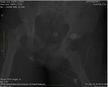
Is It Safe to Perform an Early Arthroscopy After a Traumatic Hip Dislocation With an Associated Pelvic Ring Injury? Report of Our Technique
6
0
0
Texto
Imagem


Documentos relacionados