TINNITUS, BIOMARKERS AND QUALITY OF LIFE IN AN
OLDER POPULATION
HAÚLA FARUK HAIDER
Thesis for the degree of Doctor in Medicine in Specialty of Otorhinolaryngology
in Nova Medical School | Faculty of Medical Sciences
TINNITUS, BIOMARKERS AND QUALITY OF LIFE IN AN
OLDER POPULATION
Haúla Faruk Haider Supervisors name: João C. Paço MD, MSc, PhD, Agg, Prof Catedrático FCML,
Luís M. Borrego, MD, MSc, PhD, Agg, FCML , Maria Helena Caria MSc, PhD, Prof Coordinator, Agg ESS/IPS, Prof Invited FCUL, Nuno Trigueiros MD, MSc, PhD, Agg
Thesis for obtaining the degree of Doctor in Medicine in Specialty of Otorhinolaryngology
The protocol of this PhD Thesis was approved by the Ethics Committee of the Hospital Cuf Infante Santo and the NOVA Medical School, Faculdade de Ciências Médicas and
Publications
This thesis contains partially or totally data/or methodologies published in the following peer reviewed articles or book chapters:
Hall, D. A., Szczepek, A. J., Kennedy, V., & Haider, H. (2015). Current-reported outcome domains in studies of adults with a focus on the treatment of tinnitus: protocol for a systematic review. BMJ open, 5(11), e009091.
Haider, H., Fackrell, K., Kennedy, V., & Hall, D. A. (2016). Dimensions of tinnitus-related complaints reported by patients and their significant others: protocol for a systematic review. BMJ open, 6(10), e0091.
Hall, D. A., Haider, H., Szczepek, A. J., Lau, P., Rabau, S., Jones-Diette, J., ... & Fuller, T. (2016). Systematic review of outcome domains and instruments used in clinical trials of tinnitus treatments in adults. Trials, 17(1), 270.
Haider H.F. (2016). Tinnitus therapy in Ménière’s disease. In: Indica. Learning to treat and live with Ménière’s disease, 1st edição, 193-208.
Haider, H. F., Hoare, D. J., Costa, R. F., Potgieter, I., Kikidis, D., Lapira, A., ... & Paço, J. C.
(2017). Pathophysiology, diagnosis and treatment of somatosensory tinnitus: A scoping review. Frontiers in neuroscience, 11, 207.
Fuller, T. E.*, Haider*, H. F., Kikidis, D., Lapira, A., Mazurek, B., Norena, A., ... & Brueggemann, P. G. (2017). Different teams, same conclusions? A systematic review of existing clinical guidelines for the assessment and treatment of tinnitus in adults. Frontiers in psychology, 8, 206.
Haider, H. F., Flook, M., Aparicio, M., Ribeiro, D., Antunes, M., Szczepek, A. J., ... & Caria,
H. (2017). Biomarkers of presbycusis and tinnitus in a Portuguese older population. Frontiers in aging neuroscience, 9, 346.
Haider H. F., Oliveira V., & Trigueiros N. (2017). Chronic subjective Tinnitus: diagnosis and treatment, CírculoMédico. In: Audiologia, Som e Audição das bases à clínica. 1st edition.
Fackrell, K., Smith, H., Colley, V., Thacker, B., Horobin, A., Haider, H. F., ... & Hall, D. A. (2017). Core Outcome Domains for early phase clinical trials of sound-, psychology-, and pharmacology-based interventions to manage chronic subjective tinnitus in adults: the COMIT’ID study protocol for using a Delphi process and face-to-face meetings to establish consensus. Trials, 18(1), 388. Hall, D. A.*, Fackrell, K.*, Li, A. B., Thavayogan, R., Smith, S., Kennedy, V., Tinoco, C.,
Rodrigues, E. D., Campelo, P., Martins, T. D., Lourenço, C. M., Ribeiro, D., &
Haider, H. F.* (2018). A narrative synthesis of research evidence for
tinnitus-related complaints as reported by patients and their significant others. Health and quality of life outcomes, 16(1), 61.
Michiels, S., Ganz Sanchez, T.*, Oron, Y.*, Gilles, A.*, Haider, H. F.*, Erlandsson, S., ... & Oiticica, J. (2018). Diagnostic Criteria for Somatosensory Tinnitus: A Delphi Process and Face-to-Face Meeting to Establish Consensus. Trends in Hearing, 22, 2331216518796403.
The thesis contains partially or totally the following data/ or methodologies that are submitted:
Haúla Haider, Diogo Ribeiro, Helena Caria, Nuno Trigueiros, Luís Miguel Borrego, Agnieska J Szczepek, Ana Luisa Papoila, Asma Elarbed, Sandra Smith, João Paço, Derek J Hoare. Evidence for biological markers of tinnitus: A systematic review.
Submitted in Trends in Hearing
Haúla F. Haider, Tijana Bojić, Sara F. Ribeiro, João C. Paço, Deborah A. Hall, Agnieszka J Szczepek. Pathophysiology of subjective tinnitus: triggers and maintenance.
Submitted to Frontiers in Neuroscience
Emily J. Watts, Kathryn Fackrell, Sandra Smith, Jacqueline Sheldrake, Haúla Haider, Derek J. Hoare. Why is Tinnitus a Problem? A Qualitative Analysis of Problems Reported by Tinnitus Patients. Submitted to Trends in Hearing
Haúla F. Haider, Diogo Ribeiro, Sara F. Ribeiro, Nuno Trigueiros, Helena Caria, Luís Miguel Borrego, Iola Pinto, Ana L. Papoila, Derek J Hoare, João Paço. Audiological markers of tinnitus in an older Portuguese population. Submitted to Trends in
Hearing.
Haúla F. Haider, Sara F. Ribeiro, Vasco Oliveira, Nuno Trigueiros, Derek J Hoare, Luis Miguel Borrego, João Paço. Characteristics, psychological problems, quality of life, and tinnitus in a Portuguese Older Population. Submitted to Trends in
Hearing.
Haúla F. Haider, Sara F. Ribeiro, Catarina Martins, Diogo Ribeiro, Nuno Trigueiros, Agnieszka J. Szczepek, Helena Caria, Derek J. Hoare, João Paço, Luís Miguel Borrego. Tinnitus, hearing loss and inflammatory processes in an older Portuguese population. Submitted to Journal of the Association for Research in
Otolaryngology.
Hall, D.A., Hibbert, A., Smith, H., Haider, H.F., Londero, A., Mazurek, B., & Fackrell K. One size does not fit all: developing common standards for outcomes in early-phase clinical trials of sound-, psychology-, and pharmacology-based interventions to manage chronic subjective tinnitus in adults. Submitted to Trends in Hearing. *Equal contributions
“In life there is nothing to fear, but to understand.”
(Marie Curie)
“What we know is a drop; What we do not know is an ocean.”
Sumário
O acufeno é um sintoma referente à perceção de um som nos ouvidos ou na cabeça, sem que exista um estímulo acústico externo correspondente. Está presente em diferentes patologias (otológicas ou não) e tem um impacto importante na qualidade de vida da pessoa afetada. Atualmente, o seu diagnóstico e monitorização são baseados em medidas subjetivas audiométricas e psicométricas, sendo que não existem métodos objetivos para a identificação do acufeno. Além disso, os mecanismos fisiopatológicos subjacentes ao acufeno subjectivo permanecem desconhecidos.
O objetivo da presente tese é estudar os mecanismos subjacentes ao acufeno subjectivo e a sua relação com a surdez, visto que a surdez é a co-morbilidade mais frequentemente associada ao acufeno. Pretende-se também avaliar a contribuição dos fatores genéticos, audiológicos e imunológicos na etiologia do acufeno.
Para isso, foram realizadas revisões sistemáticas (RS) sobre esta temática de forma a conhecer o estado de arte, primeiramente em relação à forma como os pacientes e os familiares percecionam o acufeno e também sobre os ensaios clínicos existentes acerca da eficácia do tratamento do acufeno. Ambas as RS contribuíram para a identificação de um conjunto de domínios relacionados com o acufeno, usado pelo COMIT’ID (Core outcome measures in tinnitus international Delphi), num método de consensos Delphi, baseado na Internet, com o objetivo de identificar um ‘Core Outcome Set’ (ou seja definir quais as queixas relacionadas com o acufeno que são imprescindíveis para a sua avaliação) recomendado para ensaios clínicos de eficácia terapêutica para o acufeno assim como para o seu diagnóstico. Estas recomendações são específicas para as três modalidades terapêuticas principais: sonora, psicológica e farmacológica uma vez que cada modalidade tem fundamentos específicos e por isso visam avaliar diferentes aspetos do acufeno.
Com o objetivo de contribuir para a padronização da avaliação e do tratamento clínico do acufeno, foi constituída a TINNET, uma rede europeia para a investigação científica do acufeno. Considerando o objetivo do presente estudo e a hipótese de integrar esta rede europeia, foram desenvolvidas um conjunto de atividades que em muito contribuíram para o conhecimento sobre o acufenos. Entre as diferentes atividades realizadas com o apoio da TINNET destaca-se a realização de uma revisão sistemática sobre as ‘guidelines’ clínicas existentes para o diagnóstico e tratamento do acufeno. Esta revisão foi uma das bases que conduziu ao desenvolvimento das ‘guidelines’ europeias multidisciplinares para o acufeno: diagnóstico, avaliação e tratamento. Estas ‘guidelines’ foram apresentadas na conferência final do TINNET e estão atualmente em fase de disseminação.
Outro foco de interesse da presente tese foi a realização de trabalhos de revisão sobre o acufeno somatosensorial (nomeadamente sobre a fisiopatologia, diagnóstico e
tratamento), bem como a participação num grupo de consenso internacional sobre o diagnóstico deste subtipo do acufeno, de forma a contribuir para uma melhor compreensão deste subtipo do acufeno. Também estas atividades contribuíram para o desenvolvimento de competências cientificas essenciais ao desenvolvimento do presente estudo, dado que permitiram uma melhor compreensão deste subtipo do acufeno, demonstrando-se a heterogeneidade e diversidade do acufeno.
De forma a alcançar os objetivos deste estudo de doutoramento, recrutaram-se 114 voluntários da população portuguesa com idade dos 55 aos 75 anos. Os indivíduos desta amostra permitiam a realização de diferentes estudos nomeadamente os laboratoriais, tendo a analise dos resultados envolvido a amostra dividida em quatro grupos consoante a presença/ausência do acufeno e de surdez. Dos resultados desta tese fazem parte quatro artigos originais que e incluem uma caracterização demográfica, aspetos relevantes a nível psicológico e de qualidade de vida, marcadores audiológicos do acufeno, perfil imunológico da população e biomarcadores da presbiacusia e do acufeno.
Os resultados obtidos sugerem a perda auditiva como fator de risco para o desenvolvimento do acufeno e as queixas a nível psicológico como fator de risco para o acufeno mais grave e consequentemente associado a menor qualidade de vida nos pacientes com este sintoma.
A nível da caracterização dos marcadores audiológicos, verificou-se que a presença de antecedentes de exposição ao ruído e a perda auditiva aumentam a probabilidade de desenvolver acufeno. Também, os participantes com um início abrupto do acufeno e que apresentam um efeito negativo ou ‘rebound’ na inibição residual têm maior probabilidade de desenvolver acufeno grave ou catastrófico. Encontrou-se nos Potenciais Evocados Auditivos, uma redução da amplitude na onda I em pacientes com acufeno, bem como valores maiores no ‘Ratio de amplitude das ondas V e I de ambos ouvidos’ estando associados a maiores probabilidades de desenvolver acufeno severo ou catastrófico.
O perfil inflamatório da nossa população mostra diferenças significativas entre o grupo com e sem acufeno quando comparados para a IL10. Quanto à relação entre os parâmetros imunológicos e a acufenometria, verificou-se uma correlação entre o aumento da IL1α e acufeno tonal, bem como entre o aumento da IL2 e a inibição residual do acufeno. Foi também encontrada uma correlação negativa para a IL10 e a duração do acufeno e para o HSP70 e a intensidade do acufeno. Estes resultados são muito originais e suscitam a necessidade de estudos futuros que permitam esclarecer os mecanismos subjacentes às correlações encontradas.
Em relação aos biomarcadores, foi efetuada uma revisão sistemática com a finalidade de sintetizar evidências para a existência e utilidade clínica dos
biomarcadores para o desenvolvimento ou gravidade do acufeno. Foi também realizado um estudo acerca do papel do GRM7 e do NAT2 na nossa amostra. Os resultados apontam para uma maior prevalência do alelo T no gene GRM7 (60,3% T/T e 33,3% A/T). Os participantes com um genótipo T/T parecem ter um maior risco para o desenvolvimento de ARHL e 33% apresentam menor risco para o desenvolvimento do acufeno, em comparação com indivíduos com A/A e genótipo A/T. Em relação ao fenótipo NAT2, o acetilador lento (53%) foi o mais comum seguido pelo intermediário acetilador (35,9%). Os nossos resultados sugerem que o genótipo A/T de GRM7 e o fenótipo acetilador lento de NAT2 como potenciais biomarcadores da severidade do acufeno.
Os resultados obtidos são originais e no seu conjunto são muito interessantes, apontando para a necessidade de estudos futuros em larga escala de forma a aprofundar as conclusões aqui obtidas. Por outro lado, os estudos translacionais poderão ser a chave para esclarecer os dilemas da fisiopatologia do acufeno.
Summary
Tinnitus is a symptom involving the perception of sound in the ears or head, without a corresponding external acoustic stimulus. It is related to many different conditions and has a major impact on quality of life of the affected person. Currently, its diagnosis and monitoring are based on subjective audiometric and psychometric measures. There are no objective methods for tinnitus identification. In addition, the pathophysiological mechanisms underlying tinnitus remains unknown.
The purpose of this thesis was to study the mechanisms underlying tinnitus and their relationship to hearing loss, being that hearing loss is the comorbidity most frequently associated with tinnitus. It also aimed to evaluate the contribution of genetic, audiological and immunological factors to the etiology of tinnitus.
For this purpose, systematic reviews (SR) were performed, in order to account the state of art, the perspectives of the patient and their relatives, and previous clinical trials of tinnitus treatments. SRs contributed to the identification of a pool of tinnitus-related complaint domains used by COMIT’ID (Core outcome measures in tinnitus international Delphi) in a 3-round internet-based Delphi survey to identifying core outcome sets (COS), i.e., which complaints related to tinnitus are essential for evaluation in clinical trials. These recommendations are specific to the three main therapeutic modalities: sound, psychological, and pharmacological.
In order to contribute to the standardization of tinnitus clinical evaluation and treatment, TINNET, a European network for scientific tinnitus research, was created. Among the different activities carried out in were a systematic review of existing national clinical practice guidelines for the diagnosis and treatment of tinnitus. This review contributed to the development of a multidisciplinary European guideline for tinnitus: diagnosis, evaluation and treatment. This guideline was presented at TINNET final meeting and it is being disseminated widely.
Another aim of the present thesis was to review work on somatosensory tinnitus (pathophysiology, diagnosis, treatment and the participation in an international Delphi consensus group on the diagnosis of this subtype of tinnitus), to contribute to a better understanding of this subtype of tinnitus.
In order to achieve the objectives of this PhD study, 114 participants aged 55 to 75 years were recruited from the Portuguese population. Participants were divided into four groups according to the presence/absence of tinnitus and hearing loss. The completion of the study protocol gave rise to four original research articles, including a demographic characterization, relevant psychological and quality of life aspects comparing the studied population and the published literature, audiologic markers of
tinnitus, and immunological profile of population and biomarkers of presbycusis and tinnitus.
The results point to hearing loss as a risk factor for the development of tinnitus and psychological complaints as a risk factor for more severe tinnitus and consequently less quality of life in patients with this symptom.
In characterizing audiological markers, the presence of previous noise exposure and the hearing loss increased the probability of developing tinnitus. Also, participants with an abrupt onset of tinnitus and who had a negative effect or rebound on residual inhibition were more likely to develop severe or catastrophic tinnitus. For the population with tinnitus, a reduction in amplitude of auditory evoked potentials wave I and a higher values in the 'Ratio of Waves V/I for both ears' were associated with a greater probability of developing severe or catastrophic tinnitus.
The inflammatory profile of the study population showed significant differences in IL10 levels between the group with and without tinnitus. IL1α was significantly higher in patients with tonal tinnitus, while IL2 was higher in participants who reported negative or rebound effect on residual inhibition of tinnitus. A negative correlation was also found between IL10 and tinnitus duration, and between HSP70 and tinnitus intensity.
Biomarkers were explored in this thesis. A systematic review was performed to synthesize evidence for the existence and clinical usefulness of biomarkers. GRM7 and
NAT2 were evaluated in the thesis population. The results indicate a higher prevalence
of the T allele in the GRM7 gene (60.3% T/T and 33.3% A/T). Participants with a T/T genotype appeared to be at a higher risk for ARHL development, and 33% have a lower risk of developing tinnitus compared to participants with A/A and A/T genotype. Regarding the NAT2 phenotype, the slow acetylator (53%) was most common, followed by the intermediate acetylator (35.9%). These results suggest that the AT allele of GRM7 and the slow acetylating phenotype of Nat2 are potential biomarkers of tinnitus severity.
The results in this thesis are very interesting and original, showing us the need for future research in larger samples, and employing rigorous methodological design in order to control for confounding variables. On the other hand, translational studies may be the key to clarifying the pathophysiologic dilemmas of tinnitus.
Dedication
Acknowledgment and motivation
Tinnitus has been the area of my special interest since at least two decades of my professional life. So, the opportunity of developing PhD studies in this area gave me the exceptional opportunity to increase knowledge regarding tinnitus pathophysiology, management and treatment.
The chance of working with other colleagues of different professional grounds sharing the same interest on tinnitus in TINNET, a European research network, was a unique occasion for improvement of research skills and learn to see different angles of the same problem, team working and also create friendships.
Those were also hardworking years, trying to reconcile research and clinical activity was sometimes difficult. But, the passion for learning, improving research skills and having more insight about scientific critical thinking, as well knowledge about tinnitus and related comorbidities has driven me through the whole process of PhD.
Nevertheless, the development of the studies related to this thesis were only possible with the structured team work of several professionals with whom I had the honor to work with, and this is the moment to express them my profound and sincere appreciation for their contributions.
To Professor Joao Paço for the encouragement, dynamism and support throughout this project, since always. His scientific enthusiasm is so great that before finishing one project he’s already thinking about a new one. He has encouraged my PhD study and also of many other colleagues from Jose de Mello Saúde.
To my supervisors, Professor Nuno Trigueiros, Professor Helena Caria and Professor Luís Borrego, for teaching me their research skills, for their constructive criticism, opportunities and ideas for scientific and clinical collaborative work and their unconditional support and patience.
To my colleagues at ENT department for referring me their patients, Dr Carlos Garcia for his research ideas and scientific ‘spirit’, Dr Maria da Luz Martins my partner at operating room and also at some of the studies like the systematic review of tinnitus biomarkers. To Dr Tereza Cardoso for listening to me. To all co-workers at Hospital Cuf Infante Santo for their support and collaboration.
To all our residents Paula Campelo and Catarina Tinoco for their collaboration at TINNET training school, and at the various stages of the systematic review of ‘A narrative synthesis of research evidence for tinnitus-related complaints as reported by patients and their significant others’.
To the audiologists of Hospital Cuf Infante Santo: Dr. Diogo Ribeiro, his collaboration was essential for the audiological evaluation work in this thesis, and also Dra. Vera Lourenço and Dra. Tania Martins who from the beginning accompanied the work. They all have participated in the patient’s systematic review.
To Dr Ana Campos, "my" resident in specific training of ENT that has read the thesis.
To Jose de Mello Saúde, Hospital CUF Infante Santo and CUF Alvalade Clinic for believing in this project and contributing to its realization.
At Nova Medical School Professor Paulo Vera Cruz for his extremely valid considerations and suggestions. To Dr Fernando Vilhena Mendonça for his magnificent anatomical images.
From the statistics I have to thank Professor Marília Antunes and Dr Mariana Aparício for their statistical analysis for the article about GRM7 and NAT2 genes. Professor Sara Dias for the articles Quality of life and Immunology. And Professors Ana Luisa Papoila and Iola Pinto for the article of audiological markers.
From TINNET, I have to mention Professor Derek Hoare, Professor Deborah Hall, Professor Agnieszka J Szczepek, Dr Kathryn Fackrell, Sandra Smith and Dr Asma Elarbed. From clinical working group Professor Rilana Cima, Professor Dimitris Kikidis, Dr Alec Lapira, Dr Arnoud Noreña and Professor Birgit Mazurek and all other colleagues speacially Dr Winfried Schlee and Professor Berthold Languth.
To Maria Dulce Martins and Rui Nunes for believing and supporting this project. To all colleagues, friends in- and outside the Hospital for being supportive. To BioISI from Faculty of Sciences – University of Lisbon, with Professors Graça Fialho and Marília Antunes.
To my Muhammad, for your love.
To my Mother (I know you are still taking care of me where you are), Father, Brother and Sisters for all the support provided to this study and letting me reaching my dreams.
To all my family and friends for being there.
The list of thanks does not end… there are a huge set of people and entities that actively or passively contributed to this work, making this job a real TEAMWORK!
Index
Publications ... ii
Sumário ... v
Summary ... viii
Dedication ... x
Acknowledgment and motivation ... xi
Index of Tables ... xv
Index of Figures ... xvii
List of abbreviations ... xix
1. Introduction ... 1
2. State of art ... 3
2.1.Ear Anatomy ... 3
2.1.1. Summary of ear anatomy and physiology of hearing ... 3
2.1.2. Central Auditory Nervous System (CANS) ... 13
2.1.3. The close correlation between hearing and cognition ... 16
2.1.4. Hearing Loss ... 18
2.2.Pathophysiology of subjective tinnitus: triggers and maintenance ... 28
2.3.Tinnitus in adults, a health problem, implications for the patients and clinicians . 48 2.3.1. Symptoms, causes and global burden ... 48
2.3.2. Diagnosis ... 50 2.3.3. Treatment ... 54 3. Objectives ... 62 3.1.General Objective ... 62 3.2.Specific Objective ... 62 4. Methods ... 62
4.1.Ethic Commission and National Committee for Personal Data Protection ... 62
4.2.Study methodology ... 63
4.2.1. Study population and sample size calculation ... 63
4.2.2. Inclusion/Exclusion criteria ... 64
4.2.3. Outcomes variables ... 64
4.2.4. Study planning and Institutions involved ... 64
5. Tinnitus as a health problem ... 66
5.1.Tinnitus in the patient’s perspective ... 66
5.1.1. Why is Tinnitus a Problem? A Qualitative Analysis of Problems Reported by Tinnitus Patients ... 67
5.1.2. A narrative synthesis of research evidence for tinnitus-related complaints as
reported by patients and their significant others ... 80
5.2.Diagnosis and treatment of Tinnitus ... 96
5.2.1. TINNET as an European research network for tinnitus ... 96
5.2.2. Different Teams, Same Conclusions? A Systematic Review of Existing Clinical Guidelines for the Assessment and Treatment of Tinnitus in Adults... 102
5.2.3. Systematic review of outcome domains and instruments used in clinical trials of tinnitus treatments in adults ... 118
5.2.4. Pathophysiology, diagnosis and treatment of somatosensory tinnitus ... 138
5.2.5. Diagnostic criteria for Somatosensory tinnitus: A Delphi consensus and Face-to-Face Meeting to Establish Consensus ... 150
5.3.Biomarkers of tinnitus ... 161
5.3.1. Evidence for biological markers of tinnitus: A systematic review ... 161
5.4.Tinnitus in an older Portuguese population ... 201
5.4.1. Characteristics, psychological problems, quality of life, and tinnitus in a Portuguese Older Population ... 201
5.4.2. Audiological markers of tinnitus in an older Portuguese population ... 219
5.4.3. Tinnitus, hearing loss and inflammatory processes in an older Portuguese population ... 241
5.4.4. Biomarkers of Presbycusis and Tinnitus in a Portuguese Older Population .... 259
6. Discussion and Conclusions ... 271
7. Future Directions ... 280
Bibliography ... 284
Index of Tables
Table 2.1-1 - Guidelines for interpreting hearing loss according to Bureau International
d’Audiophonologie (BIAP). ... 19
Table 2.1-2 - Etiology of SNHL. ... 22
Table 2.3-1 - TRT-Categories of Tinnitus and Hyperacusis patients. ... 56
Table 5.1-1 - The 18 domains of tinnitus handicap, the number of and examples of relevant codes ... 74
Table 5.2-1 - Five interactive working groups: objectives. ... 97
Table 5.3-1 - Categories of included records. ... 168
Table 5.3-2 - Descriptive Analyses of Full Blood Count ... 171
Table 5.3-3 - Descriptive Analyses of Lipid Profile. ... 173
Table 5.3-4 - Descriptive Analyses of Cortisol and Products. ... 175
Table 5.3-5 - Descriptive Analyses of other hormones (melatonin). ... 176
Table 5.3-6 - Descriptive Analyses of inorganic ions. ... 177
Table 5.3-7 - Descriptive Analyses of Oxidative Stress. ... 178
Table 5.3-8 - Descriptive Analyses of Vitamins. ... 182
Table 5.3-9 - Descriptive Analyses of Interleukins. ... 184
Table 5.3-10 - Descriptive Analyses of Neurotransmissors. ... 188
Table 5.3-11 - Descriptive Analyses of Neurotrophic and Protective Factors. ... 192
Table 5.3-12 - Descriptive Analyses of Ion Channels. ... 194
Table 5.3-13 - Descriptive Analyses of Single studies: Angiotensin converting enzyme and Alpha- Adducin. ... 195
Table 5.4-1 - Participant characteristics. ... 207
Table 5.4-2 - Clinical characterization of tinnitus sample. ... 209
Table 5.4-3 - Psychoacoustic tinnitus assessment. ... 211
Table 5.4-4 - Brief Symptoms Inventory: Median and quartiles distributed by the population with and without tinnitus. ... 212
Table 5.4-5 -Medical Outcomes Study: Median and quartiles distributed by the population with and without tinnitus. ... 213
Table 5.4-6 - Analysis of BSI Additional Items - Sleep disturbance, Thoughts of death, Feelings of guilt and Lack of appetite - for the population with and without tinnitus. 214 Table 5.4-7 - Logistic regression model of quality of life and hearing loss applied to severe tinnitus. ... 214
Table 5.4-8 - Logistic regression model in the BSI and MOS applied to severe tinnitus. ... 215
Table 5.4-9 - Logistic regression model in the BSI and MOS applied to tinnitus presence.
... 215
Table 5.4-10 - Distribution of the individuals of the sample by 4 subgroups. ... 228
Table 5.4-11 - PTA and HF_PTA according tinnitus presence. ... 229
Table 5.4-12 - Clinical characterization of tinnitus sample. ... 229
Table 5.4-13 - Psychoacoustic tinnitus assessment. ... 230
Table 5.4-14 - Comparison of Auditory Brainstem Response between patients with and without tinnitus. ... 232
Table 5.4-15 - Comparison of Distortion Product Otoacoustic Emissions in patients with and without tinnitus. ... 233
Table 5.4-16 - Univariable analysis: logistic regression model for the presence of tinnitus. ... 234
Table 5.4-17 - OEA results in patients with tinnitus, according to noise exposure condition (n=91). ... 234
Table 5.4-18 - Univariable analysis: Patient Characteristics by group (High versus lower THI score). ... 235
Table 5.4-19 - Univariable analysis logistic regression model: Tinnitus Characteristics by group (Higher versus lower THI score). ... 235
Table 5.4-20 - Distribution of the individuals by subgroups. ... 247
Table 5.4-21 - Clinical characterization of tinnitus sample. ... 249
Table 5.4-22 - Psychoacoustic tinnitus assessment. ... 250
Table 5.4-23 - Descriptive analyses of inflammatory parameters for tinnitus. ... 251
Table 5.4-24 - Descriptive analyses of inflammatory parameters for hearing loss. .... 252
Table 5.4-25 - Descriptive analyses of inflammatory parameters for deafness grade. 252 Table 5.4-26 - Correlations: inflammatory parameters and tinnitus loudness. ... 253
Table 5.4-27 - Mean and standard deviation of the inflammatory markers in the morning and afternoon. ... 254
Table 5.4-28 - Inflammatory parameters for the presence of tinnitus in the afternoon time. ... 254
Table 5.4-29 - Logistic regression model applied to presence of tinnitus. ... 255
Table 5.4-30 - Logistic regression model applied to severity of tinnitus and residual inhibition. ... 255
Index of Figures
Figure 2.1-1 - Schematic view of the structures of the ear. ... 3
Figure 2.1-2 - Schematic view of the structure of the outer ear. ... 4
Figure 2.1-3 - Schematic view of the anatomy of the pinna. ... 5
Figure 2.1-4 Anatomy of the tympanic membrane. ... 6
Figure 2.1-5 - The layers of tympanic membrane. ... 7
Figure 2.1-6 - Schematic view of the structures of the middle ear ossicles... 8
Figure 2.1-7 - Schematic view of the structures of Inner Ear. ... 8
Figure 2.1-8 - Cochlea. a) Structures of cochlear channel; b) Cross-section of a single cochlear turn. ... 9
Figure 2.1-9 - Transduction channels: opening and adaptation to potassium levels. ... 12
Figure 2.1-10 - Primary auditory pathways. ... 14
Figure 2.1-11 - Main nuclei involved in binaural hearing. ... 16
Figure 2.1-12 - Relationship between cognition, life experience and economic state with hearing. ... 17
Figure 2.1-13 - Vicious circle between hearing loss and cognitive decline. ... 18
Figure 2.1-14 - The three different types of hearing loss. ... 20
Figure 2.1-15 – Age related hearing loss alterations... 24
Figure 2.1-16 - Different structures involved in the types of presbycusis. ... 26
Figure 2.2-1 - Flowchart of the literature search and selection process. ... 31
Figure 2.2-2 - Potential mechanisms involved in tinnitus pathophysiology. ... 39
Figure 2.2-3 - Some extra auditory regions involved in tinnitus pathophysiology. ... 40
Figure 2.3-1 - Diagnosis and treatment of tinnitus. ... 51
Figure 2.3-2 - Principal structures of the limbic system involved in the pathophysiology of tinnitus, identified through imaging. ... 53
Figure 2.3-3 - Transcranial Magnetic Stimulation in a patient with chronic tinnitus compared to a control group. ... 60
Figure 5.1-1 - Cluster analysis indicating relatedness of tinnitus problems within the responses from individual patients. Distances are Euclidean distances in an 18 dimensional space. ... 75
Figure 5.2-1 - Organization and objectives for TINNET Working Groups. ... 97
Figure 5.2-2 - Network Management and Organization. ... 98
Figure 5.2-3 - Roadmap of WG5 activities. ... 100
Figure 5.3-2 - World Map with the countries of origin of the included records in our
review. ... 167
Figure 5.4-1 - Tonal Audiometry in the subgroups with and without tinnitus. ... 208
Figure 5.4-2 - Speech Audiometry in the subgroups with and without tinnitus. ... 209
Figure 5.4-3 - THI scores for participants in the tinnitus subgroup. ... 210
Figure 5.4-4 - Tonal Audiometry (average curves) in each of the 4 subgroup. ... 228
Figure 5.4-5 - Tonal Audiometry according to the presence of tinnitus. ... 229
Figure 5.4-6 - THI scores for the tinnitus individuals of the sample. ... 231
Figure 5.4-7 - Amplitude of wave I and V in patients with tinnitus and no tinnitus. ... 232
Figure 5.4-8 - Absolute latencies for waves I, III and V, and interpeak latency intervals I-III, III-V and I-V for the four subgroups. ... 233
Figure 5.4-9 - Distortion Product Otoacoustic Emissions according to the presence of tinnitus. ... 233
Figure 5.4-10 - Pure Tone Audiometry (average curves) in each of the 4 subgroups. 248 Figure 5.4-11 - Pure tone audiometry profiles based on the presence of tinnitus. ... 248
List of abbreviations
ABR Auditory brainstem response
AC Auditory Cortex
ACE Angiotensin-Converting Enzyme
ADD1 Alpha-Adducin
AEPs Auditory-evoked potentials AMPA Alfa-amine propionic acid
ANX Anxiety
ARHL Age related hearing loss
ASSIA Applied Social Sciences Index and Abstracts AVCN Antero-ventral Cochlear Nucleus
BAER Brainstem auditory evoked response BDNF Brain-derived neurotrophic factor
BF Basal forebrain
BIAP Bureau International d’Audiophonologie BioISI Biosystems & Integrative Sciences Institute BOLD Blood oxygenation level-dependent
BP Bodily Pain
BZR Benzodiazepine receptor distribution
C Control Group
CANS Central Auditory Nervous System CBC Complete blood count
CBT Cognitive Behavioral Therapy
CDSR Cochrane Database of Systematic Reviews
CENTRAL Cochrane Central Register of Controlled Trials
CINAHL Cumulative Index to Nursing and Allied Health Literature CIs Cochlear implants
CN Cochlear nuclei
CNS Central Nervous System
COMET Core Outcome Measures in Effectiveness Trials COMiT Core Outcome Measures in Tinnitus
COMIT’ID Core outcome measures in tinnitus international Delphi CONSORT Consolidated Standards of Reporting Trials
COS Core outcome set
COST Cooperation in Science and Technology
COX Cyclooxygenase
COX-2 Cyclooxygenase type-2
CR Chromium
cRP C - reactive Protein
CSOL Complex Superior Olivary Lateral CSOM Complex Superior Olivary Medium
CT Computer tomography
Cu Copper
dACC Dorsal anterior cingulate cortex DCN Dorsal Cochlear Nucleus
DEP Depression
DHEA Dehydroepiandrosterone
DPOAEs Distortion product otoacoustic emissions DSA Digital subtraction angiography
DSM American Diagnostic and Statistical of Mental Disorders EBSCO Host Elton Bryson Stephens COmpany
EEG Electroencephalography EMBASE Excerpta Medica dataBASE ENT Ear nose and throat
EPOC Effective Practice and Organization of Care;
Fe Iron
fMRI Functional magnetic resonance imaging
FR Free radical
FTQ Fear of Tinnitus Questionnaire GABA Gamma-Aminobutyric Acid
GDNF Glial cell-derived neurotrophic factor GGA Geranylgeranylacetone
GHP General Health Perceptions GP General practitioner GPNs Global perceptual networks GPX Glutathione peroxidase
GRM7 Metabotropic glutamate receptor subtype 7 GST Glutathione S-transferase
GSH-PX Plasma glutathione peroxidase GSI General Severity Scale
GWAS Genome-wide association study HADS Hospital Anxiety and Depression Scale
HC Homocysteine
HF_PTA High frequency pure-tone average
HG Heschl's gyrus
HHL Hidden hearing loss
HL Hearing Loss
HOS Hostility
HPA Hypothalamic-pituitary-adrenal
HPLC High performance liquid chromatography HR-QoL Health-related quality of life questionnaire HSP-70 Heat shock protein 70
IHC Inner Hair Cells
IAC Internal auditory channel IC Inferior colliculus
ICF International Classification of Functioning, Disability and Health ICTRP International Clinical Trials Registry Platform;
IFN- γ Interferon-gamma IL1α Interleukin-1 alfa IL1 β Interleukin-1 beta IL-1b Interleukin-1b IL-2 Interleukin-2 IL-6 Interleukin-6 IL-10 Interleukin-10
IPC Inferior parietal cortex I-S Interpersonal sensitivity
ISRCTN International Standard Randomized Controlled Trial Number registry
ISSNHL Idiopathic Sensorineural Sudden Hearing Loss
K+ Potassium
LDL Loudness discomfort level
MCS Mental Component Summary scale
MDA Malonaldehyde
MEG Magnetoencephalography MeSH Medical subject headings MFT Myofascial trigger MGB Medial geniculate body
MH Mental Health
MMA Methylmalonic Acid
MML Minimum masking level
Mn Manganese
MPO Myeloperoxidase MPV Mean Platelet Volume
MRA Magnetic Resonance Angiography MRI Magnetic resonance imaging
Na+ Sodium
NIHL Noise-induced hearing loss
NIPT Nutritional intervention program to Tinnitus NLR Neutrophil-to-Lymphocyte Ratio
NMDA N-methyl-D-aspartate
NMDAR N-methyl D-aspartate receptor
NOx Nitrite plus nitrate orofacial movements OAE Otoacustic emission
O-C Obsessive–compulsive
OR Odds Ratio
OFM Orofacial movements OHC Outer Hair Cells PAR Paranoid ideation
PC Platelet count
PCS Physical Component Summary scale PDW Platelet distribution width
PET Positron emission tomography PF Physical Functioning
PFL Paraflocculus lobe of the cerebellum PHC Parahippocampal cortex
PHOB Phobic anxiety
PICOS Patient, Intervention, Comparison, Outcome, Setting
PREC Precuneus
PRISMA Preferred Reporting Items for Systematic reviews and Meta-analyses;
PROSPERO International Prospective Register of Systematic Revisions
PSDI Positive Symptom Distress Index PST Positive Symptom Total
PSY Psychoticism
PsychINFO Database of abstracts of literature in the field of psychology, produced by the American Psychological Association
PTA Pue Tone Average
PubMed Database maintained by the United States National Library of Medicine at the
National Institutes of Health.
PVCN Postero-ventral Cochlear Nucleus QoL Quality of life
RAAS Renin-angiotensin-aldosterone system
RE Role-Emotional;
RI Residual inhibition ROS Reactive oxygen species
RP Role-Physical
rTMS repetitive Transcranial Magnetic Stimulation S Specific (lemniscal) auditory thalamus
SC Standard Care
SF Social Functioning
SG/G/IG Supragranular/granular/infragranular neuronal layers SLC12A2 Solute carrier family 12, member 2
SLC6A4 Serotonin transporter gene polymorphism SNHL Sensorineural hearing loss
SNP Single Nucleotide Polymorphism SOC Superior Olivary Complex SOD Superoxide Dismutase
SOM Somatization
SPECT Single-photon emission computed tomography SPM Statistical parametric mapping
SR Systematic Review
SRT Speech recognition threshold
ST Somatosensory tinnitus/somatic tinnitus STG Superior temporal gyrus
STMQ Self-efficacy for Tinnitus Management Questionnaire SV Sedimentation Velocity
TCDS Transcranial direct stimulation T-Cog Tinnitus Cognitions Scale
TEOAE Transient evoked optoacoustic emission TEQ Tinnitus effects Questionnaire
T-FAS FTQ Tinnitus Fear-Avoidance Cognitions and Behaviors Scale TFI Tinnitus Functional Index
TGF-β Transforming growth factor Th1 Type 1- derived cytokine THI Tinnitus Handicap Inventory THQ Tinnitus Handicap Questionnaire TINNET TINnitus research NETwork
TMD Temporomandibular joint disorders TMJ Temporomandibular joint
TMS Transcranial magnetic stimulation TMT Tinnitus management therapy TNF-α Tumor necrosis factor alpha
TP Tinnitus Patients
TPFQ Tinnitus Primary Function Questionnaire TQ Tinnitus Questionnaire
TRI Tinnitus Research Initiative TRQ Tinnitus Reaction Questionnaire TRN Thalamic reticular nucleus TRT Tinnitus retraining therapy TSI Tinnitus Severity Index TSST Trier Social Stress Test UCL Unconfortable listening level VAS Visual analog scale
VBM Voxel-based morphometry
VIT Vitally
vl/vmPFC Ventrolateral/ventromedial prefrontal cortex VNTR Variable number tandem repeats
VS Vestibular Schwannoma
vWF:Ag von Willebrand factor antigen
WG Working Groups
WHO World Health Organization
WHOQOL-BREF World Health Organisation Quality of Life (brief version)
1. Introduction
Tinnitus derives from the latin word tinnire (to ring). Subjective tinnitus is a symptom involving the perception of a sound without an external source, which is only heard by the affected person. Since ancient times, we can find records reporting the condition and its treatment. For example, an Egyptian medical document originated in 2500 BC called the Ebers Papyrus refers to the ‘bewitched ear’ and recommends intra-aural infusions as a treatment (Heller, 2003; Sandlin & Olsson, 2000).
Tinnitus can be a symptom of various diseases, is described in a variety of ways (e.g., buzzing, ringing, roaring) and can be a single sound or combination of different sounds (Coles, Vernon, & Moller, 1995; Stouffer & Tyler, 1990). It can also be perceived in one ear, both ears, or in the head, as a constant sound or fluctuating in intensity (loudness) or pitch. The sound level can vary from barely noticeable to very disturbing, and this perception also varies among individuals and within an individual over time. Tinnitus most commonly occurs bilaterally (Andersson et al., 2005).
Due to our progressively aging population it is estimated that in 2050 there will be 2 billion people older than 65. Results from the most recent World Health Organization (WHO) Global Burden of Diseases (2015) reports hearing loss as the fourth leading cause of years lived with disability. Given the strong link between hearing loss and tinnitus, we can assume that tinnitus follows this growing trend. It is estimated that one in ten people has tinnitus, and so the global burden of tinnitus is very high (Bhatt, Lin & Bhattacharyya, 2016).
Tinnitus has a variety of etiological factors and may be associated with other diseases. It often accompanies hearing loss or hyperacusis, but neither is necessary for its presence (Eggermont, 2013, 2015; Eggermont & Roberts, 2004). There are two broad categories of tinnitus; objective and subjective. Tinnitus can be objective when it is audible by others, but these account for less than 1% of all cases. In the majority of cases tinnitus is subjective and only heard by the affected person. The prevalence of tinnitus in adult population is around 10% to 15% (Henry, Dennis & Schechter, 2005) and rises to 59 to 86% whenever there is associated hearing loss (Spoendlin, 1987). In 20% of people tinnitus has a significant impact on their quality of life, with repercussions for sleep, concentration, emotional stability, and social activities (Davis & Refaie, 2000).
The majority of tinnitus cases are associated to hearing loss (Roberts 2010), which is considered to be the major risk factor for the development of tinnitus (Chung, Gannom, &Mason, 1984; Sindhusake et al., 2003, 2004). Some previous studies with audiological markers, such as the high frequency thresholds, allow us to perceive differences when tinnitus is accompanied by hearing loss, and when it is not. Also,
tinnitus psychoacoustic assessment allows us to draw a different picture from individuals with hearing loss and individuals without hearing loss. For example, it was found that tinnitus pitch is higher among individuals without hearing loss, whereas the opposite is true for regarding to loudness (Prestes & Gil, 2009).
The heterogeneity of tinnitus causes a substantial problem in its classification, which has hampered both basic and clinical research. A major challenge for the field is to identify the underlying causes of tinnitus for developing specific treatments that address the distinct manifestations (Noreña, 2015). Although much research is underway, the precise pathophysiology of tinnitus remains unclear.
For this purpose, TINNET a pan-European multidisciplinary network (COST action) has been gathering efforts to standardize the methodology for assessment, diagnosis and treatment of tinnitus. This initiative comprised five working groups with different objectives in order to facilitate the standards for clinical assessment and outcome measurement, by large-scale multi centric data assessment and by data management in a quality-controlled database. Moreover, it has the main goal of better understanding the underlying tinnitus mechanisms in order to achieve better treatments.
Although there are multiple management options for tinnitus, the majority lack high quality scientific evidence to support claims of benefit. Perhaps of all therapeutic options, Cognitive Behavioral Therapy (CBT) delivered by a qualified clinical psychologist has the most support for its effectiveness in reducing tinnitus symptom severity (Cima et al., 2012; Hesser, Weise, Westin & Andersson, 2011; Martinez Devesa, Waddell, Perera & Theodoulou, 2007).
The present thesis aims to contribute with systematic reviews of the literature in order to increase knowledge regarding clinically relevant tinnitus subtyping, the tinnitus management standardization, the patient’s perspective and the underlying pathophysiological tinnitus mechanisms, as well exploring the contribution of audiological, immunological and genetic factors to tinnitus etiology in an older population. This study has revealed new and extremely interesting results regarding tinnitus etiology and factors associated with more severe grades of tinnitus, also pointing us to future research studies.
2. State of art
2.1. Ear Anatomy
2.1.1. Summary of ear anatomy and physiology of hearing
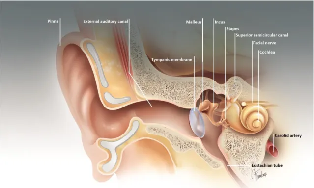
The ear is a mechano-receptor organ that acts as a link between the outer environment and the nervous system, referring to complex auditory functions. Humans can hear frquencies from 20 to 20.000Hz. The ear is composed by three primary parts: the outer ear, middle ear and inner ear. Each section is comprised of structures that play distinct roles in the process of converting sound waves into electric signals that go into the brain. The external ear collects sound waves from the external environment and funnels them toward the tympanic membrane. The middle ear ossicles transmit the sound waves to the inner ear (Figure 2.1-1).
Figure 2.1-1 - Schematic view of the structures of the ear.
The outer ear and middle ear collect, amplify, and conduct sound waves to the inner ear, where the auditory receptors are to be stimulated.
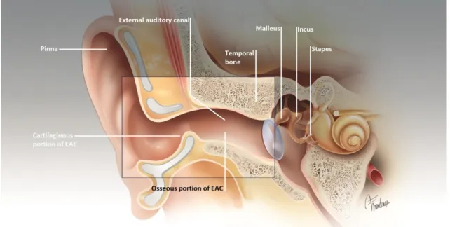
The outer ear consists of the pinna and the external auditory canal and has the function of collecting and transmitting the sound to the tympanic membrane (Figure 2.1-2). It also protects from parasitic sounds.
Figure 2.1-2 - Schematic view of the structure of the outer ear.
Legend: EAC, External Auditory Canal. (Illustration provided by Fernando Vilhena de Mendonça, MD)
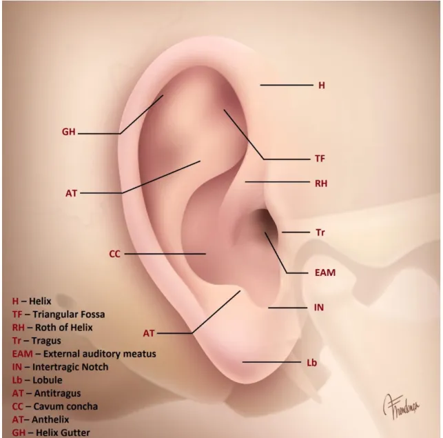
The pinna, which part protrudes from the side of the skull, attached to the temporal bone, captures sound and channels to the ear canal. The pinna is shaped like a cone that allows amplifying the sound, differently depending on its origin (Figure 2.1-3) (Geisler, 1988).
The most amplified sounds come from about 45 ° (forwards and backwards) of the ear in the horizontal plane and about 60 ° above the plane of the ear. Less-amplified sounds originate on the opposite side of the head through the mascara effect (Rosowski, 2012). Anatomically the auricle or pinna is constructed as an organ for "catch" incoming sound waves and then funnel them down the external auditory canal. The main structures are the tragus and anti-tragus, helix and anthelix (Figure 2.1-3) (Weber, Deschler, & Sokol, 2006).
Figure 2.1-3 - Schematic view of the anatomy of the pinna.
(Illustration provided by Fernando Vilhena de Mendonça, MD)
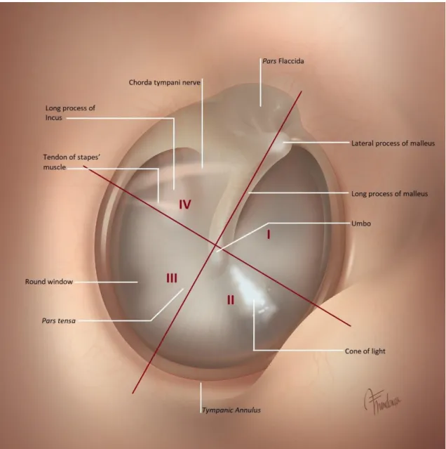
The shape of outer the ear allows a gain of approximately 20dB for sounds from 2kHz to 4kHz. The tympanic membrane separates the outer and middle ear (Alberti, 2001) (Figure 2.1-4). It is a thin (0.1 mm), cone-shaped membrane that has three layers. The external layer derived from the ectoderm is a stratified squamous epithelium. The internal layer comes from endoderm and is a cuboidal mucosal epithelium. The intermediate layer comes from mesenchyme and, is the fibrous layer (Figure 2.1-5). It is divided into the pars flaccida and the pars tensa. The pars tensa has a central fibrous layer (lamina propria), while the pars flaccida is slightly thinner (without the intermediate layer). The three layers are important in maintaining the strength of the tympanic membrane as well as in aiding the proper vibration with different frequency sounds (Lalwani, 2007). The tympanic membrane acts as a mirror of the interior of the
middle ear, and knowledge of this structure is fundamental to understand the multiple dysfunctions that affect the middle ear (Paço, 2003). According to Paço (2003), the tympanic membrane can be divided topographically into six quadrants, of which four are referred to as the pars tensa (postero-superior, postero-inferior, antero-superior and antero-inferior) and two are referred to as the pars flaccida (Figure 2.1.4).
Figure 2.1-4 Anatomy of the tympanic membrane.
Figure 2.1-5 - The layers of tympanic membrane.
Legend: CAE external auditory canal, OM middle ear. (Lalwani, 2007)
The middle ear comprises the tympanic cavity, mastoid air cells, and Eustachian (auditory) tube. The air filled space, also known as the tympanic cavity, is slightly concave and suspended by a bony ring. Posteriorly this includes the mastoid cells that communicate to tympanic cavity through the aditus ad antrum. It is connected to the back of the nose by a 35-45 mm long thin tube called the auditory tube (necessary for equalization of pressure between exterior and middle ear and drainage of mucous produced in the middle ear) (Moller, 2006). The auditory tube has an osseous portion (posterior 1/3) that opens into the tympanic cavity and a fibro-cartilaginous portion that opens into the nasopharynx. There are three important muscles for its function - tensor
veli palatini, levator veli palatini and salpingopharyngeus (Correa & Gómez, 2007).
Sound is conducted from the tympanic membrane to the inner ear by three bones that constitutes the ossicular chain – the malleus, incus and stapes (Figure 2.1-6). The ossicular chain is attached to one side of the tympanic membrane by the malleus,
which inserts into the vestibular window of the inner ear by the footplate of the stapes.
When a sound wave hits the tympanic membrane, it propagates through the ossicular chain and into the vestibular window of the inner ear. There are two muscles in the middle ear, namely the stapedius and tensor tympani, which insert into the stapes and malleus, respectively. The middle ear amplifies sound via two mechanisms: The proportion of tympanic membrane and stapes platinum surface is 14:1 and the lever effect (malleus and incus assured by the malleus and incus ligaments respectively anterior and posterior) is 18.3:1 which provides a gain of 20-35dB. The ossicular chain maximizes transference of sounds from 1-10kHz (Erminy, Skanavi, Van Den Abbeele, Avan, & Bonfils, 1995). The stapes arch reflex causes contraction of stapedius when the ear is exposed to sounds louder than 70dB, raising rigidity to ossicular chain and protecting the inner hear from noise induced damage.
Figure 2.1-6 - Schematic view of the structures of the middle ear ossicles.
(Illustration provided by Fernando Vilhena de Mendonça, MD)
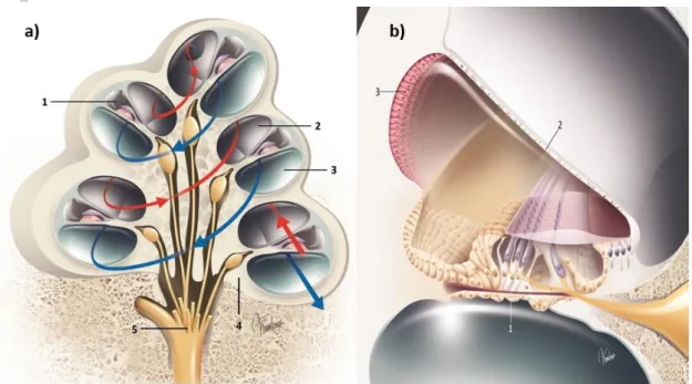
The inner ear contains a group of interconnected, fluid-filled chambers. The snail-shaped chamber, called the cochlea, is an approximately 30-mm-long coiled tube, of 2 and 3/4 turns along an osseous axis called modiolus, located within the petrous bone and divided by membranes into three chambers (Figure 2.1-7).
Figure 2.1-7 - Schematic view of the structures of Inner Ear.
The cochlea is one of the smallest organs of the human body (Figure 2.1-8). Its small size is important because if it was larger the necessary mechanical force to vibrate its structures would have to be higher and humans would only be able to hear very loud sounds. The cochlea acts as active and passive sound filter.
Figure 2.1-8 - Cochlea. a) Structures of cochlear channel; b) Cross-section of a single cochlear
turn.
Legend: a) 1 – the scala media; 2 – the scala vestibuli; 3 – the scala tympani; 4 – the spiral
ganglion neurons; 5 – Auditory nerve VIII; b) 1 – Reticular Lamina; 2 –scala vestibule ; 3 – Lateral wall (stria vascularis). (Illustration provided by Fernando Vilhena de Mendonça, MD).
The three fluid-filled chambers of the innear ear are: the scala vestibuli, the scala
tympani, and the scala media (Figure 2.1-8). The scala media or cochlear, located in the
center, is separated from the scala vestibuli (superiorly) by vestibular membrane and from the scala tympani (inferiorly) by the basilar membrane (Moller, 2006). Sound vibrations from the bones of the middle ear are transferred to the fluids of the cochlea through the vestibular window to the scala vestibuli. Near the apical termination of the bony labyrinth there is an opening called the helicotrema. This allows communication between the scala vestibuli and scala tympani that are filled by a fluid rich in Sodium (Na+) called perilymph, which is similar to cephaloraquidian fluid. The Scala tympani ends at the round (tympanic) window, and serves as an escapement of the sound wave. The scala vestibuli receives the sound wave from the vestibular window (connection to ossicular chain). The scala media narrows towards the apex of the cochlea ending just short of the apical termination of the bony labyrinth. It is filled with a fluid rich in Potassium (K+) called endolymph, similar to intracellular fluid, possesses a potential of +80mV. The basilar membrane has the ability to separated sounds according to their frequency (tonotopy spectrum). It becomes larger and less tense near the apex and so,
more sensitive to higher frequencies at the basal area where the cochlea has lower mass and higher rigidity. It has higher mass and lower rigidity at the apex where it is more sensitive to lower frequencies- This process is called passive tonotopy (Figure 2.1-9). The sound vibration propagates through cochlear scala and is maximal in the area corresponding to the sound frequency.
Figure 2.1-9 - Travelling wave.
(Illustration provided by Fernando Vilhena de Mendonça, MD).
The spiral organ (of Corti), located along the basilar membrane, contains the sensory cells (hair cells) (Moller, 2006) (Figure 2.1-10). These tiny sensors (hair cells) are mechanoreceptors that convert the sound vibrations of the basilar membrane into electrical impulses that are transmitted along the auditory nerve to the brain through the auditory pathway – this mechanism is called transduction. The Inner Hair Cells (IHC) are the principal auditory receptors. They constitute the most internal row along the spiral organ (of Corti) (approximately 3500 cells). Their function is to transform the mechanical sound stimulus transmitted from the outer and middle ear into and electrical message to be send to auditory nervous centers (in temporal lobe) through the auditory pathway (Abbas & Miller, 1993).
IHCs are sensory cells that transform the hydromechanical, vibratory energy of cochlear liquids into bioelectric energy. About 80% of the time the transducer channels are closed and only open in the excitatory phase of the stimulus. The process of transduction begins when the energy is sufficiently intense, and the stimulation leads to the depolarization of IHCs, which leads to the release of neurotransmitters, causing one or more afferent fibers of the auditory nerve to fire (Monteiro & Trigueiros, 2018).
Figure 2.1-10 - Schematic of the organ of Corti.
Legend: 1-Inner hair cell (IHC); 2-Outer hair cells (OHC); 3-Tunnel of Corti; 4-Basilar membrane; 5-Habenula perforate; 6-Tectorial membrane; 7-Deiters' cells; 8-Nuel’s space; 9-Hensen's cells; 10-Inner spiral sulcus. (Illustration provided by Fernando Vilhena de Mendonça, MD).
The three outer rows along the spiral organ (of Corti) corresponds to the Outer Hair Cells (OHC), nearly 12000 cells (Figure 2.1-10). Surrounded by supporting cells – Hensen and Claudius cells also have a role in the cycle of K+ and Glutamate (Monteiro & Trigueiros, 2018).
OHCs are pressurized and have a central core without a cytoskeleton which gives them strength, flexibility and electromotility according to the stimulus, providing a refinement in frequential sensitivity and selectivity. Hence OHC are called the cochlea amplifiers. OHCs length can vary from 12µm in the basal region to more than 90µm in the apex of the cochlea (Brownell, Spector, Raphael, & Popel, 2001).
Because only the apical part of ciliated cells are in contact with endolymph cochlea found a process to transport K+ from or to cells without ATP consumption, this electrochemical gradient is achieved through the stria vascularis, located at external wall of scala media and lying in the basilar membrane. It is a highly vascularized and metabolically active organ (Figure 2.1-8b).
Within the cochlea, vibratory energy results in an interaction called the traveling wave, which is a wave of fluid vibration along the basilar membrane along the cochlear coil. Wave energy is placed at a particular site of the basilar membrane, depending on the frequency of vibration. Through the depolarization of cells, the action of mechanical vibratory energy is used more effectively in the transduction of bioelectric energy. This gradient acts like a battery whose energy permits hearing. Ciliated cells have synapses
at their basal pole. The delivery of neurotransmitters to the synaptic cleft (transforming electrical energy in chemical) is regulated by alterations in cellular membrane Differentiated vibration of the basilar membrane and tectorial membrane causes stereocilia flection. The OHCs respond to stimulation when their stereocilia are flexed in an external direction. When the stereocilia are flexed in the excitatory direction (towards the top), the links between tops are stretched, which increases the likelihood of calcium channel opening. Calcium plays a determinant role in the intracellular balance, being that it maintains cellular homeostasis through the mechanotransducing channels (Figure 2.1-11). The energy of acoustic stimulus induces the movement of the basilar membrane synchronized with the deflection of the stereocilia that triggers transducer currents. This allows the entrance of K+ into the OHCs and leads to their depolarization. The resulting action potentials produce a motor response caused by the properties of the prestin protein. This vibratory energy is returned to the basilar membrane (Monteiro & Trigueiros, 2018).
Figure 2.1-9 - Transduction channels: opening and adaptation to potassium levels.
(http://www.cochlea.eu/po/)
The middle portion of OHCs is where electrical energy is converted into mechanical – electromotility. If electrical energy resulting from sound vibration coincides with the natural frequency of that cochlear portion then the magnitude of vibration augments. Otherwise it decreases. Accordingly, there is a release of neurotransmissor in the inferior pole of the cell.
The OHCs produce a sound that can be detected through a microphone in the outer ear – Otoacoustic emissions (OAE). This is the demonstration of the active cochlea mechanisms that are exclusive of OHCs. No other cells in the human body have this electromotility, a biological form of piezoelectricity.
Hearing occurs by air conduction or bone conduction. In air conduction sound reaches the inner ear by propagating in the air reaching tympanic membrane through the external auditory channel. Movement of the tympanic membrane is transmitted to the ossicular chain that propagates to the cochlea through the oval window connected to stapes footplate. The structures of middle ear serves as an impedance-matching mechanism, improving the efficiency of energy transfer from the air to the fluid-filled inner ear. Hearing by bone conduction happens when the sound source is in physical contact with the head, causing vibration of the bones of the skull that generate a travel wave in the cochlea’s basilar membrane (Brownell et al., 2001; Dallos & Fakler, 2002).
The nervous fibers that come from IHCs converge to form the spiral ganglion. From here originates the cochlear part of VIII cranial nerve, the anterior portion of which goes to the internal auditory channel (IAC).
2.1.2. Central Auditory Nervous System (CANS)
The cochlear nerve contains a total of 30,000 afferent nerve fibers (Spoendlin, 1987). The cell bodies of these fibers are found in the spiral ganglion, the cochlear part (auditory) of VIII cranial nerve. The anterior portion goes to the IAC and enters the brain at the spinal bulb (Bonaldi, Lago, Crema, Fukuda, & Smith, 2004; Ruah, 2002)
At low frequencies individual auditory nerve fibers can respond more or less synchronously with the stimulating tone. At higher frequencies phase-locking occurs so that neurons can alternate in response to particular phases of the sound wave cycle. Three aspects encode the intensity of the sound wave cycle: 1) the amount of neural activity in individual neurons, 2) the number of neurons that are active, and 3) the specific neurons that are activated.
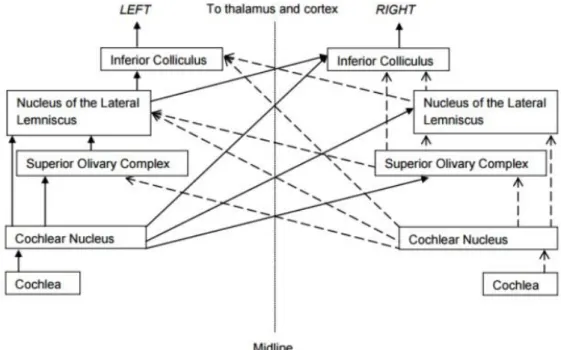
The central auditory pathways are composed of various structures, beginning in the auditory nerve, followed by the cochlear nuclei, the superior olivary complex, the lateral lemniscus, the lower colliculus, the middle geniculate body, terminating in the auditory cortex (Figure 2.1-12 and 2.1-13). The auditory nerve divides in two pathways. The ascending/afferent auditory pathway (afferent fibers (myelinated bipolar type I neurons) carries sound information to the brain mostly come from IHCs, and the descending/efferent auditory pathway (efferent fibers, non-myelinated bipolar type II neurons) carries information back from the brain to the OHCs (Malmierca & Hackett, 2010).
Ascending or afferent auditory pathway, is a fast pathway, which begins in the cochlear nucleus – first integrator centre, are composed by three subnucleus: Dorsal Cochlear Nucleus (DCN), the postero-ventral Cochlear Nucleus (PVCN) and the antero-ventral Cochlear Nucleus (AVCN) (Malmierca & Hackett, 2010; Marinho, 2011; Moller, 2006; Phillips, 2007), where the central auditory system
begins its course to the auditory cortex (Aquino, Chandra, Haines, & Micco, 2002; Ruah, 2002) located at spinal bulb, the fibers keep the cochlear frequencial selectivity. From here some projections go to ascendant reticular pathway – non- primary auditory pathway, the main function of these pathways, also connected to wake and motivation centers as well as to vegetative and hormonal systems, is to select the type of sensory message to be treated first. This pathway is inactive during sleep (Cochlea, 2013).
Figure 2.1-10 - Primary auditory pathways.
(Illustration provided by Fernando Vilhena de Mendonça, MD).
Second integrator centre is Superior Olivary Complex (SOC) also located at spinal bulb, majority of fibers cross to the opposite side, SOC is divided in three nucleuses: the Complex Superior Olivary lateral (CSOL), the Complex Superior Olivary medium (CSOM) and the medial nucleus of do trapezoid body (Martínez & Nieto, 2003; Musiek & Baran, 1986; Neijenhuis, 2003).
The third integrator centre is Inferior Colliculus, located at midbrain. Together with SOC have important role in the localization of sound and integration of sounds with a complex temporal pattern, also receives fibers from the Cochlear
Nucleus.
The fourth integrator centre before cortex is medial geniculated body located in thalamus, here starts the integration of information. Can be subdivided in three regions: the ventral projects to the primary auditory region of temporal lobe, the middle projects to the other temporal lobe regions and the dorsal projects afferent to cerebellar associative areas (Seikel, King, & Drumright, 2009).
Primary auditory cortex is located in the temporal lobe, hidden by the lateral sulcus incisures. Auditory message arrives here largely decoded by the previous nucleus, is memorized, and possibly integrated in a motor response like vocalization. The primary auditory cortex has an important role in phoneme discrimination, and is also involved in temporal and spectral discrimination (Bellis, 2003). Posteriorly, angular girus, represents the Wernicke region, responsible for linguistic stimuli recognition and speech understanding (Specht, 2014).
The descending or efferent auditory pathway extends from the auditory cortex to the hair cells. The majority of the bodies of the efferent fibers are located in the SOC of the brainstem.
The anatomical description and role of this pathway is still a matter of debate, however it is agreed that it begins at the auditory cortex and associative areas. It is divided in two segments. The rostral segment, involves auditory cortex, associative secondary areas, medial geniculated body, inferior colliculus and the lateral lemniscus. It is possible that this segment has a regulator role of afferent pathway, namely cochlea, the auditory nerve, and the inferior nucleus of the brainstem (Baran, Brooke Shinn, & Musiek, 2006). The caudal segment comprises the SOC (lateral and middle), cochlear nucleus, and ends at the cochlea (IHCs and mainly at OHCs). This segment is also called medial olivo-cochlear bundle. In the efferent pathway the main neurotransmitter is acetylcholine but dopamine, dynorphins and encephalin are also present. It is considered that the physic-acoustic model closest to reality should be based on oscillators tuned in frequencies (OHCs) that would be regulated through the medial olivo-cochlear bundle.
Figure 2.1-11 - Main nuclei involved in binaural hearing.
(Porter, 2012)
2.1.3. The close correlation between hearing and cognition
As far as hearing is concerned we do not hear using a single part of our brain. Instead hearing stimuli ‘switch on’ several brain areas. Even a simple word has the capacity to activate not only the auditory cortex, but also several other areas where it is ‘understood’ or semantically or cognitively connected. Evidence demonstrates a very close association between hearing and cognition. Recent studies on people aged 50 to 79 included tests to evaluate hearing capacity, central auditory processing and cognitive skills. The most predictive factor of speech discrimination in noisy environment was central processing of sound, followed by cognitive skills (such as working memory and short-term memory), and by life experience such as socio-economic status. Hearing sensitivity evaluated by tonal audiometry was the ‘weakest’ contributor to performance of this task (Figure 2.1-14).