Hugo Miguel Lambuça Ferreira
Graduated in Cell and Molecular Biology
Overcome challenges in influenza virus-like
particles downstream process
Dissertation to obtain the Master Degree in Biotechnology
Supervisor: Dr. Cristina Maria da Costa Peixoto Lisboa
i
Hugo Miguel Lambuça Ferreira
Graduated in Cell and Molecular Biology
Overcome challenges in influenza virus-like
particles downstream process
Dissertation to obtain the Master Degree in Biotechnology
Supervisor: Dr. Cristina Maria da Costa Peixoto Lisboa
Examination Committee
Chairperson: Prof. Dr. Pedro Miguel Ribeiro Viana Baptista
Supervisor: Dr. Cristina Maria da Costa Peixoto Lisboa
Member of the Committee: Prof. Dr. José Paulo Barbosa Mota
iii
Overcome challenges in influenza virus-like particles bioprocessing
Copyright © Hugo Miguel Lambuça Ferreira, Faculdade de Ciências e Tecnologia, Universidade Nova de Lisboa.
A Faculdade de Ciências e Tecnologia e a Universidade Nova de Lisboa têm o direito, perpétuo e sem limites geográficos, de arquivar e publicar esta dissertação através de exemplares impressos reproduzidos em papel ou de forma digital, ou por qualquer outro meio conhecido ou que venha a ser inventado, e de divulgar através de repositórios científicos e de admitir a sua cópia e distribuição com objetivos educacionais ou de investigação, não comerciais, desde que seja dado crédito ao autor e editor.
v
vii
A
CKNOWLEDGEMENTS
À Professora Paula Alves, pelo conhecimento, interesse e paixão pelo que faz e pelo que transmitiu nas aulas durante o mestrado, que de certa forma contribuiu para ter realizado a minha tese nesta unidade.
Ao Professor Manuel Carrondo, pelo seu conhecimento, espírito de liderança e rigor na Ciência que inspira qualquer jovem estudante.
À minha orientadora, Dr. Cristina Peixoto, pela oportunidade que me deu de poder realizar a minha tese. Agradeço também pela paciência, apoio e por todos os conselhos ao longo deste ano.
À equipa do DSP aqui vão os agradecimentos. Ao Dr. Ricardo Silva por tudo o que me ensinou, todos os conselhos e discussão sobre este trabalho, assim como pela disponibilidade de ajuda. À Sofia Carvalho pelo que me ensinou, assim como pela partilha de resultados e discussão dos mesmos. À Mafalda Moleirinho por estar sempre pronta a ensinar e ajudar. À Sara Rosa pela paciência que teve para ter aturado todas as minhas perguntas e pedidos. À Sofia Moreira pelo apoio, conselhos e danças ao longo do ano. À minha colega de secretária, Ana Quendera por todas as risadas e conselhos. Ao João Mendes por todas as gargalhadas sobre tudo e de tudo. E ao Tiago pela prontidão em ajudar e ensinar. Obrigado a todos pelo bom ambiente e disposição.
Agradeço também aos restantes membros do TCA pela ajuda nos momentos em que precisei ao longo de todo este ano.
Quero agradecer também aos meus colegas de mestrado, Bernardo, Gonçalo, Vânia, Patrícia, Mariana, entre outros pelos momentos que por muito curtos que fossem dava sempre para “dois dedos de conversa”. Pelo apoio e desabafos ao longo do ano. Estamos juntos!
Agradeço também a todos os meus amigos, padrinhos e afilhados de faculdade por estarem sempre dispostos a ouvir-me e dispostos sempre a ajudar.
Agradeço à família anTUNiA por todos os momentos de folia que me fizeram espairecer este ano e pelos fins-de-semana bem passados.
Obrigado especial ao TM, Flávio e Ticas por tudo. Pelos desabafos, pelas lamentações e pelas infinitas gargalhadas. Muito Obrigado por estarem lá sempre e por serem os amigos que muitos gostariam de ter.
viii
Agradeço também às minhas manas, Mónica e Inês. Por serem tudo e tudo. Por não estarem por perto, mas eu ter 100 % certezas de que estão. Muito Obrigado por me ouvirem e por estarem comigo sempre e para sempre.
Agradecimento muito, muito especial à minha Bárbara, por tudo o que partilhámos, pela força, incentivo, motivação e gargalhadas. Obrigado por todo o teu apoio. Sem ti, teria sido uma história bem diferente.
Agradeço à minha família, a de sempre e a nova. Aos meus padrinhos, primos, avô Lambuça e avó Quina por estarem lá sempre em cada fim-de-semana de visita e por me receberem sempre de braços abertos. Aos pais, tios e primos da Bárbara, em especial à afilhada e minha sobrinha Matilde.
À minha avó Lucinda e ao meu avô Ferreira. Onde quer que estejam estão sempre ao meu lado, dia-a-dia e sei que fizeram de tudo para esta tese acabasse bem.
Agradeço, por fim aos meus pais por terem confiado em mim ao longo destes 5 anos. Um especial obrigado pelo que me puderam proporcionar e por serem pessoas excecionais e espetaculares. Muito, muito obrigado. (Apesar de estar em inglês), É a vós que dedico este meu trabalho.
ix
A
BSTRACT
The development of new vaccines for influenza virus introduced a new generation of vaccines using virus-like particles (VLPs). The lack of genetic material, possibility of production on cell lines and presence of antigens with immunogenicity are the main advantages over the traditional vaccines. The development of a cost-effective downstream process while maintaining the high purity, potency and quality of VLPs is a challenge. In this thesis, several purification steps – clarification, concentration, chromatography, polishing and sterile filtration – were studied to develop a new downstream proves for influenza VLPs.
In clarification step, a strategy using D0HC followed by Opticap XL SHC filters presented the best result. For concentration step, the cassette with cut-off of 300 kDa presented a higher yield on hemagglutinin recovery and the lowest process time. For chromatography step, the membrane Sartobind Q and the resin HiTrap Q HP were evaluated, concluding that resin HiTrap presented higher dynamic binding capacity and better resolution on elution. For polishing step, size-exclusion chromatography and multimodal chromatography operate in flow-through mode were compared. The last presented higher recovery yield on hemagglutinin and it was select due to the non-limitation for scale-up. Different materials were analysed for the final sterile filtration.
A proof of concept run was performed were the optimized conditions and best devices were evaluated. In the end of process, it was obtained influenza VLPs with concentration and quality enough to advance for animal in vivo studies and for clinical phase I.
Additionally, a new tool – magnetic sulphated cellulose particles – was evaluated with the goal to obtain purified and concentrated samples to use in characterization techniques.
Overall, this thesis contributes to introduce a new tool and a novel cost-effective downstream purification process with high purity, potency and quality for the next generation of influenza vaccines - VLPs.
Keywords: purification process; influenza vacines; vírus-like particles; magnetic sulphated cellulose particles.
xi
R
ESUMO
O desenvolvimento de novas vacinas para o vírus de influenza introduziu uma nova geração de vacinas utilizando partículas semelhantes a vírus (VLPs). A ausência de material genético, possibilidade de produção em linhas celulares e presença de antigénios com imunogenicidade são as principais vantagens em relação às vacinas tradicionais. O desenvolvimento de um processo de purificação de baixo custo mantendo a elevada pureza, potencia e qualidade das VLPs é um desafio. Nesta tese, alguns passos de purificação – clarificação, concentração, cromatografia, polimento e filtração estéril final – foram estudados para desenvolver um novo processo de purificação de VLPs de influenza.
Na clarificação, a estratégia usando os filtros D0HC seguido do Opticap XL SHC apresentaram os melhores resultados. Na concentração, a cassete com cut-off de 300 kDa apresentou um maior rendimento na recuperação de hemaglutinina e o mais baixo tempo de operação. Na cromatografia, a membrana Sartobind Q e a resina HiTrap Q HP foram avaliadas, concluindo-se que a resina apresenta maior capacidade de ligação dinâmica e maior resolução na eluição. No polimento, a cromatografia de exclusão molecular e a cromatografia multimodal, operada em flow-through comparadas. Esta última apresentou valores superiores de recuperação de hemaglutinina sendo escolhida por não conter limitações no escalamento. Diferentes materiais foram analisados na filtração estéril final.
Na realização da corrida de prova de conceito as condições ótimas e os melhores materiais foram estudadas. No final do processo, obteve-se VLPs de influenza na concentração e qualidade suficiente para avançar para estudos em animais in vivo e para fase clínica I.
Adicionalmente, uma nova ferramenta – partículas magnéticas de celulose sulfatada – foram estudadas com objetivo de obter VLPs purificadas e concentradas para utilização em técnicas de caracterização.
Em geral, esta tese contribuiu para introduzir uma nova ferramenta e um novo processo de purificação mais económico com elevada pureza, potência e qualidade, para a nova geração de vacinas - VLPs.
Palavras-chave: processo de purificação; vacinas de influenza; partículas semelhantes a vírus; partículas magnéticas de celulose sulfatada.
xiii
L
IST OF
C
ONTENTS
List of Figures ... xv
List of Tables ... xvii
List of Abbreviations ... xix
Chapter 1: Introduction ... 1
1.1. Vaccines ... 1
1.1.1. Influenza vaccines ... 2
1.1.2. Influenza virus-like particles ... 3
1.2. Downstream process of virus-like particles ... 4
1.2.1. Clarification ... 6
1.2.2. Concentration ... 7
1.2.3. Intermediate purification ... 8
1.2.4. Polishing step ... 8
1.3. Aim of the thesis ... 9
Chapter 2: Materials and Methods ... 11
2.1. Cell maintenance ... 11
2.2. Recombinant baculovirus expansion ... 11
2.3. Recombinant baculovirus titration ... 12
2.4. Influenza virus-like particles production ... 12
2.5. Influenza virus-like particles downstream processing ... 13
2.5.1. Clarification studies ... 13
2.5.2. Concentration studies ... 13
2.5.3. Chromatography studies ... 14
2.5.4. Polishing studies ... 15
2.5.5. Final sterile filtration studies ... 16
2.5.6. Proof of concept ... 17
2.5.7. Magnetic Sulphated Cellulose Particles studies ... 17
2.6. Analytical methods ... 18
2.6.1. Hemagglutination assay (HA assay) ... 18
2.6.2. Total protein quantification ... 19
2.6.3. Total dsDNA quantification ... 19
2.6.4. Particles concentration and size distribution ... 19
2.6.5. Immunoblotting... 20
2.6.6. Baculovirus quantification ... 20
2.6.7. Transmission Electron Microscopy analysis ... 21
Chapter 3: Results and Discussion ... 23
3.1. Clarification studies ... 23
3.1.1. Influenza VLPs recovery ... 23
3.1.2. Impurities removal ... 24
xiv
3.2.1. Hollow-Fibers ... 26
3.2.1.1. Scouting Transmembrane Pressure ... 26
3.2.1.2. Shear rate studies ... 26
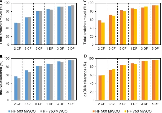
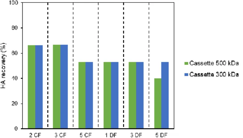
3.2.1.3. Impurities removal ... 28 3.2.2. Cassettes ... 29 3.2.2.1. Influenza VLPs recovery ... 29 3.2.2.2. Impurities removal ... 30 3.3. Chromatographic studies ... 31 3.3.1. Membrane Chromatography... 31
3.3.1.1. Dynamic Binding Capacity determination ... 31
3.3.1.2. Elution condition evaluation ... 32
3.3.1.3. Run and load condition evaluation ... 33
3.3.2. Resin Chromatography ... 34
3.3.2.1. Elution condition evaluation ... 34
3.4. Polishing and Sterile Filtration studies ... 36
3.4.1. Multimodal Chromatography ... 36 3.4.2. Size-Exclusion Chromatography ... 37 3.4.3. Sterile Filtration ... 38 3.5. Proof of concept ... 38 3.5.1. Clarification... 39 3.5.2. Concentration ... 39
3.5.3. Resin Chromatography – Anion Exchange Chromatography ... 40
3.5.4. Polishing (multimodal chromatography) and Sterile Filtration ... 43
3.5.5. Characterization of Product ... 45
3.6. Magnetic Sulphated Cellulose Particles studies ... 47
3.6.1. Desalted buffers evaluation ... 47
3.6.2. Beads mass evaluation ... 48
3.6.3. Impurities removal ... 48
Chapter 4: Conclusions and Future Perspectives ... 51
xv
L
IST OF
F
IGURES
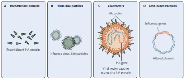
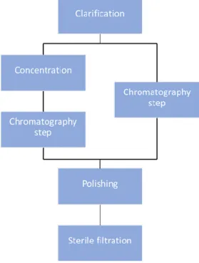
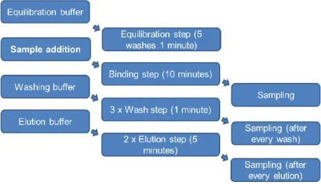
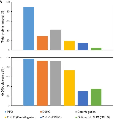
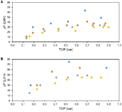
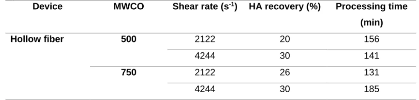
Figure 1.1 – New generation of influenza vaccines. (A) Recombinant proteins, (B) Virus-like particles (VLP), (C) Viral vectors, (D) DNA-based vaccines. (Adapted from literature3) ... 3 Figure 1.2 – Downstream process train evaluated for influenza VLPs. ... 5 Figure 2.1 - Magnetic Sulphated Cellulose Particles (MSCPs) experiment steps. ... 18 Figure 3.1 – HA recovery (%) after each filter and centrifugation evaluated for clarification studies. PP3, D0HC and centrifugation was evaluated for first step. 2 XLG and Opticap XL SHC was evaluated for second step. ... 23 Figure 3.2 – (A) Total protein removal (%) and (B) dsDNA clearance (%) after each filter and centrifugation evaluated for clarification studies. PP3, D0HC and centrifugation was evaluated for first step. 2 XLG and Opticap XL SHC was evaluated for second step. . ... 24 Figure 3.3 – Determination of optimal TMP for concentration studies with HFs, at different flow rates, determining the yP. (A) 500 MWCO HFs and (B) 750 MWCO HFs. ... 26 Figure 3.4 – HA recovery (%) as a function of CF and DF volume for both evaluated HFs with shear rate values of (A) 2122 s-1 and (B) 4244 s-1. ... 27 Figure 3.5 – (A) Total protein removal (%) and (B) dsDNA clearance as a function of CF and DF volume with 2122 s-1 of shear rate value. For 4244 s-1 of shear rate, (C) total protein removal (%) and (D) dsDNA clearance as function CF and DF volume. ... 28 Figure 3.6 – HA recovery (%) as a function of CF and DF volume for both evaluated cassettes. ... 29 Figure 3.7 – (A) Total protein removal (%) and (B) dsDNA clearance as a function of CF and DF volume for each evaluated cassettes. ... 30 Figure 3.8 – FT section of membrane chromatography for DBC determination through the breakthrough curve (calculated by Cout/Cin of each fraction collected). ... 31 Figure 3.9 – Elution condition evaluation for membrane chromatography. Steps of 100 mM of NaCl were performed. Fractions of peaks were collected and analysed. (A) total chromatogram and (B) elution step chromatogram. ... 33 Figure 3.10 – Elution condition evaluation for resin chromatography. Fractions of peaks were collected and analysed. (A) total chromatogram and (B) elution step chromatogram with reference to which concentration of salt (NaCl) exist on each step. ... 35 Figure 3.11. – Quantity of HA (µg) and impurities (dsDNA (µg) and total protein (mg)) along the elution evaluation step of resin chromatography. ... 35 Figure 3.12 – Chromatogram representative of multimodal chromatography run. The FT and elution fractions were collected for analysis. ... 36 Figure 3.13 – Quantity of HA (µg) and impurities (dsDNA (µg) and total protein (mg)) along the run of multimodal chromatography. dsDNA has not representation due to samples had less than 0.0078 µg mL-1 (minimal detection limit by method). ... 36 Figure 3.14 – Chromatogram representative of SEC run. The peak 1 was collected in the void volume of the column. Both fractions (peak 1 and 2) were collected and analysed. ... 37 Figure 3.15 – Quantity of HA (µg) and impurities (dsDNA (µg) and total protein (mg)) of SEC samples. ... 37 Figure 3.16 – Downstream train of influenza VLPs with all selected strategies and devices. ... 38 Figure 3.17 – HA recovery and impurities removal (%) on both clarification filters used (D0HC and Opticap XL SHC). ... 39 Figure 3.18 – HA recovery and impurities (dsDNA and total protein) removal as function CF and DF volumes of concentration step. ... 40 Figure 3.19 – (A) FT section of resin chromatography for dynamic binding capacity determination through the breakthrough curve (calculated by Cout/Cin of each fraction collected) and (B) selected area zoom. .... 41
xvi
Figure 3.20 – Elution of resin chromatography step - proof of concept run. The 3 steps of conductivity correspond to the optimal NaCl concentration for elution step. All peaks identified were collected and evaluated. ... 42 Figure 3.21 – Quantity of HA (µg) and impurities (dsDNA (µg) and total protein (mg)) from the elution step of resin chromatography. ... 42 Figure 3.22 – Multimodal chromatography chromatograms – proof of concept run. (A) Corresponds to peak 1 prevenient from resin chromatography step and (B) correspond peak 3 prevenient from resin chromatography elution step. ... 44 Figure 3.23 – (A) Analysis of total protein through the SDS-PAGE present on SF(A) and SF(B) filter 3 samples. Immunoblotting analysis (B) for identification of HA protein and (C) for identification of the M1 protein. TEM images of Influenza VLPs prevenient from (D) polishing step – peak 1 from chromatography step and (E) final sample prevenient from SF(B) of filter 3. (F) TEM image of baculovirus present on the final sample from SF(B) of filter 3. Scale bars represent 100 nm. ... 46 Figure 3.24 – Scheme of all samples collected from MSCPs experiments with an indication where HA is loss or recovered. ... 47 Figure 3.25 – (A) Percentage of total protein removal and (B) Percentage of dsDNA clearance in the three steps (Supernatant, Elution 1 and Elution 2) of MSCPs mass of beads evaluation experiments. ... 49
xvii
L
IST OF
T
ABLES
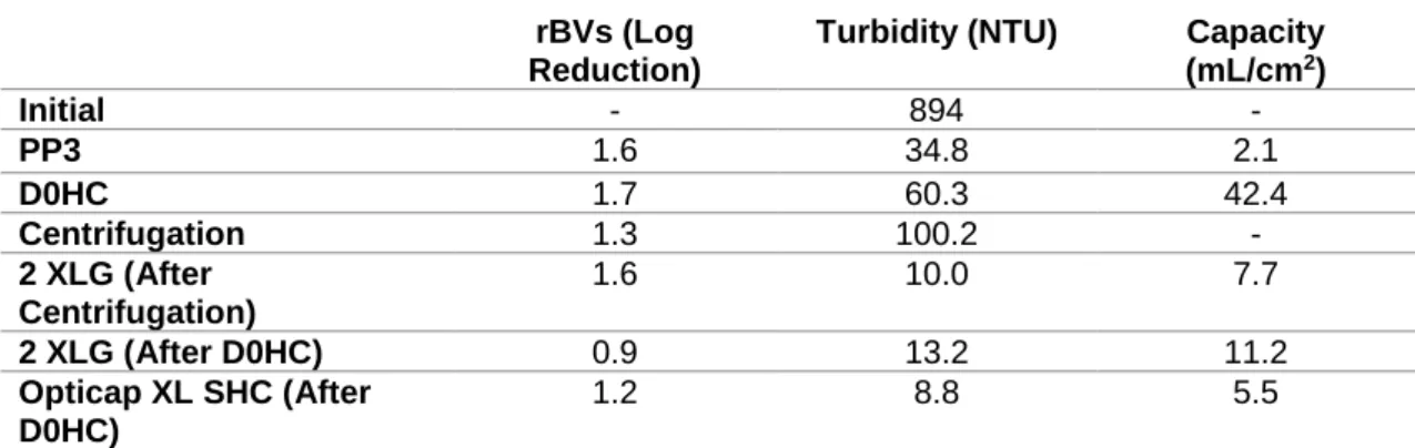
Table 2.1 – Description of the area, pore and feed flow rate of all used filters on clarification studies. ... 13 Table 2.2 – Syringe filters devices used with correspondent filter area. ... 16 Table 2.3 – Buffers for equilibration, washing and elution steps for Magnetic Sulphated Cellulose Particles experiments. ... 18 Table 3.1 – Baculovirus reduction (value by log reduction), turbidity and capacity of each filter and centrifugation evaluated on clarification studies. PP3, D0HC and centrifugation was evaluated for first step. 2 XLG and Opticap XL SHC was evaluated for second step. ... 25 Table 3.2 – HA recovery (%) and processing time (min) of all UF/DF process in function the different processed shear rate of each HFs evaluated. ... 27 Table 3.3 – Baculovirus removal (value by log reduction) comparable with the initial value from all experiments of evaluated HFs samples. ... 28 Table 3.4 – HA recovery (%) and processing time (min) of all UF/DF process in function the different cassette devices evaluated. ... 30 Table 3.5 – HA loss on flow-through (%) and HA recovery on elution (%) due to the evaluated different binding and load conditions of NaCl concentration (mM). ... 34 Table 3.6 – Representation of all filters utilized for sterile filtration steps with the correspond HA recovery (%). ... 38 Table 3.7 – Baculovirus reduction (value by log reduction) for each peak of chromatography step – proof of concept run. ... 43 Table 3.8 – Filters selected for evaluation on proof of concept run. HA recovery of both experiments. SF (A) corresponds to FT fraction of chromatogram A from polishing step. SF (B) corresponds to FT fraction of chromatogram B from polishing step. ... 45 Table 3.9 – HA recovery yields (%) of each step for each buffer evaluated. (HA recovery was calculated to have in consideration the initial quantity of HA). ... 47 Table 3.10 – HA recovery yields (%) in each step for each different mass of beads evaluated. Bulk desalted for 10 mM Tris pH 7.4 + 50 mM NaCl. ... 48
xix
L
IST OF
A
BBREVIATIONS
CCI – Cell concentration at infection CF – Concentration factor
CIP – Cleaning-in-place CV – Column volumes
DBC – Dynamic Binding Capacity DEAE – Diethylaminoethyl DF – Diafiltration
DLS – Dynamic light scattering FDA – Food and Drug Administration FT – Flow-through
GMP – Good manufacturing practices HA – Hemagglutinin
HBV – Hepatitis B virus HCP – Host-cell protein HEV – Hepatitis E virus HFs – Hollow fibers
HIC – Hydrophobic interaction chromatography hpi – hours post-infection
HPLC – High performance liquid chromatography HPV – Human papilloma virus
IC/BEVS – Insect cells/Baculovirus expression vector system IEX – Ion exchange chromatography
MF – Microfiltration
MOI – Multiplicity of infection
MSCPs – Magnetic sulphate cellulose particles MTT – Microculture tetrazolium assay
MWCO – Nominal molecular weight cut-off NA – Neuraminidase
NFF – Normal flow filtration
NMR – Nuclear magnetic resonance NTA – Nanoparticle tracking analysis PES – Polyethersulfone
xx
PS – Polysulfone
PVDF – Polyvinylidene fluoride Q – Quaternary amine
qPCR – real-time quantitative polymerase chain reaction rBVs – Recombinant baculovirus
RC – Regenerated cellulose RT – Room temperature S – Sulfonic acid
SEC – Size exclusion chromatography SFCA – Surfactant-free cellulose acetate TEM – Transmission electron microscopy TFF – Tangential flow filtration
TMP – Transmembrane pressure UF – Ultrafiltration
VLPs – Virus-like particles WHO – World Health Organization yP – Permeate flux
1
C
HAPTER
1:
I
NTRODUCTION
1.1. Vaccines
According to World Health Organization (WHO), a vaccine “is a biological preparation that improves immunity to a particular disease.” It ”contains an agent that resembles a disease-causing microorganism, and is often made from weakened or killed forms of the microbe, its toxins or on of its surface proteins.”1. Improvement of immunity is one of the most important roles of
vaccines. When administrated on humans or animals allow their immune system to recognize these particles and answer with a specific immune response. If a pathogenic microorganism causes an infection, the immune system, that already recognizes the pathogen, will be faster to induce the specific and right response to eliminate it and prevent any kind of pathology.
Edward Jenner gave the first concept of the vaccine in the 18th century when smallpox virus
was a serious world threat. With the advance of technologies all over the years, vaccines had an evolution on manufacturing. Most of all are based on live-attenuated or chemical-inactivated infectious viral strains2. However, the new generation of vaccines are raising: recombinant
proteins, viral vectors, DNA-based vaccines and virus-like particles (VLPs)3. This last one is
described as the promising alternative to actual vaccines4–6. Three vaccines, based on VLPs, are already on market with successful application for hepatitis B virus (HBV), human papilloma virus (HPV)2,6–9 and hepatitis E virus (HEV)9. They were accepted by Food and Drug Administration
(FDA) in 1986, 2006 and 2011, respectively4,9.
Vaccines are very important as a market health-care tool due to: (i) the emerging of markets that search for new and more efficient vaccines; (ii) the significant importance to solve problems where medical need are unperformed; and (iii) the possibility of treating illness without cure that can be prevented with vaccination because of the advance on health areas like immunology and microbiology10,11.
The magnitude of infectious diseases’ challenge calls the attention of pharmaceutical industry for investment on vaccines development – considered the major tool to prevent it. However, the stringent guidelines from the regulatory authorities (FDA) demand that the product respect some
C
HA
P
TER
ítu
lo
1
2
criteria as purity, potency, quantity, and others12. For purity, several tests need to be performed
along the manufacture of the product. Two types of impurities exist, product-related (product aggregates, molecular variants, degraded products) and process-related (Host-cell protein (HCP), host cell DNA, media components, enzymes/chemicals). For potency, the tests are related to the cell-based, which is measure the biochemical or physiological response at the cellular level or animal-based which the measure the biological response from organism to the product respectively. The quality is related to the protein content (in mass) that should be determined using appropriate assays12.
1.1.1. Influenza vaccines
Native influenza virus belongs to the Orthomixoviridae family that presents segmented RNA genome and it is composed of surface glycoproteins, Hemagglutinin (HA) and Neuraminidase (NA). It is surrounded by lipid membrane – coming from virus budding process on infected cells – and that’s why it is characterized as an enveloped virus. The core proteins are M1 protein – confer structure to the envelope and virus capsid – and M2 protein – an essential ion-channel.
Every season, influenza virus causes around 5-15% of infection on northern hemisphere’s population, according to WHO13. Therefore vaccination plays a major role in the prevention of
influenza virus’ infection8,14. For several decades (and even nowadays), influenza vaccines were
made from live-attenuated or chemical-inactivated virus obtained by infection of chicken fertilized eggs2. This manufacturing process is so complex and slow that in case of a pandemic event it
cannot answer in time to prevent or fight the virus infection3,6,8,11,15.
Influenza vaccines are compromised by the weak initial immunogenicity and efficacy, mainly because of two facts: the virus strains present on vaccine (do not have a good match with the circulating virus) and the antigenic drift of influenza virus6,8. This last one promotes the change of
the proteins of virus’ surface specially the most abundant – HA5,8. Consequently, a new strain of
influenza virus appears every year5,9. One example is the pandemic H1N1 influenza virus in 2009
that was caused by genetic reassortment between different species of avian, swine and human origin9,16,17.
The challenge is developing vaccines and producing-platforms with more efficiency than the actual ones3,8 never forgetting the main goal on vaccinology – getting an “universal” influenza
vaccine with the capacity to prompts an immunity response, and that can respond to all influenza strains, be administrated just one time in life and can be produced fast and in controlled conditions3. In answer to that, a new generation of vaccines are approaching, and some examples
3
Figure 1.1 - New generation of influenza vaccines. (A) Recombinant proteins, (B) Virus-like particles (VLP),
(C) Viral vectors, (D) DNA-based vaccines. (Adapted from literature3)
These technologies achieve a revolution step because of their manufacturing process, that are made in cell-based technology, replacing the egg-based one3,11. Some new generation of
vaccines are already in early stages of development with the aim of manufacturing time-reduction and cost-effective as well as efficacy and safety improvement.
1.1.2.
Influenza virus-like particles
VLPs are self-assembly structures that can be similar especially on structure namely in the organization and conformation to virus particles18. However, VLPs shows many advantages: (i)
do not have viral genome inside the particle – preclude the replication; (ii) can be produced in cell lines – bacteria, yeasts, plants, mammalians and insects; (iii) own active antigens – have immunogenicity response that does not need adjuvants8,19. The focus of this thesis will be
influenza VLPs.
Influenza VLPs presents the same size (80-120 nm18) and a lipid envelope alike the virion.
On this envelope are contained the antigens - HA and NA - that stimulate the antigenic response from immune system to VLPs. That is one of the reasons why VLPs can be used for vaccine application. However, on VLPs structure just HA exists due to the low immunity response of NA on vaccines5,20. Besides that, the combination between HA and M1 is sufficient to generate a
functional VLPs with immunogenic properties21.
The influenza VLPs’ envelope allows these ones to follow the methodology of releasing from cell – by the budding process. Instead of a lytic process, where virus lysis the cell where it was produced, a budding process where cells are not lysed, and VLPs will get lipid bilayer derived from cells plasma membrane22. This allows VLPs to get some characteristics like the recombinant
4
Some new tries to produce VLPs were studied with different cells (Since Sf-9, High Five, CHO, HEK293, Escherichia coli, Saccharomyces cerevisiae to Potato and Tobacco), expression systems (Insect Cells/Baculovirus Expression Vector System (IC/BEVS), Yeast, Mammalian cells, Bacteria and Transgenic plants), and specifics recombinant proteins (several antigens from different virus)4. Focus on production of influenza VLPs are, in all founded examples, described
on Insect cells (Sf-9 and High Five cell lines)4.
The high complexity of influenza VLPs, the presence of lipid envelope and the need of post-transcriptional modifications requires a eukaryotic host23,24. Sf-9 insect cells are from Spodoptera
frugiperda pupal ovarian tissue and are very useful for the generation and amplification of rBVs. In another hand, High Five™ insect cells are from cabbage looper ovary of parental Trichoplusia ni and are more efficient to produce recombinant proteins using IC/BEVS with higher yields25 and
higher complexity24. About the influenza recombinant proteins, this production is based just on
the required proteins to generate an immune response – HA and M1 proteins4,19,26,27.
IC/BEVS is a powerful weapon used for influenza VLPs production28. It presents advantages:
not able to replicate in mammalian cells and are approved by regulatory authorities8,28,29; easy to
scale-up on influenza VLPs production in answer to a pandemic scenario or to a new emerging strain every year25; able to perform post-transcriptional modifications4,19 (however not similar to
the mammalian cells6,8,15); able to grow in serum- and protein-free media without CO27 and have
capacity to accommodate and express large and multiple foreign protein genes30,31. However, as
bottlenecks are the viral stock maintenance where infectious particles titer decreases gradually with time32, as well as, after the downstream process, the presence of rBVs on the final vaccine
product that is still a big challenge to overcome15,19,25,27.
1.2. Downstream process of virus-like particles
The downstream process is an important step for biopharmaceutical however and compare with the upstream step, it is the most cost-intensive step due to the values that can reach 70 % of total process33–35, what make the combination with cost and yields/impurities reduction an important aspect to be in count. The main goal is to remove several product-related and process-related impurities, with several examples: media-components, host cells impurities (as cells debris, proteases, DNA, proteins, among the others), other components added during the upstream or downstream process (as for example, nucleases) and rBVs. Essentially some efforts were done to minimize the difficulty on the downstream process, as the usage of serum-free media mainly because of animal-derived supplements4 even as the usage of a nuclease
(Benzonase endonuclease) on culture bulk4,23,36,37.
Nonetheless, the utilization of disposable membrane technology (including tangential flow filtration (TFF)), as well as traditional techniques like size-exclusion, ion exclusion and affinity
5
chromatography is the new trends for utilization on downstream process4. However, when it is
necessary to design a train for downstream process, the main goal should remain on get a product with high purity (without all impurities related to the production and process), high potency (reach a high concentration sufficient for a vaccine dose) and high quality (all particles are in suitable conditions for inducing immunogenic response). With that, the process should be robust and scalable.
In order to get the desired product and process for influenza VLPs, a downstream process train (see figure 1.2.) is evaluated, on this thesis, where new approaches are performed and characterized for this kind of biological material (VLPs). Due to the centrifugation scale-up and high-cost limitations, the train starts with the clarification step where is evaluated depth filtration, microfiltration and centrifugation (for comparison). On concentration step is evaluated the concentration factor and diafiltration volumes optimal for getting a concentrated sample. Afterwards, chromatography step is performed by ion exchange chromatography (IEX) with a resin chromatography column. In another hand, and instead of concentration step, influenza VLPs can be purified by an IEX performed by membrane adsorber (chromatography step – right side on figure 1.2). After each chromatographic step, a polishing step will be performed with two different approaches - size-exclusion chromatography (SEC) and multimodal chromatography. For the last, the sterile filtration step where will be evaluated different types of filters.
6
1.2.1. Clarification
Clarification is considered for some authors a part of upstream process36 and for others is
considered the first step on a downstream train38, so it is important that some bulk characteristics
(as cell density and viability, nature of product release through budding process or cell lysis15,36)
should be considered to get satisfactory yields and high impurities removal. This step has on its base the separation of the final product from all cells debris, solids and some process impurities (DNA and HCP) present on harvest bulk23. In clarification, some technologies can be used as
examples, centrifugation, depth filtration and membrane filtration (including microfiltration (MF))23,36,39. Normally, this step is composed by two phases: the first one is essential to remove
cells debris and large solids from the product; the second one is important to remove some others small impurities36.
The productions of VLPs by IC/BEVS have some characteristics: (1) the rBVs has rod-shaped form (60-80 nm of width and a length of 300-400 nm) – they are similar in size to VLPs; (2) the production can reach high cell culture densities; (3) the large quantities of host cell nucleic acids; (4) the large quantities of proteins come from the culture medium and cells. All of these characteristics present some challenges in clarification step. Nevertheless, it should guarantee the possibility of removal of some impurities (HCP, DNA and rBVs)36.
The addition of Benzonase – an engineered endonuclease – is normally used on cells culture to digest nucleic acids existents into smaller ones, with the aim to facilitate the clarification23,36,40,41, despite that it is a very expensive step. At the same time, the addition of
antiproteases is essential, mainly for the safety of the desired product, in this case, the influenza VLPs39.
In cases of bulks with high cell density36 and high viscosity (like influenza virus prevenient
from chicken embryos eggs36,42), centrifugation is sometimes performed on the first phase of
clarification step23. The good recoveries of product and the good removal of DNA and proteins
are one of value point of this methodology. However, the high costs and the difficulty to implement this technology on scale-up are the main disadvantages for this technology36.
MF is an alternative to centrifugation43,44. The greatest advantages of this technology are the
easiest scale-up, very cost-effective and the high product recoveries. This technology operates in TFF with a pore size range of 0,1-0,65 µm and it is preferable for primary clarification step36.
Some devices as flat membranes and hollow fibers (HFs) is normally used for this step39,43, as
performed and indicated for HPV VLPs23. Moreover, MF can be operated in continuous mode44.
In another hand, depth-filtration operate in normal flow filtration (NFF) (dead-end mode) and it was utilized for rotavirus-like particles45 and canine adenovirus37. Some advantages of this
filtration method are: it is a disposable technology; it is easily to scale-up and it has the potential to retain some impurities like DNA because of the membrane is positive-charged36,43,46.
7
Furthermore, some of these membranes are constituted as multi-layer structures with a pore size range of the filter in a way to optimize the removal of cell debris and other impurities already mentioned43 and confer a high capacity to the filter that can present an advantage for this
technology36.
1.2.2. Concentration
After clarification, a large volume is obtained and it needs to be processed and purified. For concentrated clarified bulk, “traditional” methods like utilization of ammonium sulphate and polyethylene glycol23 are described for several VLPs. But, because of the several disadvantages
presented, like time-consuming, cost-intensive, the difficulty to scale-up and others, the volume reduction – ultrafiltration (UF) – is normally performed. UF is followed by a change for formulation buffer – diafiltration (DF) – that is important to guarantee that VLPs are on the desired buffer to be utilized in the further downstream steps. UF/DF can be made by using devices like flat sheet cassettes and HF cartridges. The membranes of these devices can be constructed from a wide variety of polymers: polyethersulfone (PES), polyvinylidene fluoride (PVDF), polysulfone (PS) and regenerated cellulose (RC)47.
UF/DF is performed in TFF, it means that the flow is perpendicular to the membrane (cross-flow filtration), which avoid the retention of retained particles48. This step is normally performed
with transmembrane pressure (TMP) (see equation 2.1) constant. However, permeate flux (yP) and pore size are parameters to be in count when the goal remains on to get high yields on VLPs concentration. The pore size of membranes is mainly given as nominal molecular weight cut-off (MWCO) defined as the molecular weight of solute that has a retention on membrane with a coefficient of 90%. A large range – between 100 and 1000 kDa – are available on market and described for several VLPs purification/concentrations as HIV gag-VLPs, rota-VLPs, cytomegalo-VLPs and enterovirus 71 cytomegalo-VLPs23. To scale-up this step it is important to remain constant the TMP,
feed flux and concentration profiles along the length of the filtration module43,49.
Considering that VLPs and viruses are sensitive particles, the shear rate is a parameter to be in consideration to get an efficient and higher-yield process40. The higher shear rate can increase
the risk of deformation or disassembly of particles23,50,51 due to it is a characteristic that is directly
associated to the cross-flow on the membrane and to the feed flow paths40,43. So, comparing HFs
to cassettes, the lowest shear rate is described on HFs40,43, what makes it preferable to process
this step on “live vaccines”40. But, cassettes can present a less processing time and a large flow
channel that allow a fast-clean step and avoid (in comparison with HFs) gel layer formation and concentration polarization40,43.
8
1.2.3. Intermediate purification
Intermediate purification is accomplished by using chromatographic methods based on adsorption methods that include IEX and hydrophobic interaction chromatography (HIC). These kinds of chromatography operate in bind-and-elute or a new approach – in FT mode52,53. With
that, and as an alternative to concentration step and after it, an IEX is an option to consider. IEX and HIC matrices have functional groups positively charged that promote the adsorptions of the virus and VLPs. Some examples present on market are quaternary amine (Q), diethylaminoethyl (DEAE) and sulfonic acid (S). In literature, IEX is described as an excellent method to capture (bind-and elute mode) the influenza virus54,55. Based on that, functional group Q is evaluated on
this thesis, not only on membrane adsorbers (in the alternative to concentration step) but also on resin (after concentration step) (see figure 1.2.).
In the market exists some devices as membrane adsorbers, monoliths and resin beads that can be used for capture VLPs. Some characteristics are important to guarantee a high-yield purification, highlight the morphology (membrane adsorbers, monoliths and resin beads) and pore size (that can be from 13 nm to 6 µm – depending on manufacturer – on membrane adsorbers and monoliths; to 10-100 nm in case of resins beads)40 . Another characteristic is the mode of
operation that can be in positive mode – VLPs adsorbing to the medium – or negative mode – VLPs do not adsorb to the medium and are present on flow-through (FT)54.
For scale-up an IEX step, it is necessary to determine the dynamic binding capacity (DBC). DBC is the amount of VLPs that binds to the medium – at the moment when the outlet reaches the initial concentration of VLPs – per volume of resin or membrane56. This characteristic allows
to get minimal losses of VLPs on FT, that can be described in 1 % or 10 %. In membrane adsorbers and monoliths it is an advantage mainly because of the high value of DBC due to the convective material transport (that make DBC independent of flow rate value57) with no diffusion
limitations40. Even so, the DBC of monoliths is in general higher than membrane adsorbers40.
However, due to the high diffusion limitation and internal surface area, for virus particles, beads have lower DBC values57.
A new approach for capture the influenza virus particles with magnetic sulphate cellulose particles (MSCPs) was developed and further compare with other techniques such as centrifugation58 and previously tested for inducing immunization on mices59. In this thesis, this
methodology will be evaluated as capture step for influenza VLPs after clarification step.
1.2.4. Polishing step
In the last step of the downstream train for influenza VLPs the polishing and the sterile final filtration steps are included. The main goal is getting the most purify product with high quality to
9
clinical applications. The several impurities that still remain on influenza VLPs (proteins, DNA and rBVs) must be removed on this step. Chromatography or UF/DF is usually performed. These steps include the conditioned to final formulation buffer, where VLPs are more stable and potency and quality are not compromise.
In chromatography, SEC42,54 or multimodal chromatography37 operating in negative mode
could be used. As influenza VLPs are large molecules54, the purification on SEC is determined
by the differences in the size of VLPs and impurities. In multimodal chromatography, where a SEC combined with hydrophobic and positively charged octylamine ligands is perform, the VLPs will be eluted in FT while impurities will link to the ligands. However, due to the similar sizes and/or surface charge at given pH, the rBVs are still a challenge in the downstream process of VLPs23,39.
One challenge of SEC is the difficulty to scale-up, mainly because with increasing the sample volume, higher column volume is used (because the feed volume should not exceed 10% of total column volume). Furthermore, it is high time-consumption and low cost-effective. However, continuous mode of operation with SEC is finding on literature for adenovirus60 and influenza
virus61. Instead of SEC, UF/DF is easy to scale-up due to in case of an increase of sample volume,
it is possible to upgrade membrane area (with a continuous process or with other devices with large membrane area existents on market). In case of antibodies purification, the UF/DF is where the final conditions (final concentration and formulation buffer) are achieved62.
Multimodal chromatography operates in FT mode (negative mode), where the exclusion limit and the core composed of functionalized beads with octylamine ligands are the main characteristics of these devices63. They are mainly described on literature related to purification
steps of virus and VLPs38,53,64,65. In the examples, the impurities are separated from the final
product mainly because that can access to the core and are adsorbed to the ligands until virus and VLPs pass in FT63.
The final sterile filtration is composed of 0,22 µm filters that confer the sterility of the final product. This is a demand of FDA, that in several cases just can be achieved through this step. The polymer that composes these filters can be PES, PVDF or RC43.
1.3. Aim of the thesis
The optimization, design and development of a downstream process for influenza VLPs are still a challenge to overcome, mainly on chromatographic steps that are consider the major bottleneck. Nevertheless, on this thesis a new purification train for influenza VLPs was developed, from clarification to final sterile filtration, using different strategies and different devices.
Starting in clarification it was evaluated different approaches, mainly depth filtration and centrifugation. In concentration step, it was evaluated different devices existents on market (like
10
HFs and flat sheet cassettes) to achieve a fast, high-yield and cost-effective step. In both cases, recovery of HA, impurities removal and others parameters were determined. To overcome the bottleneck on chromatography step, two different devices (membrane adsorbers and resins) were evaluated in different stages of VLPs purification. In both cases, some critical parameters were determined, as DBC, elution and load condition. For the lasts steps, polishing by SEC was compared with a new strategy – by multimodal chromatography. On sterile filtration, the evaluation of different filters was evaluated have in consideration the different polymers that compose the filters and the filtration area. All over the process the removal of process-associated impurities was monitored by different analytical tools, in a way to guarantee the purity of the final product.
In parallel, a new tool using MSCPs was evaluated for influenza VLPs. The goal was getting a final sample that can be used for several analytical tools for characterization, as dynamic light scattering (DLS), transmission electron microscopy (TEM), high performance liquid chromatography (HPLC), nuclear magnetic resonance (NMR) and others. For that, it was evaluated the buffer that confers better recovery yields and the better ratio between beads mass and influenza VLPs concentration.
This thesis aims to develop and evaluate a new downstream train for purification of influenza VLPs, passing through developing new strategies and determine the better conditions to improve productivity, reduce costs of the whole downstream train, focus on getting a process scalable and obtain a final product with high purity, potency and quality.
11
C
HAPTER
2:
MATERIALS AND METHODS
2.1. Cell maintenance
Gibco Sf9 cells cloned from parental IPLBSF-21 (Sf21) cell line that derived from the pupal ovarian tissue of Spodoptera frugiperda (Sf9 cells) (11496015, Gibco™) were used for rBVs expansion in Sf-900™ II SFM medium (10902104, Gibco™). Cells were passaged at a concentration between 2-3 x 10⁶ cells mL-1 and were inoculated at 5 x 10⁵ cells mL-1.
For influenza VLPs production, High Five™ Cells (BTI-TN-5B1-4) from parental Trichoplusia ni cell line (B85502, Gibco™) were used in Insect-XPRESS™ Protein-free Insect Cell Medium with L-glutamine (BE 12-730Q, Lonza Group Ltd). Cell passage occurs when cells achieve cell concentration between 2-3 x 10⁶ cells mL-1 and were reinoculated at 3 x 10⁵ cells mL-1.
For cell counting and viability determination, a Fuchs-Rosenthal haemocytometer (Brandt, Germany) was used, following the trypan blue exclusion dye method with 0.1 % trypan blue (111732, Merck Millipore). Cedex HiRes Analyzer (Roche Diagnostics GmbH, Germany) was also used for the same purpose above mentioned.
2.2. Recombinant baculovirus expansion
The rBVs used was kindly provided by EDUFLUVAC partners. rBVs stock was expanded using Sf9 cells in Sf-900™ II SFM medium (10902104, Gibco™) in a working volume of 50 mL. Cells were infected with rBVs at a cell concentration at infection (CCI) of 1 x 10⁶ cells mL-1 and
multiplicity of infection (MOI) of 0.1 infectious particles per cell. The harvest was performed by centrifugation using Centrifuge 5810 R (Eppendorf AG, Germany) at 200x g, 4 ºC for 10 minutes. The rBVs stock was obtained after supernatant collection and stored at 4 ºC covered with aluminium foil to be protected from the light.
C
HA
P
TER
ítu
lo
2
12
2.3. Recombinant baculovirus titration
rBVs titration was performed by Microculture tetrazolium assay (MTT) following the protocol described elsewhere66. Briefly, it was used Falcon 96-well Cell Culture Microplate (353072,
Corning Inc.) for two different purposes: to be cultured with cells and implemented in the assay; The others microplates were utilized to prepare the rBVs dilutions. Triplicates of each plate were performed. Firstly, on cells microplates, it was inoculated 5.0 x 10⁴ of Sf9 cells per well. These
plates were incubated at 27 ºC for 1 hour to guarantee that cells adhered to the bottom. Serial dilutions in a range between 10-1 to 10-11 of rBVs stock were made with Sf-900™ II SFM medium
(10902104, Gibco™). On cells microplates, the culture medium was removed and added 100 µL of corresponding rBVs dilution. In the end, plates were incubated at 27 ºC for 6 days.
Thiazolyl blue tetrazolium bromide solution was prepared by dissolution of the powder (M5655, SIGMA-ALDRICH) in ultrapure H2O (Mili Q water - 18.2 MΩ cm-1 at 25 ºC) at a
concentration of 5 mg mL-1 and filtrated with Acrodisc Syringe Filter 0.2 µm (PN4612, Pall
Corporation). 10 µL were added to each well. The plates were incubated for 4 hours at 27 ºC. Then, the medium was removed and 150 µL of dimethyl sulfoxide (D4540, SIGMA-ALDRICH) were added to each well. The plates were agitated in Wellmix shaker WM-506 (Denley, Needham) for 10-20 minutes to dissolve the crystals and the absorbance at 570 nm and 690 nm was read by Infinite 200 PRO NanoQuant (Tecan Group Ld, Switzerland) multimode microplate reader. Data were analysed with GraphPad Prism software (GraphPad Software, Inc) to obtain the rBVs titration.
2.4. Influenza virus-like particles production
For influenza VLPs production, High Five™ cell culture was used. The cells were thawed in Insect-XPRESS™ Protein-free Insect Cell Medium with L-glutamine (BE 12-730Q, Lonza Group Ltd) and maintained in culture. The production of influenza VLPs was performed on a 3000 mL Erlenmeyer (431252, Corning Inc.) with a working volume of 1250 mL. To achieve this volume, a pre-inoculum in 2000 mL Erlenmeyer (431255, Corning Inc.) with a working volume of 300 mL was used. The cell culture was infected with rBVs from virus stock as a CCI of 2 x 10⁶ cells mL-1
and a MOI of 1 infectious particles per cell.
At 60 hours post-infection (hpi), cOmplete™ EDTA-free Protease Inhibitor Cocktail (05056489001, Roche Diagnostics GmbH) at a final concentration of 1 tablet per 50 mL of cell culture were dissolved in 25 mL of culture media Insect-XPRESS™ Protein-free Insect Cell Medium with L-glutamine (BE 12-730Q, Lonza Group Ltd). This mixture was filtered in 0.22 µm Stericup-GP (SCGPU02RE, Merck Millipore) and then Benzonase (101656, Merck Millipore) was added at a final concentration of 50 U mL-1.
13
The harvest occurred when cell viability reached 50-60 % and the concentration of HA was higher or equal to 0.7 µg mL-1.
2.5. Influenza virus-like particles downstream processing
2.5.1. Clarification studies
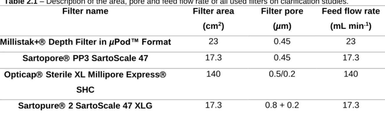
The bulk (1250 mL) was clarified in two different steps. For the first step, Millistak+ Depth Filter in µPod™ Format (MD0HC23CL3, Merck Millipore), Sartopore PP3 SartoScale 47 (5055306PS--FF--MM, Sartorius) and centrifugation were used. For the second step, Opticap Sterile XL Millipore Express SHC (KHGES015FF3, Merck Millipore) and Sartopure 2 SartoScale 47 XLG (5445307GS--FF--M, Sartorius) were utilized.
Centrifugation was performed on Centrifuge 5810 R (Eppendorf AG) at 200x g and 4 ºC for 10 minutes. Regarding filtration, a Tandem 1081 Pump (Sartorius Stedim Biotech) was used to control the feed flow rate, as well as a MasterFlex 16 tubes (MasterFlex Group) with 3.1 mm internal diameter. To control pressure through the filters, in-line pressure transducer (080-699PSX-5, SciLog) was used. The filtered volume was monitored over time using a technical scale (TE4101, Sartorius Stedim Biotech). All of the filtrations were performed at constant feed flow rate, as described in table 2.1.
Table 2.1 – Description of the area, pore and feed flow rate of all used filters on clarification studies.
Filter name Filter area (cm2)
Filter pore (µm)
Feed flow rate (mL min.1)
Millistak+ Depth Filter in µPod™ Format 23 0.45 23 Sartopore PP3 SartoScale 47 17.3 0.45 17.3 Opticap Sterile XL Millipore Express
SHC
140 0.5/0.2 140
Sartopure 2 SartoScale 47 XLG 17.3 0.8 + 0.2 17.3
It is important to emphasize that first, all filters were wet with ultrapure H2O (Mili Q water -
18.2 MΩ cm-1 at 25 ºC). The equilibration step and clarification operation were performed using a
buffer (50 mM HEPES, pH 7.4). Also, in the beginning, and at the end of the process, turbidity was measured by 2100Qis Portable Turbidimeter (2100QIS01, HACH).
2.5.2. Concentration studies
Concentration and diafiltration of influenza VLPs were evaluated by two types of membranes modules designs: HFs and cassettes. The HFs used were MidGee Ultrafiltration Cartridge with 1.0 mm internal diameters fibers and 0.0016 m2 fibers area (500000 MWCO pore size
(UFP-500-14
E-MM01A) and 750000 MWCO pore size (UFP750EMM01A), GE Healthcare). The cassettes used were Pellicon XL Ultrafiltration Module Biomax 0.005 m2 membrane area (300 kDa
(PXB300C50) and 500 kDa (PXB500C50), Merck Millipore). Both cassettes and HFs were set up accordingly with the manufacturer’s instructions. A Tandem 1081 Pump (Sartorius Stedim Biotech) was used, with MasterFlex 16 tubing (MasterFlex Group). The retentate was recycled to the feed container and, at the same time, the permeate was collected separately. To control and set up the TMP (see equation 2.1), an in-line pressure transducer (080-699PSX-5, SciLog) was used. The TMP was controlled over time by adjusting the retentate flow rate using a flow restrictor valve. Feed/retentate and permeate volumes were monitored over time using technical scale (TE4101, Sartorius Stedim Biotech).
TMP = (Pfeed+ Pretentate)
2 − Ppermeate
Every device was firstly washed with ultrapure H2O (Mili Q water - 18.2 MΩ cm-1 at 25 ºC) to
eliminate trace preservatives. Then, each one was equilibrated with buffer (50 mM HEPES, pH 7.4). The initial volume to perform UF and DF of influenza VLPs clarified bulk was carried out considering a ratio of 3 mL of feed volume per each cm2 of membrane area. In all experiments,
the volume of bulk was reduced 5-fold and diafiltrated other 5 times. DF was performed with the same buffer of equilibration (50 mM HEPES, pH 7.4).
Before starting, bulk recirculated (capped the permeate side of membranes) for 5 minutes. Over the process of UF/DF, the TMP was adjusted to 0.3 bar for HFs and 0.8 bar for cassettes, as well as, some samples were taken to analyse the recovery of HA quantity and the removal of impurities. At the end of the process, all the devices were submitted to cleaning-in-place (CIP) procedure. For HFs 0.5 M NaOH was added, followed by a 60 minutes incubation, and for cassettes, 0.1 M NaOH was added and subjected to the same time of incubation.
All these procedures were made at room temperature (RT) (20-22 ºC) and the final product of UF/DF was injected at chromatography column.
2.5.3. Chromatography studies
The chromatography studies were performed by two different devices, a membrane and a packed-resin.
Membrane chromatography step was performed by Sartobind Q nano 1 mL (96IEXQ42DN-11--A, Sartorius) membrane adsorber operated in positive mode (elution) at RT (20-22 ºC). This membrane adsorber was functionalized with a Q ligand. The runs were performed on ÄKTA Explorer 100 liquid chromatography system (GE Healthcare) equipped with UV and (2.1)
15
conductivity/pH monitors. System operation and data gathering/analysis were done using the UNICORN™ 5.01 software (GE Healthcare).
For DBC determination, the FT was analysed to guarantee that membrane adsorber was saturated. For this analysis, a buffer (50 mM HEPES, pH 7.4) was used for equilibration step and for running of experiments. The flow rate was set to 2 membrane volume per minute. To ensure the recuperation of all particles, an elution buffer (50 mM HEPES + 2 M NaCl, pH 7.4) was used. VLPs concentration along these fractions was determined by HA assay (see section 2.6.1.). Regeneration step was performed by 1 M NaOH.
In packed-resin chromatography step, HiTrap Q HP (17-1154-01, GE Healthcare) with 5 mL bed volume was used. It was functionalized with a Q ligand and operated at positive mode (elution). The runs were performed on ÄKTA Explorer 100 liquid chromatography system (GE Healthcare) equipped with UV and conductivity/pH monitors. System operation and data gathering/analysis were performed using the UNICORN™ 5.01 software (GE Healthcare).
UF/DF product was injected to determine DBC of the column. The runs were performed at a flow rate of 150 cm h-1 and at RT (20-22 ºC). Buffer (50 mM HEPES, pH 7.4) was used in
equilibration step and during the experiment, while elution buffer (50 mM HEPES + 1 M NaCl pH 7.4) was used to recover all HA linked to the resin.
The FT fractions were collected over time and HA assay was performed (see section 2.6.1.) to understand when the resin was already fully saturated. Elution was executed in gradient over 20 column volumes (CV) and fractions were analysed to evaluate at which NaCl concentration influenza VLPs start to elute off the resin. All those fractions were analysed to quantify the major impurity present. Regeneration of HiTrap Q HP was made with 1 M NaOH at same feed flow rate and temperature performed on runs.
2.5.4. Polishing studies
Polishing studies were evaluated through Sepharose™ 4 Fast Flow resin (17014901, GE Healthcare) based on SEC and HiScreen™ Capto™ Core 700 (17-5481-15, GE Healthcare) with the principle of multimodal chromatography. The runs were performed on ÄKTA Explorer 100 liquid chromatography system (GE Healthcare) equipped with UV and conductivity/pH monitors. System operation and data gathering/analysis were performed using the UNICORN™ 5.01 software (GE Healthcare). Sepharose 4 Fast Flow was packed on XK 16/20 Column (28988937, GE Healthcare) with 33.9 mL of bed volume.
The samples used for injection on both polishing column were collected from HiTrap Q HP column or Sartobind Q nano 1 mL (see section 2.5.4.). The equilibration and experiment run buffer was the same (50mM HEPES, pH 7.4) for both columns. The flow rates performed were
16
different and column dependent. For Sepharose 4 Fast Flow a flow rate of 119 cm.h-1 was
performed while for HiScreen™ Capto™ Core 700 a flow rate of 258 cm.h-1 was used. Due to the
characteristics of this last column, an elution buffer was used (50 mM HEPES + 1 M NaCl, pH 7.4).
All fractions (from FT and elution) were collected and analysed for HA quantification and impurities presence through corresponding analytical methods (see section 2.6.).
2.5.5. Final sterile filtration studies
On the final sterile filtration step, several syringe filters were evaluated (table 2.3). The various filters devices are described by filter area and filter material: RC, hydrophilic PES, hydrophilic PVDF and surfactant-free cellulose acetate (SFCA).
Table 2.2 – Syringe filters devices used with correspondent filter area.
Syringe filters devices Filter Material Filter area (cm2)
Whatman SPARTAN RC 30 syringe filters pore size 0.2 µm (10 462 960, GE Healthcare)
RC 5.7
Millex-GP Syringe Filter Unit, 0.22 µm (SLGP033RS, Merck Millipore)
Hydrophilic PES 4.5
Millex-GV Syringe Filter Unit, 0.22 µm (SLGV033RS, Merck Millipore)
Hydrophilic PVDF 4.5
Minisart NML Syringe Filter, 0.2 µm (16534---GUK, Sartorius)
SFCA 6.2
Minisart High Flow Syringe Filter, 0.22 µm (16532---K, Sartorius)
PES 6.2
Acrodisc Syringe Filters, Sterile, 0.2 µm (PN4602, PALL)
PES 1.0
Acrodisc PF Syringe Filters, 0.8/0.2 µm (PN4658, PALL)
PES 5.8
Acrodisc Syringe Filters, Sterile, 0.2 µm (PN4907, PALL)
PVDF 2.8
For these studies, the HA recovery evaluation and impurities removal were performed with samples from polishing step. In each filter, the tested volume was correspondent to the ratio of 1 mL cm-2.
17
2.5.6. Proof of concept
On proof of concept run, some modifications were performed. It was produced 2500 mL of influenza VLPs and for clarification step Millistak+ HC Pod Depth Filter (MD0HC021H1, Merck Millipore) with 270 cm2 of filter area was used with the difference on tubes, where MasterFlex
35 tubes (MasterFlex Group) with 7.9 mm internal diameter was utilized. The last modification was on concentration step, a Pellicon 2 Mini Ultrafiltration Module Biomax 0.1 m2 membrane
area and 300 kDa (P2B300C01, Merck Millipore) was used in Sartocon Slice 200 Holder (17525--01, Sartorius Stedim).
On final sterile filtration step, the filters used were Millex-GV Syringe Filter Unit, 0.22 µm (SLGV033RS, Merck Millipore), Millex-GP Syringe Filter Unit, 0.22 µm (SLGP033RS, Merck Millipore), Acrodisc Syringe Filters, Sterile, 0.2 µm (PN4602, PALL) and Acrodisc Syringe Filters, Sterile, 0.2 µm (PN4907, PALL) with specifications described on table 2.2.
2.5.7. Magnetic Sulphated Cellulose Particles studies
MSCPs were kindly provided by Max Planck Institute for Dynamics of Complex Technical Systems Magdeburg, Germany. The production of MSCPs is described elsewhere58.
All experiments were done in 50 mL tubes and the agitation of them was performed by hand following the protocol illustrated at figure 2.1. MSCPs were equilibrated 5 times (duration of 1 minute each) with equilibration buffer. The addition of sample – Influenza VLPs – was followed by a binding step of 10 minutes. The MSCPs were washed 3 times (duration of 1 minute each) with the equilibration buffer ensure that unlinked particles were completely removed. The elution step was performed with 2 different concentrations of NaCl. Each one had the duration of 5 minutes. In all those steps (except equilibration), a sample was collected for analysis. In the end, the MSCPs were regenerated with 1 M NaOH for 10 minutes, then washed with equilibration buffer until pH was the same as the buffer pH. Lastly, they were stored at 4 ºC in 20 % ethanol. All mentioned buffers are referred in table 2.3.
18
Figure 2.1 – Magnetic Sulphated Cellulose Particles (MSCPs) experiment steps.
In these experiments, the ratio between HA quantity and mass of beads was evaluated. The used mass of MSCPs was (i) 0.8 g; (ii) 1.2 g; (iii) 1.5 g; (iv) 2 g, but the initial quantity of HA was always the same. The sample was previously prepared for these experiments by being desalted on HiPrep 26/10 Desalting (17508701, GE Healthcare Life Science) on ÄKTA Explorer 100 liquid chromatography system (GE Healthcare). The applied flow rate was 170 cm h-1 and running buffer
was the same as the one used on MSCPs experiments at both equilibration and washing steps.
Table 2.3 – Buffers for equilibration, washing and elution steps for Magnetic Sulphated Cellulose Particles
experiments.
Equilibration and washing step Elution steps Phosphate Buffer Saline (PBS)
(-/-), pH 7.4 PBS (-/-) + 1 M NaCl, pH 7.4 PBS (-/-) + 2 M NaCl, pH 7.4 PBS* (-/-), pH 7.4 PBS* (-/-) + 1 M NaCl, pH 7.4 PBS* (-/-) + 2 M NaCl, pH 7.4 10 mM TRIS + 50 mM NaCl, pH 7.4 10 mM TRIS + 1 M NaCl, pH 7.4 10 mM TRIS + 2 M NaCl pH 7.4
PBS* - Phosphate Buffer Saline made with low NaCl concentration (20mM).
2.6. Analytical methods
2.6.1. Hemagglutination assay (HA assay)
HA concentration on each sample was obtained by HA assay, based on the protocol described elsewhere67 with some changes. Briefly, in 96-wells microtiter plates with V bottom
(611V96, Thermo Scientific) the samples were initially diluted (1:2 and 1:3) in D-PBS (14190-094, Gibco™). Two-fold serial dilutions to take a range of 11 concentration values, with a final volume of 50 µL, were made. As a positive control, a commercially available vaccine (ISTIVAC, Sanofi