PANDEMIC INFLUENZA AND THE SWINE INFLUENZA VIRUS1
M.A.W. Hattwick,2 R. J O’Brien,’ C. H. Hoke, Jr.,’ and W. R. Dowdle4
Evidence points to a repeating, cyclic pattern in the appearance of antigenic shifts in the influenza A virus. Development of the
swine influenza strain isolated at Fort Dix, New Jersey, similar to the one that sparked the 1918 pandemic, had been predicted three years earlier.
Influenza Virus
Influenza virus was first isolated from swine in 1930 (I) and from man in 1933 (2). The virus consists of a nucleoprotein core
composed of single-stranded RNA with a
segmented genome coding for seven recog- nized viral proteins (3). The nucleoprotein is encapsulated by a double membrane con- taining a virus-coded inner protein layer and an outer lipid layer of host cell origin.
Two proteins, the hemagglutinin (HA) and
the neuraminidase (NA), project from the
virus surface. Influenza viruses isolated
from man have contained four distinct
hemagglutinins and two distinct neurami- nidases. At least 11 additional types of
hemagglutinins and seven additional
‘Extract from a longer paper, “Pandemic influenza, the swine influenza virus, and the national influenza program,” prepared under the auspices of the Center for Disease Control, U 3. Public Health Service; also appearing in BoE Of Sanit Panam, 1977.
‘Chief, Respiratory and Special Pathogens Branch, Viral Diseases Division, Bureau of Epidemiology, Center for Disease Control, U.S. Public Health Service, Atlanta, Georgia, U.S.A.
SRespiratory and Special Pathogens Branch, Viral DiseasesDivision, Bureau of Epidemiology, Center for Disease Control, U.S. Public Health Service, Atlanta, Georgia, U.S.A.
4Director. Virology Division, Bureau of Labora- tories, Center for Disease Control, U.S. Public Health Service, Atlanta, Georgia, U.S.A.
neuraminidases have been found in viruses
isolated from avian, equine, and other
animal hosts (4).
Nomenclature for the influenza virus is standardized (4). Each virus is identified by a distinct signature consisting of: (1) the type of nucleoprotein core (A, B, or C); (2) the animal host from which the virus was isolated; (3) the location of the laboratory in
which the isolation was made; (4) the
laboratory number for that particular
isolate; and (5) the year in which the
isolation was made. The designation for
influenza A viruses also includes (6) the type of hemagglutin and (7) the type of
neuraminidase present. For example, an
influenza virus isolated from a swine in Michigan in 1965 is designated A/swine/
Michigan/2/65(HswlNl). For viruses iso-
lated from man, the host designation is not
included in the name. For example,
A/Victoria/3/75(H3N2) refers to the strain which caused the 1975-1976 epidemic. The virus responsible for the recent “swine-type influenza” in man is identified as A/New Jersey/8/76(HswlNl).
One of the unique characteristics of the influenza virus is that the hemagglutinin
and the neuraminidase undergo frequent
antigenic changes. Minor changes, which
occur relatively often, are referred to as
284 PAHO BULLETIN l vol. X, no. 4, 19 76
antigenic drifts. Antigenic drift is thought to result from the selection of mutants through increasing antibody pressure in the
human population. At longer intervals,
major changes, referred to as antigenic shifts, take place in the hemagglutinin or neuraminidase. The origin of these shifts remains uncertain: they might be caused by the direct transmission of antigenic variants
from reservoirs in human or nonhuman
hosts: they could be the result of genetic
recombination of human and animal
strains of influenza; or they might simply be the end product of a series of mutations (5, 6).
Epidemiology of Influenza
The occurrence of influenza epidemics
depends upon a poorly understood interac- tion of virus, population susceptibility, and
environmental conditions. When condi-
tions are suitable, epidemics may occur
rapidly, affecting large numbers of people and causing many deaths.
Both the influenza A and B viruses have
been associated with major epidemics in
human populations, but only influenza
A has been consistently associated with
pandemics. Pandemics are major epidemics
due to a single virus type which sweep
around the world in a short period of time and cause marked increases in mortality. They are associated with major antigenic shifts. Human populations with no previous exposure to newly appearing antigens have
no immunity and hence no protection
against the-new strain.
Following an antigenic shift and the as- sociated pandemic, a period of interpan- demic influenza ensues. During these years a series of smaller epidemics takes place due to viruses which are related to the previous pandemic strain but which have slight dif- ferences in the surface antigens (antigenic drifts). Morbidity and mortality associated with these epidemics are much lower than in the preceding pandemic.
The rapidity with which pandemic
influenza can spread is a phenomenon
unique in the history of infectious diseases. In the well-studied pandemics of 1957 and 1968 the time lapse from the initial report- ing of isolation of a new strain of influenza to the dissemination of disease to all major areas in the world was under six months. The first outbreak of disease in 1957 was reported in China in late February (7, 8). By early J une cases of illness due to this new strain, subsequently called “Asian flu,” had already begun to occur in the United States of America, and by the end of October
outbreaks were being reported from all
parts of the country. A similar pattern was seen 11 years later with the Hong Kong strain: it was first isolated from cases in Asia on 17 July 1968 (9), outbreaks in the United States began in September, and by December the disease had extended to the entire country (IO).
The reasons for the rapid spread of influ- enza are only partly understood. Doubtless- ly important are: the short incubation period (24-48 hours), the large size of the population susceptible to infection, and the virus’ easy transmissibility. The fact that epidemics occur most often in the winter suggests that cold weather may also play a
role. Since it has been observed that
widespread simultaneous outbreaks develop
following a period of sporadic cases, it has been postulated that virus is seeded in the susceptible population by mildly ill indi- viduals.
Morbidity and Mortality
Hattwick et al. l SWINE INFLUENZA VIRUS 285
F&m 1. Excess mortality during epidemic periods, United States of America, 1911-1976.
Source: Data from United States Public Health Service, Center for Disease Control, Bureau of Epidemiology
patients place a heavy burden on available
medical and emergency-room services.
Moreover, the illness-associated absenteeism has a nationwide effect. Finally, complica- tions, such as bacterial superinfections, often result in hospitalization.
The impact of influenza pandemics has
been measured since the early 1800’s by the
number of deaths occurring during the
epidemic in excess of the number normally expected from all causes during the period in question (Figure 1). Especially evident are excess deaths in the cause category “pneumonia and influenza.” In the United States the excess deaths in this category are almost always due to influenza. Mortality is usually highest among the elderly and in
persons with such chronic conditions as
pulmonary or cardiovascular disease. Dur-
ing the pandemics-as in 1918, 1957, and
1968-mortality has been most marked at
the beginning. Excess mortality has also
been observed in interpandemic years
(Table 1).
The worst influenza pandemic on record occurred in 1918. It caused approximately half a million deaths in the United States of America and an estimated 20 million deaths worldwide (II). Estimated death rates in the United States ranged between 400 and 598 per 100,000 population (II, 12, 23), and rates as high as 6,000 per 100,000 were
reported from some countries and in
concentrations of military personnel (II, 14). An unusual feature of this pandemic was that excess mortality was most marked
in persons aged 20 to 40. The recent
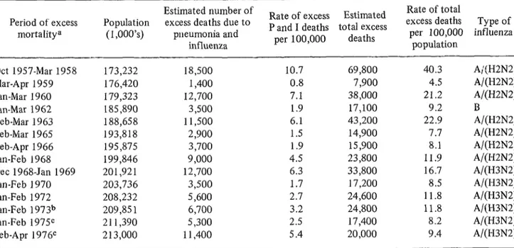
Table 1. Excess mortality due to pneumonia and influenza, 1957-1976.
Period of excess mortalitya
Estimated number of Rate of total
Population excess deaths due to Rate of excess Estimated P and I deaths total excess excess deaths Type of (I ,000’s) pneumonia and per 100,000 influenza
influenza per 100,000 deaths population Ott 1957-Mar 1958 173,232 18,500
Mar-Apr 1959 176,420 1,400 Jan-Mar 1960 179,323 12,700 Jan-Mar 1962 185,890 3,500 Feb-Mar 1963 188,658 11,500 Feb-Mar 1965 193,818 2,900 Feb-Apr 1966 195,875 3,700 Jan-Feb I968 199,846 9,000 Dee 1968-Jan 1969 201,921 12,700 Jan-Feb 1970 203,736 3,500 Jan-Feb 1972 208,232 5,600 Jan-Feb 1973b 209,85 1 6,700 Jan-Feb 1975C 211,390 5,300 Feb-Apr 1 976c 213,000 11,400
10.7 69,800 40.3 A;(H2N2) 0.8 7,900 4.5 A/(H2N2) 7.1 38,000 21.2 A/(H2N2)
1.9 17,100 9.2 B
6.1 43,200 22.9 A/(H2N2) 1.5 14,900 7.7 A/(H2N2) 1.9 15,900 8.1 A/(H2N2) 4.5 23,800 11.9 A/(H2N2) 6.3 33,800 16.7 A/(H3N2) 1.7 17,200 8.5 A/(H3N2) 2.7 24,600 11.8 A/(H3N2) 3.2 24,800 11.8 A/(H3N2) 2.5 17,400 8.2 A/(H3N2) 5.4 20,000 9.4 A/(H3N2) aNo excess mortality observed in 1961, 1964, 1967, 1971, or 1974.
bBased on a 10% sample of mortality data from the National Center for Health Statistics. The mortality data for the earlier periods are based on final NCHS data.
Hattwick et al. . SWINE INFLUENZA VIRUS 287
In the United States during the winter of
1957-1958 there were some 69,800 excess
deaths, and in 1968 the figure calculated was 33,800 excess deaths.
Virologic Characterization
The understanding of influenza virology and epidemiology has increased as laborato-
ry techniques have improved. Precise
virologic characterization of the influenza
A viruses according to type of surface
antigen can be made only for those viruses that have been present since the 1930’s, when viral isolation first became possible. However, from serologic studies of indi- viduals born since the mid-1800’s, infer- ences have been made about the antigenic character of earlier strains of virus. These studies have been possible because persons infected with an influenza virus maintain antibody throughout life against the surface antigens of the strains by which they were first infected, an immunologic phenomenon referred to as the “doctrine of original antigenic sin” (16).
Evidence now suggests that antigens
which were prevalent at one time may again
become prevalent when population immu-
nity wanes. Some investigators have in-
terpreted serologic data to indicate that
viruses related to the 1957-1958 Asian
strains (H2) were prevalent in the late nineteenth century (17). These strains were apparently followed in turn (18, 19, 20) by
ones whose hemagglutinin type (H3) was
similar to that of A/Hong Kong/l/68
(HSN2), although the neuraminidase was
quite distinct (21), being more similar to that of the equine Neq2. Similar serologic studies indicate that the 1918 pandemic was
due to an HswlNl virus which had been
isolated only from swine until its first isola- tion from man two years ago (22).
As shown in Table 2, four major strains
of human influenza virus have been
isolated since the 1930’s: A/Puerto Rica/8/
34 (HONl), A/Fort Monmouth/l/47
Table 2. History of influenza A in man.
1880-
1910?
1918
1933 1934 1946
1957
1958- 1967 1968- 1969 1969- 1976 Feb 1976
HZ and H3 hemagglutinins may have
circulated sequentially as determined from serologic studies of antibody preva- lence in persons who were children during these years.
Pandemic probably due to (HswlNl)-like virus as indicated by later serological studies.
A/Walter Smith/33 (HONl). First influ- enza virus isolated from man.
A/Puerto Rico/B/34 (HONl) isolated.
A/Fort Monmouth/l/47 (HlNl). Not a
true antigenic shift from HONl. Not associated with a pandemic.
A/Japan/305/57 (H2N2). Antigenic shift in both antigens. Caused a major pan- demic.
Several epidemics due to (H2N2) viruses associated with antigenic drifts.
A/Hong Kong/l/68 (H3N2). Antigenic
shift in hemagglutinins only. Caused a moderate pandemic.
Several epidemics due to (HSN2) viruses associated with antigenic drifts.
A/New Jersey/B/76 (HswlNl) isolated.
(H lNl), A/J span/305/57 (H2N2), and
A/Hong Kong/l/68 (H3N2). Virologic
characterization at the time of the 1957- 1958 pandemic (Japan strain) indicated that the hemagglutinin and the neuraminidase were totally different from those of the earlier viruses. Smaller epidemics during the next 10 years were caused by viruses related to the HZN2 reference strain. The most recent pandemic occurred in 1968 and was caused by a strain whose reference was
designated A/Hong Kong/l/68 (H3N2). In
this strain an antigenic shift in the
hemagglutinin, but not the neuraminidase, had occurred. Since 1968 there have been six epidemics due to related H3N2 viruses,
the most recent being due to strains
designated A/Port Chalmers/l/73 (H2N2)
288 PAHO BULLETIN l vol. X, no. 4, 1976
The fact that H2 and H3 have occurred in sequence over the past 20-year period has given rise to the hypothesis that H2, H3, and Hswl may have also circulated sequen- tially in the late nineteenth and early twentieth century. It has been proposed that
a limited number of hemagglutinin anti-
gens exist and that they occur in a recycling pattern (17, 23, 24). Indeed, Masurel and Marine predicted in 1973 that a swine-like
influenza A virus might recur in man
during the latter part of this century (25).
Swine Influenza Virus
Swine influenza virus (e.g., A/swine/
Tennessee/75 (HswlNl) is distinguished by
its surface antigens (4). Some antigenic variation among strains is recognized (26).
Isolation is usually made in eggs. The
clinical disease in swine was first recognized in 1918, and the virus was first isolated in 1930 from swine (I). The available evidence suggests that the virus spread from man to pigs during the 1918 pandemic (1). Exten- sive studies since 1930 have confirmed that
this virus commonly produces disease in
swine every year and may be present today in about half of the swine herds in the United States (27, 28). The illness in swine consists of fever and upper respiratory
symptoms; death in younger pigs and
abortions in sows may occur.
Serologic Studies in Man
Although the virus is common in swine, there has been little evidence that it was
reentering the human population (29).
Serologic studies of the general human
occasional sporadic human infection with
mild symptoms, if any, and no secondary spread. Occupational exposure to the virus may also have resulted in immunization without infection. Interpretation of these serosurvey data must take into account that during the 1950’s and early 1960’s military personnel were immunized with a vaccine containing swine antigen and that infection
or vaccination with H3N2 viruses may
produce heterologous antibody responses in some cases (G. R. Noble, personal commu- nication).
Infection in Man
Until recently there was no direct con- firmation that A/swine virus antibodies in younger individuals resulted from clinical- ly apparent infections. Since 1974, however,
evidence has begun to accumulate that
human influenza due to infections with
A/swine-like viruses may in fact occur
(Table 4). That year a swine-like influenza
virus, A/Mayo Clinic/103/74 (HswlNl),
was isolated postmortem from the lungs of a 16-year-old boy who died of pneumonia- complicated progressive respiratory insuffi- ciency due to Hodgkin’s disease. Prior to his terminal illness, the patient lived on a farm
that had pigs which were subsequently
shown to have antibody against swine
influenza virus (22).
In October 1975 a previously healthy
8-year-old boy in rural Wisconsin developed
a febrile illness of unknown etiology.
Paired sera showed seroconversion to the
A/swine virus. This child also lived on a
farm where pigs with swine influenza
antibodies were present. Investigation of the
population show a high prevalence of family showed that of seven household
antibodies to the A/swine virus in persons members, five (ages three, four, seven, nine, old enough to have been exposed to and 33) had titers against the A/swine virus, influenza prior to the late 1920’s (Table 3). suggesting past infection. None had evi- The surveys also show increased prevalence dence of recent H3N2 infection. None of 24
of antibodies in younger individuals who classmates of the boy had evidence of
have been occupationally exposed to swine. infection, and the prevalence of A/swine
Hattwick et al. l SWINE INFLUENZA VIRUS 289
Table 3. Summary results of serologic surveys to determine the prevalence of A/swine influenza antibody in US. populations.
Year Location Population Age at time blood was drawn
No. with titers/No. tested (% of population)
1966= Illinois General public (Statewide) Persons occupationally
exposed to swine 1971b Atlanta, Community members
Georgia
1976c Atlanta, Community members Georgia
1976 Sheboygan, Community members Wisconsin
1976 Fayetteville, Community members Pennsylvania
(15 16-29 30-45 745
1/2oo(s)d l/247(.4) 39/242( 16) 116/151(77)
- 17/182(9.3) 81/413(20) 251/345(72)
116 17-31 3246 752
l/161(.6) 14/163(8.6) 43/112(38) 217/250(87)
520 21-30 31-40 41-50 750
l/37(3) m5w 9/33(27) 9/25(36) 27/27( 100)
515 16-29 3049 >50
4/156(3) 2/25(S) 2/19(11) 33/88(87)
115 16-29 30-49 750
‘J/60(0) 5/38(13) 9/52( 17) 46/52(88) akhnurrenberger, et al. (29).
bCourtesy of Dr. William Marine. CCourtesy of Dr. Gary Noble.
dNumber having titers/number tested (per cent population).
Table 4. Summary of cases of influenza due to A(HswlNlj-Iike viruses and of investigations of man-to-man transmission.
Location
Investigation of spread Age Sex Onset Diagnostic
evidence Household Local Other parts
contacts contacts of community
-~- ----~_
Mayo Clinic, Rochester, Minnesota
16= M
Sheboygan, Wisconsin
8a M
Fort Dix, New Jersey
19 M
Charlottesville, 40a F Virginia
55 M
July- Sept 1974
Ott
1975
Feb 1976
4 cases 6 cases 5 Dee 1975 27 Dee 1975
Virus isolation on postmortem examination Seroconversion and flu-like illness
Virus isolation on postmortem examination Isolations Seroconversion Pneumonia and seroconve*Slon Pneumonia and seroconversion
2 breeder sows had antibodies; boy’s parents were negative 5 of 7 household members had Hl titers ? 20; none had titers for A/Kc. 34% positive titers in platoons with confirmed cases
-
O/5 chddren positive O/4 cases in household
O/24 classmates Age Post/tesred % had titers I50 33138 87 16-49 4144 9 L15 4/156 3 2731132 1
(21% ) single sera positive
6% positive titers in platoons without confirmed cases
- -
O/4 cases in -
close neighbors
290 PAHO BULLETIN l vol. x, no. 4, 1976
nity was similar to that observed in other serosurveys (30).
In January and February 1976, during
an outbreak at Fort Dix, New Jersey, of
human influenza due to A/Victoria
(H3N2), swine influenza-like viruses-
including what is now the reference strain,
A/New Jersey/W76 (HswlNl)-were iso-
lated from five recruits, oneof whom died of acute viral pneumonia. Six additional cases were confirmed by fourfold rises in titer, and a serologic survey based on single serum specimens indicated that several hundred cases of infection due to A/NJ/8/76-like viruses may have occurred. Thirty-four per cent of 110 close contacts of confirmed cases were found to have A/swine virus antibody, while only 6 per cent of those with no direct contact with known cases had antibody. A
comparison of 10 hospitalized cases of
A/New Jersey influenza with 10 cases of
A/Victoria influenza at Fort Dix during the same period indicated that their illnesses
were similar but that A/New Jersey
influenza may have been a somewhat
milder illness (P. Russell, personal commu-
nication). By early March the Fort Dix
outbreak was subsiding, and most of the
cases that had occurred since early February had been due to A/Victoria (H3N2).
Following the Fort Dix outbreak, the
U.S. Center for Disease Control investigated
family members and close contacts of 22
recruits who: had entered Fort Dix during the first two weeks in January, reported swine contact prior to entry, and demon- strated antibody against the A/swine virus
(30). Of 171 civilian contacts of these
Table 5. Investigation of close contacts of 22 recruits at Fort Dix, New Jersey, with history of swine contact and swine antibody.
Community of origin No. No. with HI 2 20
tested (ages)
Arley, Alabama Headland, Alabama Thornton, Colorado Lyons, Colorado Laurel, Delaware Kellogg, Iowa
Franklin, Massachusetts Belding, Michigan Munnsville, New York Everington, Ohio Columbus, Ohio Valencia, Pennsylvania Erie, Pennsylvania Fayetteville, Pennsylvania Newville, Pennsylvania Hemingway, South Carolina Bristol, Tennessee
Woodstock, Virginia Toppenish, Washington Tacoma, Washington Mannington, West Virginia Wheatland, Wyoming Total
4 4 9 3 4 5 4 13 9 1 2 4 7 11 4 4 2 10 20 35 11 5 171
1 (42) 1 (61)
1 (50) 0 0 0 0 0 0 0 0 0
Ia(lI, 12, 20,25) I(50)
2 (60, l&y 0
3 (47,49, 75) 2 (56,32) 3 (60, 53, 59) 1w
Hattwick et al. l SWINE INFLUENZA VIRUS 291
recruits in 22 communities, 19 were found Further surveys in 1976 have revealed a
to have antibody titers against A/swine number of individuals who have had
influenza (Table 5). In one family from contact with pigs and have elevated A/swine
Fayetteville, Pennsylvania, four indi- antibody titers. In addition, two patients
viduals (ages 11, 12, 20, and 25) had titers with pneumonia and seroconversion against
suggesting past infection with A/swine A/swine were found in Charlottesville,
influenza. None had a history of swine Virginia. One of these patients had no
contact, suggesting that person-to-person history of swine contact and no evidence of
transmission had taken place. Investiga- A/Victoria infection, Investigation did not
tion, however, did not indicate that there indicate spread to immediate contacts or the had been any spread to the community. community (31).
SUMMARY
The hemagglutinin and the neuraminidase that project from the surface of the influenza A virus undergo frequent antigenic changes. The minor changes, which occur relatively often, are referred to as antigenic drifts. They are associated with small epidemics. The major changes, antigenic shifts, involve a total modi- fication in the nature of the hemagglutinin and/or the neuraminidase. Since much of the world’s population has no previous immunity against the newly composed virus, the shift brings with it a pandemic that spreads with unique rapidity.
The influenza A viruses isolated from man since the 1930’s have contained four distinct hemagglutinins and two distinct neuraminida- ses. The observation that people maintain high antibody throughout life to the influenza virus that first infects them led to serologic studies of the older population in the United States of America. The results indicated that the hemag- glutinin prevalent in 1957-1967, known as H2, and that appearing in 1968, designated H3, may well have circulated in the same sequence over the period 1880-1910. These conclusions gave rise in turn to the hypothesis that a limited number of hemagglutinin antigens exist-per- haps four that are pathogenic for man -and that they occur in a recycling pattern. Thus it was
predicted in 1973 that an influenza A virus similar to the one that sparked the tragic 1918 pandemic (Hswl-designated with the “SW” because swine are involved in the transmission cycle) might soon appear.
Isolations of such a virus were in fact made in 1974 and 1975, but in each case the patient had had contact with pigs and careful investigation revealed no further spread in the community. The outbreak at Fort Dix, New Jersey, in 1976 was the first indication since 1918 that man-to- man transmission of Hswl had occurred. A virus containing the Hswl antigen was isolated from five recruits, six additional cases were confirmed by fourfold rises in titer, and a serologic survey indicated that there may have been several hundred such infections.
Influenza has far-reaching importance be- cause of its tremendously high morbidity. Death can ensue, especially when the disease is complicated by pneumonia, and even though the case-fatality rate as such is not high, the great numbers of cases mean that many excess deaths do in fact occur. Moreover, a pandemic causes extensive absenteeism, at cost to the national economy, and places an almost overwhelming burden on medical care, hospital, and other health services.
REFERENCES
(I) Shope, R. E. Swine influenza. III. Filtra- (3) Choppin, P. W., andR. W. Compans. The tion experiments and etiology. J Ex# Med structure of influenza virus. In: E. Kilbourne
542373-380, 1931. (ed.), The Influenza Viruses and Influenza.
292
PAHO BULLETIN lvol. X, no. 4, 1976
(5) Dowdle, W. R., M. T. Coleman, and M. B. Gregg. Natural history of influenza type A in the United States, 1957-1972. Prog Med Vi4
17:91-135, 1974.
(6) Webster, R. G., and W. G. Laver. Antigenic variation of influenza viruses. In: E. Kilbourne (ed.), The Influenza Viruses
and
ZnfZuenza. New York, Academic Press, 1975. pp, 270-314.(7) U.S. Public Health Service, Communica- ble Disease Center. Asian Influenza, 1957-1960. Atlanta, 1960.
(8) Dunn, F. L. Pandemic influenza in 1957: Review of international spread of new Asian strain.JAMA 166:1140-1144, 1958.
(9) Chang, W. K. National influenza experi- ence in Hong Kong, 1968. Bull WHO 41: 349- 351, 1969.
(10) Sharrar, R. G. National influenza expe- rience in the USA, 1968-1969. Bull WHO 41: 361-366, 1969.
(II) Jordan, E. 0. Epidemic influenza of 1918.JAMA 89:599, 1603,1689, and 1779; 1927. (12) Frost, W. H. The epidemiology of influenza. Public Health Re$ 34:1823-1836, 1919.
(13) Collins, S. D., and J . Lehmann. Excess deaths from influenza and pneumonia and from important chronic diseases during epidemic periods, 1918-1951. U.S. Public Health Service Publication 213, Public Health Monograph. Washington, Federal Security Agency, Public Health Service, 1953. p. 21.
(14) Hoehling, A. A. The Great Efiidemic. Boston, Little Brown and Company, 1961. p. 217.
(15) Kavet, J. Influenza and PubZic Policy. Doctoral Thesis, Harvard University, School of Public Health. Cambridge, 1972. p. 369.
(16) Francis, T. On the doctrine of original antigenic sin. Proc Am Philos Sot 104:572-578, 1960.
(17) Mulder, J ., and N. Masurel. Pre-epi- demic antibody against 1957 strain of Asiatic influenza in serum of older people living in the Netherlands. Lancet 1:810-814, 1958.
(18) Masurel, N. Serological characteristics of a “new” serotype of influenza A virus: The HongKongstrain. BuZZ WHO 41:461-468, 1969.
(19) Fukumi, H. Interpretation of influenza antibody patterns in man; Existence and
significance of Hong Kong antibody in old people prior to the Hong Kong influenza epidemic. Bull WHO 41:469-473, 1969.
(20) Davenport, E. M., E. Minuse, and A. V. Hennessy. Interpretations of influenza antibody patterns in man. Bull WHO 41:453-460, 1969.
(21) Fedson, D. S., M. A. Huber, J. A. Kasel, et al. Presence of A/Equi-2 hemagglutinin and neuraminidase antibodies in man. Proc Sot Exfi BioZ Med 139:825-826, 1972.
(22) Smith, T. F., E. 0. Burgert, W. R. Dowdle, et al. Isolation of swine influenza virus from autopsy lung tissue of man. N EngZ J Med 294:708, 1976.
(23) Davenport, F. M., A. V. Hennessy, and T. Francis. Epidemiologic and immunologic significance of age distribution of antibody to antigenic variants of influenza virus. J Ex# Med 98:641-656, 1953.
(24) Schoenbaum, S. S., M. T. Coleman, W. R. Dowdle, and S. R. Mostow. Epidemiology of influenza in the elderly; Evidence of virus recycling. Am J Efiidemiol 103:166-173, 1976.
(25) Masurel, N., and W. M. Marine. Recy- cling of Asian and Hong Kong influenza virus hemagglutinins in man. Am J Epidemiol 97:44-49, 1973.
(26) Meier-Ewert, H., A. J. Gibbs, and N. J. Dimmoch. Studies on antigenic variation of the hemagglutinin and neuraminidase of swine influenza virus isolates. J Gen ViroZ 6:409-419,
1970.
(27) Easterday, B. C. Immunological consid- eration in swine influenza. JAMA 160:645-648, 1972.
(28) Young, G. A., and N. R. Underdahl. An evaluation of influenza in midwestern swine. Am J Vet Res 16:545-552, 1955.
(29) Schnurrenberger, P. R., G. T. Woods, and R. J. Martin. Serologic evidence of human infection with swine virus. Am Rev Respz’r Dis 102:356-361, 1970.
(30) U.S. Public Health Service, Center for Disease Control. Influenza morbidity and mor- tality weekly report. In: Morbidity and Mortali- ty Weekly Reports 25:6-15, 1976.