Knock, knock-let the bacteria in:
enzymatic potential of plant associated
bacteria
Lorena Carro1and Esther Mene´ndez2 1
Microbiology and Genetic Department. University of Salamanca. Salamanca. Spain,2ICAAM - Instituto de Cieˆncias Agra´rias e Ambientais Mediterraˆnicas, Laborato´rio de Microbiologia do Solo, University of E´vora, E´vora, Portugal
Chapter outline
13.1 Introduction 169
13.2 Amylases 170
13.3 Cellulases, pectinases and xylanases 171
13.4 Chitinases 172
13.5 Lectins 173
13.6 Lipases 174
13.7 Proteases 174
13.8 Conclusions and future perspectives 175
References 175
13.1 Introduction
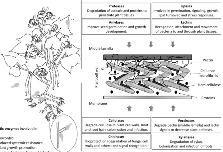
Plants are not considered as whole individual independent entities any more, being now considered as whole communities at which microorganisms play fundamental roles; thus, plant microbiomes should be studied together with their host plants (Carro and Nouioui, 2017). The interactions between plants and their surrounding habitants are in con-stant readjustments and hundreds of microorganisms reach plant tissues every day. Since a few years, it has become clear that an optimum equilibrium in these processes is fundamental to maintain a correct health status of the plants, with small distortions generating the development of diseases or sub-developed conditions. The interaction of bacteria with plants have been focused for years on three main groups: (i) nitrogen fixing interactions of rhizobia with legumes and to a lesser extent of Frankia with actinorhizal plants, (ii) symbiosis with arbuscular mycorrhizae, and (iii) pathogen-esis (Farrar et al., 2014). However, metagenomic studies opened a door to a whole diversity of interactions between microorganisms and plants, becoming the focus of study of many groups around the world (Dong et al., 2018; Navra´tilova´ et al., 2018; Wang et al., 2018). In the same line, the study of enzymes, and specifically, hydrolytic enzymes have been mainly related to plant pathogens and their colonization methods, including the degradation of plant tissues at final steps (King et al., 2011; Yarullina et al., 2016). Nevertheless, this new idea of different microorganisms able to colonize plant tissues without side effects for the plant has originated the search of other functions for these compounds, exemplified by their role as recognition molecules. Mainly when their expression in colonization and endo-phytic life, contrary to what was previously expected for these enzymes, was more frequently found up-regulated than down-regulated (Zhang et al., 2015). Endophytes are able to live asymptomatically inside plants at any stage of growth, providing additionally resources for the plants with the production of enzymes, nutrients and secondary metabolites that can be used by the plant to protect itself from biotic and abiotic stresses. Many of these enzymes produced by the bacte-ria are extracellular enzymes, generating their activities outside of their cells, with direct implications for their environ-ments, or in the case of endophytes, for their host (Khan et al., 2017). Between the most commonly studied enzymes previously detected in beneficial bacteria are amylases, cellulases, chitinases, lipases, pectinases, proteases, chitinases, and xylanases (Fig. 13.1), which will be the focus of this study.
169
Molecular Aspects of Plant Beneficial Microbes in Agriculture. DOI:https://doi.org/10.1016/B978-0-12-818469-1.00014-6
13.2 Amylases
Amylases are a class of enzymes that catalyse the hydrolysis of starch into glucose and maltose sugars. Three main groups of amylases have been described: α-amylases (EC 3.2.1.1), hydrolase enzymes that catalyse the hydrolysis of α-1, 4-glycosidic linkages producing glucose and maltose; β-amylases (EC 3.2.1.2), exo-hydrolases that hydrolyzed α-1, 4-glucan linkages into maltose units; and γ-amylases (EC 3.2.1.3), enzymes that cleaves α(1 6) and α(1 4) gly-cosidic linkages yielding glucose (Sundarram and Murthy, 2014). Amylases are constitutive enzymes which require cal-cium to stabilize its conformation and are tolerant to acidic conditions. However, its action could be inhibited by heavy metals (Yarullina et al., 2016). They could be found in several taxonomic groups, including fungi, oomycetes and bacte-ria. Within the bacteria amylases, some calcium-independent amylases have been described, although their tolerance to temperatures are more limited (Yarullina et al., 2016). Although the production of these enzymes have been mainly related to the penetration and degradation of plant tissues by pathogens, latter studies have confirmed its induction dur-ing germination of several plants by growth promotdur-ing bacteria (Islam et al., 2016). During the germination process, amylases are crucial for the hydrolysis of seed’s endosperm, rich on starch, which allow the development providing the necessary energy for root and shoot development. Therefore, amylase induction and amylase production of plant growth promotion bacteria (PGPB) can improve plant establishment and general growth of the plants (Duarah et al., 2011). Moreover, the induction ofα-amylases have been also related to the production of phytohormones, able to stimulate the plant growth, as for example the gibberellins (Kaneko et al., 2002).
It has been observed for several PGPB microorganisms belonging to distant phylogenetic taxa that amylases produc-tion is variable at different growth stages (Khan et al., 2017). Although it has been detected in many endophytes and its production is also over-expressed in contact with the plants, there are almost no studies showing its direct implication in the penetration of plant tissues. One of the hypotheses is that amylases help other enzymes in the formation of pores within roots, stems or leaves to allow bacterial colonization. However, more studies should be done to confirm at which level these enzymes are implicated in the colonization or just as helpers in tissues recycling.
13.3 Cellulases, pectinases and xylanases
Cellulase and xylan are the most abundant polysaccharides forming part of the plant cell walls. Several enzymes acting alone or in synergy are required to deconstruct those polysaccharides (Nguyen et al., 2018). The term cellulase is exten-sive to 3 different kinds of enzymes: endoglucanases (endo-1,4-β-D-glucanase; EC 3.2.1.4), which cleave ß-1,4-glucan linkages, exoglucanase (exo-1,4-β-D-glucanase; EC 3.2.1.91/176), which degrade the rest of the polysaccharide chain originating cellobiose disaccharides andβ-glucosidases (EC 3.2.1.21), which catalyse the breakdown of cellobiose dis-accharides (Mene´ndez et al., 2015). These enzymes act in synergy in order to hydrolyze cellulose. There are also 3 types of xylanases: endo-ß-1,4-xylanases (EC 3.2.1.8), which target the xylan backbone in plants and algae, endo-ß-1,3-xylanases (EC 3.2.1.32), which act just in algae and finally, ß-xylosidases (EC 3.2.1.37), which degrade xylobiose (Nguyen et al., 2018).
Pectin is a complex polysaccharide and one of the most abundant components of the middle lamella and primary cell walls (Fig. 13.1). Also, it is present in less quantity in the secondary cell wall (Zhou et al., 2017). The degradation of pectin requires the combined action of different enzymes with pectinolytic activity (Zhou et al., 2017). Pectinases (EC 3.1.1.11/6, EC 3.2.1.15/67/82 and EC 4.2.2.10/9/2), as well as cellulases, are considered as key in the bacterial endophytic lifestyle, helping them to colonize, penetrate and spread into plant host tissues (Afzal et al., 2017; Ma 2017).
These enzymes usually act in synergy among them in order to hydrolyze the plant host substrates, namely plant cell walls (Mene´ndez et al., 2015; Talamantes et al., 2016; Zhou et al., 2017; Nguyen et al., 2018). All of them are gathered in the CAZY database (Lombard et al., 2014) (www.cazy.org), in which this carbohydrate-active enzyme classes are classified in different modules: Glycosyl Transferases (GT), Glycoside Hydrolases (GH), Polysaccharide Lyases (PL), Carbohydrate Esterases (CE) and Auxiliary Activities (AA).
The breakdown of plant polysaccharides, especially the ones that can be found in the plant cell wall, require a com-plex and tightly regulated production of lytic enzymes produced by endophytic bacteria (Talamantes et al., 2016; Kandel et al., 2017; Mene´ndez and Garcı´a-Fraile, 2017; Mene´ndez et al., 2017). Bacteria presenting cellulases and other hydrolytic enzymes are usually considered as biocontrol agents (Berendsen et al., 2012; Mene´ndez and Garcı´a-Fraile, 2017); however, these enzymes are also related to the entry modes of bacterial endophytes into plants, being involved in the localized breakdown of plant cell walls and in the translocation of several compounds from the outside to the plant apoplast (Santi et al., 2013; Egamberdieva et al., 2017; Liu et al., 2017).
Bacterial endophytes with biocontrol and plant growth promoting properties use to exhibit easily detectable cellulolytic, pectinolytic and xylanolytic activities (Santoyo et al., 2019). Bach et al. examined biocontrol potential of a set of already known PGP bacteria, showing that all of the strains were able to produce hydrolytic enzymes (xylanases, cellulases, pectinases, xanthanases and amylases) in high levels (Bach et al., 2015). On the con-trary of what was expected, those strains were not pathogenic on potato tubers. In a study on biocontrol agents against Verticillium dahliae were detected several Pseudomonas strains with cellulase, one with ß-glucosidase and other with xylanase and glucanase activities with potential against this olive tree disease agent (Go´mez-Lama Cabana´s et al., 2018). However, none of the isolates were positive for pectinase activity. Another study reported that the production of cellulase enzyme in a set of chickpea root endophytes is co-related with the genus affiliation, being the genera Bacillus, Paenibacillus and Pseudomonas the ones holding the highest cellulolytic activities (Brı´gido et al., 2019). Interestingly, these authors found a higher proportion of isolates when chickpea-growing soils were inoculated with a mesorhizobial strain. These activities were also found in Gram positive plant endophytes both in vitro (Trujillo et al., 2015) and in sili-co analysis (Carro et al., 2018), showing the presence of cellulase, pectinase, amylase, and xylanase activities in Micromonospora endophytes.
The production of hydrolytic enzymes appears to be necessary in the bacterial endophytic lifestyle (Sessitsch et al., 2012; Walitang et al., 2017; Afzal et al., 2019). The presence of cellulases and related enzymes forming part of cellu-lose biosynthesis operons are crucial for the successful colonization of different plant hosts (Monteiro et al., 2012; Robledo et al., 2012; Straub et al., 2013). Studies involving NGS were able to identify that genes involved in plant cell wall degradation and invasion are enriched within the genomes from bacterial phytopathogens (Karpinets et al., 2014). These genes are also present in the genomes of many beneficial endophytic bacteria but in less number, codifying for few enzymes with plant cell walls degrading activity, such is the case of the known N2-fixing grass endophyte Azoarcus sp. BH72 (Krause et al., 2006), the plant growth promoting Burkholderia phytofirmans PsJN (Weilharter et al., 2011; Mitter et al., 2013) and the bacterial endophyte of Miscanthus biomass grass, Herbaspirillum frisingense GSF30T(Straub et al., 2013). In addition, metagenomic studies showed that genes encoding cellulases, xylanases, cello-biohydrolases, endoglucanases and cellulose-binding proteins were detected in rice root endophytic bacterial
communities (Sessitsch et al., 2012). Houfani et al. compared the associated bacterial communities of several ecosystems (agricultural, garden, desert, among others) and determined functions in silico and in vitro, highlighting the presence of a higher proportion of genes encoding cellulases, hemicellulases and pectinases assigned to forest soils, in which there is more lignocellulosic materials available for degradation (Houfani et al., 2019).
Thus, beneficial endophytes produce lower levels of plant cell wall degrading enzymes in comparison with phyto-pathogenic bacteria, which produce deleterious levels of this kind of enzymes. Several members of rhizobia and Frankia were described as producers of low amounts of cellulases, hemicellulases and pectinases (Mateos et al., 1992; Jimene´z-Zurdo et al., 1996; Igual et al., 2001). These beneficial endophytes might overcome the plant immune defense pathways, which are triggered in the presence of bacteria (Afzal et al., 2019). In the case of rhizobia, there are several studies showing how the rhizobial strain can erode the apex of the root hair, made of non-crystalline cellulose, by means of the production of cellulase CelC2 in a tightly regulated way and pass through the hole opened on the tip (Robledo et al., 2008, 2012). This rhizobial cellulase elicits some transient responses in the host plant, but those responses are not enough to unleash plant immunity and also, not enough to abort the infection and nodulation process (Robledo et al., 2011, 2018). It has been also reported that the “aggressive” infection of rice roots by H. seropedicae Z67 occasionally elicited a localized host defense response (James et al., 2002). This strain also possessed cellulase and pectinase enzymes produced in low levels. It was suggested that the expression of a gene encoding for a pectinesterase (catalysed the de-esterification of pectin into pectate and methanol) is crucial for the association of a non-photosynthetic Bradyrhizobium strain to rice roots (Piromyou et al., 2015). The expression of peces gene is also tran-sient in response to rice root exudates, suggesting that the levels of this enzyme remained low through the interaction.
Different strains from several Pseudomonas spp, are able to produce lytic enzymes, colonized root hairs of olive, barley and rice roots as main point of entry; however, strain PICF7 of P. putida appeared to prefer localized spots in the olive root hair tips (Prieto et al., 2011). In Azoarcus sp BH72, the expression of the EglA endoglucanase is impor-tant for the success of this bacterium in the colonization of rice roots (Reinhold-Hurek et al., 2007). Moreover, Herbaspirillum frisingense strains possessed endo-1,4-D-glucanases and α-glucosidase (Straub et al., 2013). Some strains of the diazotrophic bacterium Gluconoacetobacter diazotrophicus were able to produce cellulases, hemicellu-lases and pectinases, bringing the possibility of those enzymes are responsible of the penetration of this bacteria and their subsequent mobility within the root endosphere (Adriano-Anaya et al., 2005). In B. phytofirmans PsJN, an endo-glucanase, an endopolygalacturonase and other associated plant cell wall degrading enzymes were important in the entry into Vitis vinifera roots (Compant et al., 2005). The production of cellulases, pectinases and xylanases is therefore a key step in plant root colonization for many endophytes, producing these enzymes in small quantities; enough for pen-etration but not for induce plant defences against them.
13.4 Chitinases
Chitinases (EC 3.2.1.14) hydrolyze the β-1,4-glycoside bond of chitin and other N-acetylglucosamine compounds. Their molecular size is between 20 and 90 kDa and most of them belong to the glycosyl hydrolases families 18 (classes III and V) and 19 (classes I, II, and IV) (Kumar et al., 2018). They are produced by a whole range of organisms, from plant to viruses, including bacteria and fungi, and are involved in nutrition, morphogenesis, pathogenic and symbiotic processes (Kasprzewska, 2003). Classically, chitinases have been studied for their role against pathogen attack and sys-temic defence mechanisms in plants; however, they are also implicated in general growth and development status (Regalado et al., 2000). Velusamy and Kim found a strong correlation between bacterial chitinolytic activity and the lysis of the fungal mycelium under in vitro conditions (Velusamy and Kim, 2011). Some genera of endophytic bacteria, such as Enterobacter, Bacillus, Micromonospora, Streptomyces, Burkholderia and Stenotrophomonas were reported with chitinolytic activities and effective against fungal phytopathogens (Tabli et al., 2018; Gasmi et al., 2019). However, Bach et al. did not detect chitinolytic activities in tests performed with isolates, which were positive for the presence of other hydrolytic enzymes (Bach et al., 2015).
Chitinolytic enzymes are able to regulate the colonization process by generating or degrading signal molecules. Nodular factors in rhizobia are basically chitin oligomers (bacterial lipochitooligosaccharides), being chitinases an indi-rect regulator of rhizobia calls to nodule formation (Cullimore et al., 2001; Cissoko et al., 2018). Lectin-like and chitinase-like proteins have been also proposed as direct receptors for these lipo-chitooligosaccharides, having a direct role in bacteria perception is that is the case. Plant chitanase isoforms have been shown to vary depending on the bacte-ria that have penetrating the tissues; from one side the recognized bactebacte-ria induce regulating chitinases while, from the other side, not compatible or not symbiotic bacteria are thought to induce chitinases that respond to elicitors molecules and trigger plant defence mechanisms (Kasprzewska, 2003).
Plant chitinases have been also related to embryogenesis, ethylene production and tolerance to several biotic and abiotic stresses (Kumar et al., 2018), steps at which PGP bacterial chitinases could also mediate for the induction of beneficial responses. Most of the bacteria chitinases belongs to family 18 of glycosyl hydrolases. Several bacteria chiti-nases studies have been focused on its utilization as a source of energy, and lately, in its use as biocontrol agents to inhibit the growth of pathogenic fungi. However, it is shown how some mutualistic microorganisms use the production of chitin elicitors obtained after treatment with their own chitinases to prepare their host for the mutualistic relationship (Sa´nchez-Vallet et al., 2015). Similar to what was observed in pathogens; endophytic microorganisms are subjected to plant defence mechanisms during the first steps of the interaction. Although many of these steps are still undetermined, it is probably that plant endophytes generate effectors to avoid these plant immune responses (Rovenich et al., 2014). The recognition of the plants should be related to their ability to identify chitin decoration or the length of the oligomer generated by pathogens and beneficial microorganisms differentially allowing host discrimination. Nevertheless, the molecular mechanisms underlying these processes are still unknown (Broghammer et al., 2012).
13.5 Lectins
Lectins are carbohydrate-binding proteins, which are distributed as clusters of glycoproteins universally distributed in prokaryotic and eukaryotic cells with implications in insect pest resistance as well as tolerance against other microor-ganisms (Ingale and Hivrale, 2013). They can be detected in several vegetative organs like bark, bulbs, corms, flowers, fruits, latex, leaves, nectar, phloem sap, rhizomes, roots, stems and tubers. Lectins are extremely specific for their sugar molecules and plant lectins have been shown to possess analogous biological activities as well as chemical properties and are directly implicated in several plant defense mechanisms (Ingale and Hivrale, 2013). It has been also demon-strated that plant lectins are able to recognize the symbiotic bacteria that fix nitrogen in the roots and induce the trans-port of glycoproteins, sugars and hormones implicated in the symbiotic process (Bohlool and Schmidt 1974).
Between the industrial applications of lectins it can be found the agglutination, toxicity and inhibition or promotion of microorganism’s growth. Lectin antibacterial activity is key for plant defense, binding to carbohydrates of the bacte-rial cell wall or to extracellular glycans. The attachment of lectins from plants to cell wall peptidoglycans shows a strong interaction with muramic acid, and the bacteria are able to recognize these lectins that have been attached to their cell walls, triggering a whole response cascade (Ayouba et al., 1994). Between these recognition molecules, LysM receptor is one of the most studied. This receptor is in charge of the perception and recognition of microbial compo-nents including GlcNAc, exemplified by bacterial PGN and fungal chitin (Madsen et al., 2003). In Arabidopsis, the AtCERK1 LysM receptor recognizes chitin oligomers triggering the phosphorylation of intracellular kinase domains for plant defense (Petutschnig et al., 2010) and similar is happen in Oryza sativa plants (Liu et al., 2012). Recently, the GNA Lectin receptor like protein (LecRLK)-ligand interaction has been identified, being able to detect specifically lipopolysaccharides from Pseudomonas and Xanthomonas through LORE (Lipooligosaccharide specific reduced elicita-tion) molecules. However, the downstream signaling responses that induce less susceptibility to the bacterial infection are still unidentified (Ranf et al., 2015).
In addition, lectins are able to stop the flagella mechanisms, preventing the bacterial movement (Xu et al., 2016). In Azospirillum brasilense, it was demonstrated that the presence of plant lectins with specificity for N-acetyl-β-D -glucos-amine oligomers decelerate the bacteria speed and induce granular colonies as a consequence of the interactions between agglutinins and lectin-binding polymers from bacterial cell wall (Schelud’ko et al., 2009). Some lectins have been also identified as the responsible for the recognition of extracellular ATP (biotic stress), inducing a whole set of responses in the plant structures, as well as activating jamonic acid signaling and other defense genes (Tripathi et al., 2017). On the other side, the reaction of bacteria to this process has been less studied until now, but if the bacteria are able to inhibit the action of these lectins, they will be more easily penetrating plant tissues. In this line, it has been recently shown how a fungi chitin is able to interact with a rice lectin, preventing the defense mechanism and allowing the fungi infection (Han et al., 2019). Lectins are also accumulated in plant cell nucleus and cytosol after biotic stress, inducing to think they possess a role as defenders or cellular signaling for future invasions (Van Holle and Van Damme, 2018). Another type of lectins implicated in defense are the jacalins, also known as jacalin-related pectins, which are active under biotic and abiotic stress, but also have been shown to be implicated in the development of the plant (Xiao et al., 2015).
Recently, new technologies have generated whole genome sequences from several plant species, allowing the identi-fication of multitude of different lectin sequences. The family is highly complex and its biochemical properties and activities are highly diverse. Only some of them have been functionally characterized, showing implication in several steps of plant-bacteria interactions, both positive and negative, but they are still many unknown functions of these
molecules, existing an enormous potential of plant lectins underrated (Van Holle and Van Damme 2018) and should be the focus of future studies for a better understanding of their implication in plant colonization.
13.6 Lipases
Lipases (EC 3.1.1.3) are enzymes that catalyze the hydrolysis of different lipid substrates and belong to subclass II of the alpha and beta hydrolase superfamily and show a versatile collection of catalytic properties, including aminolysis, alcoholysis, decarboxylation and transesterification. They are essential lipolytic proteins that can be found in great vari-ety of organisms, from prokaryotes to eukaryotes (Lee and Park 2018). Lipases are implicated in a wide range of roles within the plants, including plant germination, signaling processes, plant growth and development, lipid turnover, and stress responses (Matos and Pham-Thi, 2009), amongst others.
Between the defense responses, it has been observed that when microorganisms from air reach leaves found a lipo-philic cuticle covering the leaf surface, if they produce lipases for cuticle degradation, plants respond with the release of lipid compounds that act as signal molecules to inform the plant about the microorganism’s arrival (Lee and Park, 2018). This liberation of lipids through lipases is a general strategy in plant defense, although the specificity of substrate and cellular responses are highly variable for the different lipases. The glycine-aspartate-serine-leucine (GDSL) lipases have been usually described as plant developers and environmental stress related enzymes. One of these secreted lipases from Arabidopsis (GLIP1) increase the expression of plant defense related genes and plant resistance to multiple patho-gens (Oh et al., 2005). This lipase, GLIP1, has been also related with directly production of antimicrobial peptides, able to generate spore disruption and hyphae fragmentation of fungi, and dependent of the ethylene pathway (Kim et al., 2013). The capacity of microorganisms to produce ACC deaminase, indirectly regulate ethylene abundance, and would be an important step to avoid plant lipase defenses when PGP bacteria are trying to get access to plant internal tissues. Some microorganisms are also able to produce a type of lipases called cutinases, able to hydrolyze the cuticular polye-sters of the plant penetrating this way the host plant. During this process, an increase of permeability has been also observed, allowing the leakage of plant fluids to be used by bacterial for growth (Tang et al., 2007).
The production of lipases by endophytic bacteria has been detected in several analysis, being also related to their ability in plant protection, being the presence of these enzymes in strains with broad antimicrobial activity (Mohamad et al., 2018; Liu et al., 2019). Lipases and other hydrolytic enzymes studied here are related to degradation of fungal cell wall or cell membrane to protect the plants against pathogens, but also with the degradation of extracellular viru-lence factors and stimulating the plant immune system (Mohamad et al., 2018).
13.7 Proteases
Proteases, also known as peptidases or proteinases, are enzymes that catabolized the breakdown of proteins by hydroly-sis of peptide bonds acting at post- and translational levels. It have been described that five main types of proteases can be found depending on the catalytic residue related to the attachment to the substrate bond, including aspartate, cyste-ine, metalloproteases, serine and threonine (Rawlings et al., 2018). The proteases from fungi and bacteria have an important role in carbon and nitrogen recycling of proteins, including nutritional signals that regulate other process in cascade (Sims, 2006). Moreover, plant proteases have been implicated in the response to microorganisms invasion, trig-gering the hypersensitive response that induce the programmed cell dead of the invaded cells and the necessary signals to activate defenses in distant areas (Salguero-Linares and Col, 2019). For example, the vacuolar processing enzymes (VPEs), a group of cysteine proteases, react to the bacterial elicitor harpin, inducing the cell dead by the hypersensitive response system in Nicotiana benthamiana (Zhang et al., 2010).
Several bacteria have been also shown the ability to produce proteases, which are capable to interact with the pro-ducts of plant resistance genes. For example, a metalloproteinase produced by an Erwinia strain splits the potato lectin produced as plant defense reaction (Feng et al., 2014).Hatsugai et al. (2009)proposed a defense mechanism that con-sists in the fusion of the central vacuole of the plant with the plasma membrane in the presence of pathogenic bacteria to allow the liberation of antimicrobial hydrolytic enzymes to the apoplast and inhibit bacterial growth in Arabidopsis. During this study, they verified that the avirulent infection with the bacteria Pseudomonas syringae pv. tomato DC3000 (with AvrRPt2 or AvrRpm1) does not trigger the fusion, and this was responsible of a caspase (type of protease) and proteasome specific inhibitors produced by the bacteria, avoiding the liberation of antimicrobial compounds (Hatsugai et al., 2009). Similar effects have been observed from the other side, existing inhibitors of proteases produced by the plants that stop the activity of these enzymes secreted by the microorganisms and also implicated in microbial growth inhibition (Gomes et al., 2011). As there is an increasing number of studies focused in the analysis of proteases in the
plant-microorganism interactions, it become clear the implication of these enzymes in the communication between both partners, being key signals in recognition process.
13.8 Conclusions and future perspectives
The plant and its microbiome interact between and within them. These interactions could be detrimental (pathogenic microbes), neutral (commensal microbes) or beneficial (mutualists or symbionts). All these associations are going to depend on the exchange of molecules and signaling processes between the partners, definitively, the success of their interaction relies in the communication networks. When microbes interact with plants, trying to colonize and to invade their surface and inner tissues, the plant activate both genetic and metabolic mechanisms of defense to protect them-selves and acquire a certain level of host resistance. In these processes, known as gene-for-gene interactions, there is a group of plant genes and/or products encoded by those genes involved in plant resistance, as well as some molecules localized at the surface and other intracellular immune receptors able to recognize microbial derived gene products and generate a response for plant protection. These defense mechanisms are strongly activated when the plant interacts with pathogens. The defense focuses directly on the ways to avoid the pathogen colonization and penetration into plant tis-sues. When beneficial plant growth promoting bacteria colonize and/or invade plant tissues, the defense response is transient and eventually not enough to abort the colonization and the infection process through the plant cell wall. Hydrolytic and carbohydrate-binding enzymes, namely cellulases, xylanases, pectinases, chitinases, amylases, proteases and lectins, are involved in biocontrol against pathogens and have key importance in those processes related with plant surfaces colonization and invasion through plant cell walls and intercellular spaces. However, most of the effects are still unknown and the implication of degradative enzymes in these processes has not been fully demonstrated. The gen-eration of data by new technologies, including genomics, transcriptomics and proteomics, has increased enormously the knowledge of new hydrolytic enzymes and their roles, as well as their differential expression in colonization and symbi-otic processes. We should keep an eye to all the hydrolytic enzymes and their implication on the molecular dialogue at first contact steps. Future studies dedicated to this topic, involving either cultivable or culture-independent approaches, will be of paramount importance in order to achieve a deep understanding of how these bacterial enzymatic toolkits are involved in the bacterial interaction with and within plants.
References
Adriano-Anaya, M., Salvador-Figueroa, M., Ocampo, J., Garcia-Romera, I., 2005. Plant cell-wall degrading hydrolytic enzymes of Gluconacetobacter diazotrophicus. Symbiosis 40, 151 156.
Afzal, I., Iqrar, I., Shinwari, Z.K., Yasmin, A., 2017. Plant growth-promoting potential of endophytic bacteria isolated from roots of wild Dodonaea viscosa L. Plant Growth Regul. 81, 399 408. Available from:https://doi.org/10.1007/s10725-016-0216-5.
Afzal, I., Shinwari, Z.K., Sikandar, S., Shahzad, S., 2019. Plant beneficial endophytic bacteria: mechanisms, diversity, host range and genetic determi-nants. Microbiol. Res. 221, 36 49. Available from:https://doi.org/10.1016/j.micres.2019.02.001.
Ayouba, A., Causse, H., Van Damme, E., et al., 1994. Interactions of plant lectins with the components of the bacterial cell wall peptidoglycan. Biochem. Syst. Ecol. 22, 153 159.
Bach, E., dos Santos Seger, G.D., Carvalho Fernandes, G. de., et al., 2015. Evaluation of biological control and rhizosphere competence of plant growth promoting bacteria. Appl. Soil Ecol. 99, 141 149. Available from:https://doi.org/10.1016/j.apsoil.2015.11.002.
Berendsen, R.L., Pieterse, C.M.J., Bakker, P.A.H.M., 2012. The rhizosphere microbiome and plant health. Trends Plant Sci. 17, 478 486. Available from:https://doi.org/10.1016/j.tplants.2012.04.001.
Bohlool, B.B., Schmidt, E.L., 1974. Lectins: a possible basis for specificity in the Rhizobium-legume root nodule symbiosis. Science 185 (80-), 269 271. Available from:https://doi.org/10.1126/science.185.4147.269.
Brı´gido, C., Singh, S., Mene´ndez, E., et al., 2019. Diversity and functionality of culturable endophytic bacterial communities in chickpea plants. Plants 8, 42. Available from:https://doi.org/10.3390/plants8020042.
Broghammer, A., Krusell, L., Blaise, M., et al., 2012. Legume receptors perceive the rhizobial lipochitin oligosaccharide signal molecules by direct binding. Proc. Natl. Acad. Sci. 109, 13859 13864. Available from:https://doi.org/10.1073/pnas.1205171109.
Carro, L., Nouioui, I., 2017. Taxonomy and systematics of plant probiotic bacteria in the genomic era. AIMS Microbiol. 3, 383 412. Available from:
https://doi.org/10.3934/microbiol.2017.3.383.
Carro, L., Nouioui, I., Sangal, V., et al., 2018. Genome-based classification of micromonosporae with a focus on their biotechnological and ecological potential. Sci. Rep. 8. Available from:https://doi.org/10.1038/s41598-017-17392-0.
Cissoko, M., Hocher, V., Gherbi, H., et al., 2018. Actinorhizal signaling molecules: Frankia root hair deforming factor shares properties with NIN inducing factor. Front Plant Sci. 9, 1 12. Available from:https://doi.org/10.3389/fpls.2018.01494.
Compant, S., Reiter, B., Sessitsch, A., et al., 2005. Endophytic colonization of Vitis vinifera L. by plant growth- promoting bacterium Burkholderia sp. strain PsJN. Appl. Environ. Microbiol. 71, 1685 1693. Available from:https://doi.org/10.1128/AEM.71.4.1685.
Cullimore, J.V., Ranjeva, R., Bono, J.J., 2001. Perception of lipo-chitooligosaccharidic Nod factors in legumes. Trends Plant Sci. 6, 24 30. Available from:https://doi.org/10.1016/S1360-1385(00)01810-0.
Dong, L., Cheng, R., Xiao, L., et al., 2018. Diversity and composition of bacterial endophytes among plant parts of Panax notoginseng. Chin. Med. (United Kingdom) 13, 1 9. Available from:https://doi.org/10.1186/s13020-018-0198-5.
Duarah, I., Deka, M., Saikia, N., Deka Boruah, H.P., 2011. Phosphate solubilizers enhance NPK fertilizer use efficiency in rice and legume cultivation. 3 Biotech 1, 227 238. Available from:https://doi.org/10.1007/s13205-011-0028-2.
Egamberdieva, D., Wirth, S.J., Shurigin, V.V., et al., 2017. Endophytic bacteria improve plant growth, symbiotic performance of chickpea (Cicer arie-tinum L.) and induce suppression of root rot caused by Fusarium solani under salt stress. Front. Microbiol. 8, 1 13. Available from:https://doi. org/10.3389/fmicb.2017.01887.
Farrar, K., Bryant, D., Cope-Selby, N., 2014. Understanding and engineering beneficial plant-microbe interactions: plant growth promotion in energy crops. Plant Biotechnol. J. 12, 1193 1206. Available from:https://doi.org/10.1111/pbi.12279.
Feng, T., Nyffenegger, C., Højrup, P., et al., 2014. Characterization of an extensin-modifying metalloprotease: N-terminal processing and substrate cleavage pattern of Pectobacterium carotovorum Prt1. Appl. Microbiol. Biotechnol. 98, 10077 10089. Available from:https://doi.org/10.1007/ s00253-014-5877-2.
Gasmi, M., Kitouni, M., Carro, L., et al., 2019. Chitinolytic actinobacteria isolated from an Algerian semi-arid soil: development of an antifungal chitinase-dependent assay and GH18 chitinase gene identification. Ann. Microbiol. 69, 395 405.
Gomes, M.T.R., Oliva, M.L., Lopes, M.T.P., Salas, C.E., 2011. Plant proteinases and inhibitors: an overview of biological function and pharmacologi-cal activity. Curr. Protein Pept. Sci. 12, 417 436. Available from:https://doi.org/10.2174/138920311796391089.
Go´mez-Lama Cabana´s, C., Legarda, G., Ruano-Rosa, D., et al., 2018. Indigenous Pseudomonas spp. strains from the Olive (Olea europaea L.) rhizo-sphere as effective biocontrol agents against Verticillium dahliae: from the host roots to the bacterial genomes. Front. Microbiol. 9. Available from:https://doi.org/10.3389/fmicb.2018.00277.
Han, Y., Song, L., Peng, C., et al., 2019. A magnaporthe chitinase interacts with a rice jacalin-related lectin to promote host colonization. Plant Physiol. 179, 1416 1430. Available from:https://doi.org/10.1104/pp.18.01594.
Hatsugai, N., Iwasaki, S., Tamura, K., et al., 2009. A novel membrane fusion-mediated plant immunity against bacterial pathogens. Genes Dev. 23, 2496 2506. Available from:https://doi.org/10.1101/gad.1825209.
Houfani A.A., Navarrete O.U., Martina ˇS., 2019. Cellulase2 Hemicellulase activities and bacterial community composition of different soils from algerian ecosystems. 713 725
Igual M., Vel E., Mateos P.F., et al., 2001. Cellulase isoenzyme profiles in Frankia strains belonging to different. PDF. 35 39.
Ingale, A.G., Hivrale, A.U., 2013. Plant as a plenteous reserve of lectin plant as a plenteous reserve of lectin. Plant Signal. Behav. 8, 12. Available from:https://doi.org/10.4161/psb.26595.
Islam, S., Akanda, A.M., Prova, A., et al., 2016. Isolation and identification of plant growth promoting Rhizobacteria from cucumber rhizosphere and their effect on plant growth promotion and disease suppression. Front. Microbiol. 6, 1 12. Available from: https://doi.org/10.3389/ fmicb.2015.01360.
James, E.K., Gyaneshwar, P., Mathan, N., et al., 2002. Infection and colonization of rice seedlings by the plant growth-promoting bacterium Herbaspirillum seropedicae Z67. Mol. Plant-Microbe Interact. 15, 894 906.
Jimene´z-Zurdo, J.I., Mateos, P.F., Dazzo, F.B., Martı´nez-Molina, E., 1996. Cell-bound cellulase and polygalacturonase production by Rhizobium and Bradyrhizobium species. Soil Biol. Biochem. 28, 917 921. Available from:https://doi.org/10.1016/0038-0717(96)00059-4.
Kandel, S., Joubert, P., Doty, S., 2017. Bacterial endophyte colonization and distribution within plants. Microorganisms 5, 77. Available from:https:// doi.org/10.3390/microorganisms5040077.
Kaneko, M., Itoh, H., Ueguchi-tanaka, M., et al., 2002. The alpha-amylase induction in endosperm during rice seed germination is caused by gibberel-lin synthesized in epithelium. Plant Physiol. 128, 1264 1270. Available from:https://doi.org/10.1104/pp.010785.1264.
Karpinets, T.V., Park, B.H., Syed, M.H., et al., 2014. Metabolic environments and genomic features associated with pathogenic and mutualistic inter-actions between bacteria and plants. Mol. Plant-Microbe Interact. 27, 664 677. Available from:https://doi.org/10.1094/mpmi-12-13-0368-r.
Kasprzewska, A., 2003. Plant chitinase regulation and function. Cell Mol. Biol. Lett. 8, 809 824.
Khan, A.L., Raheem, S., Al-Harrasi, A., Lee, I.-J., 2017. Endophytic microbes: a resource for producing extracellular enzymes. In: Maheshwari, D.K., Annapurna, K. (Eds.), Endophytes: Crop Productivity and Protection. Springer International Publishing, pp. 95 110.
Kim, H.G., Kwon, S.J., Jang, Y.J., et al., 2013. GDSL LIPASE1 modulates plant immunity through feedback regulation of ethylene signaling. Plant Physiol. 163, 1776 1791. Available from:https://doi.org/10.1104/pp.113.225649.
King, B.C., Waxman, K.D., Nenni, N.V., et al., 2011. Arsenal of plant cell wall degrading enzymes reflects host preference among plant pathogenic fungi. Biotechnol. Biofuels 4, 4. Available from:https://doi.org/10.1186/1754-6834-4-4.
Krause, A., Ramakumar, A., Bartels, D., et al., 2006. Complete genome of the mutualistic, N 2 -fixing grass endophyte Azoarcus sp. strain BH72. Nat. Biotechnol. 24, 1385 1391. Available from:https://doi.org/10.1038/nbt1243.
Kumar, M., Brar, A., Yadav, M., et al., 2018. Chitinases—potential candidates for enhanced plant resistance towards fungal pathogens. Agriculture 8, 88. Available from:https://doi.org/10.3390/agriculture8070088.
Lee, H., Park, O., 2018. Lipases associated with plant defense against pathogens. Plant Sci. 279, 51 58.
Liu, B., Li, J.-F., Ao, Y., et al., 2012. Lysin motif-containing proteins LYP4 and LYP6 play dual roles in peptidoglycan and chitin perception in rice innate immunity. Plant Cell 24, 3406 3419. Available from:https://doi.org/10.1105/tpc.112.102475.
Liu, H., Carvalhais, L.C., Crawford, M., et al., 2017. Inner plant values: diversity, colonization and benefits from endophytic bacteria. Front. Microbiol. 8, 1 17. Available from:https://doi.org/10.3389/fmicb.2017.02552.
Liu, Y.-H., Wei, Y.-Y., Abdalla Abdelshafy Mohamad, O., et al., 2019. Diversity, community distribution and growth promotion activities of endo-phytes associated with halophyte Lycium ruthenicum Murr. 3 Biotech 3, 144. Available from:https://doi.org/10.1007/s13205-019-1678-8. Lombard, V., Golaconda Ramulu, H., Drula, E., et al., 2014. The carbohydrate-active enzymes database (CAZy) in 2013. Nucleic Acids Res. 42,
490 495. Available from:https://doi.org/10.1093/nar/gkt1178.
Ma, Y., 2017. Beneficial bacteria for disease suppression and plant growth promotion. In: Singh, D.P., Singh, H.B., Prabha, R. (Eds.), Plant-Microbe Interactions in Agro-Ecological Perspectives. Springer, pp. 513 529.
Madsen, E.B., Madsen, L.H., Radutoiu, S., et al., 2003. A receptor kinase gene of the LysM type is involved in legume perception of rhizobial signals. Nature 425, 637 640.
Mateos, P.F., Jimenez-zurdo, J.I., Chen, J., et al., 1992. Cell-associated pectinolytic and cellulolytic enzymes in Rhizobium leguminosarum biovar tri-folii this article has been cited by other articles. Microbiology 58, 1816 1822.
Matos, A.R., Pham-Thi, A.T., 2009. Lipid deacylating enzymes in plants: old activities, new genes. Plant Physiol. Biochem. 47, 491 503. Available from:https://doi.org/10.1016/j.plaphy.2009.02.011.
Menendez, E., Garcia-Fraile, P., 2017. Plant probiotic bacteria: solutions to feed the world. AIMS Microbiol. 3, 747 748. Available from:https://doi. org/10.3934/microbiol.2017.4.747.
Menendez, E., Garcia-Fraile, P., Rivas, R., 2015. Biotechnological applications of bacterial cellulases. AIMS Bioeng. 2, 163 182. Available from:
https://doi.org/10.3934/bioeng.2015.3.163.
Mene´ndez, E., Martı´nez-hidalgo, P., Silva, L.R., 2017. Microbes for legume improvement. Microbes Legum. Improv. 45 74. Available from:https:// doi.org/10.1007/978-3-319-59174-2.
Mitter, B., Petric, A., Shin, M.W., et al., 2013. Comparative genome analysis of Burkholderia phytofirmans PsJN reveals a wide spectrum of endo-phytic lifestyles based on interaction strategies with host plants. Front. Plant Sci. 4, 1 15. Available from: https://doi.org/10.3389/ fpls.2013.00120.
Mohamad, O.A.A., Li, L., Ma, J.B., et al., 2018. Evaluation of the antimicrobial activity of endophytic bacterial populations from Chinese traditional medicinal plant licorice and characterization of the bioactive secondary metabolites produced by Bacillus atrophaeus against Verticillium dahliae. Front. Microbiol. 9, 1 14. Available from:https://doi.org/10.3389/fmicb.2018.00924.
Monteiro, R.A., Balsanelli, E., Tuleski, T., et al., 2012. Genomic comparison of the endophyte Herbaspirillum seropedicae SmR1 and the phytopatho-gen Herbaspirillum rubrisubalbicans M1 by suppressive subtractive hybridization and partial phytopatho-genome sequencing. FEMS Microbiol. Ecol. 80, 441 451. Available from:https://doi.org/10.1111/j.1574-6941.2012.01309.x.
Navra´tilova´, D., Tla´skalova´, P., Baldrian, P., et al., 2018. Diversity of fungi and bacteria in species-rich grasslands increases with plant diversity in shoots but not in roots and soil. FEMS Microbiol. Ecol. 95. Available from:https://doi.org/10.1093/femsec/fiy208.
Nguyen, S.T.C., Freund, H.L., Kasanjian, J., Berlemont, R., 2018. Function, distribution, and annotation of characterized cellulases, xylanases, and chitinases from CAZy. Appl. Microbiol. Biotechnol. 102, 1629 1637. Available from:https://doi.org/10.1007/s00253-018-8778-y.
Oh, I.S., Park, A., Bae, M., et al., 2005. Secretome analysis reveals an Arabidopsis lipase involved in defense against Alternaria brassicicola. Plant Cell 17, 2832 2847. Available from:https://doi.org/10.1105/tpc.105.034819.
Petutschnig, E.K., Jones, A.M.E., Serazetdinova, L., et al., 2010. The lysin motif receptor-like kinase (LysM-RLK) CERK1 is a major chitin-binding protein in Arabidopsis thaliana and subject to chitin-induced phosphorylation. J. Biol. Chem. 285, 28902 28911. Available from:https://doi.org/ 10.1074/jbc.M110.116657.
Piromyou, P., Songwattana, P., Greetatorn, T., et al., 2015. The type III secretion system (T3SS) is a determinant for rice-endophyte colonization by non-photosynthetic Bradyrhizobium. Microbes Environ. Environ. 30, 291 300. Available from:https://doi.org/10.1264/jsme2.me15080.
Prieto, P., Schiliro`, E., Maldonado-Gonza´lez, M.M., et al., 2011. Root hairs play a key role in the endophytic colonization of olive roots by Pseudomonas spp. with biocontrol activity. Microb. Ecol. 62, 435 445. Available from:https://doi.org/10.1007/s00248-011-9827-6.
Ranf, S., Gisch, N., Scha¨ffer, M., et al., 2015. A lectin S-domain receptor kinase mediates lipopolysaccharide sensing in Arabidopsis thaliana. Nat. Immunol. 16, 426 433. Available from:https://doi.org/10.1038/ni.3124.
Rawlings, N.D., Barrett, A.J., Thomas, P.D., et al., 2018. The MEROPS database of proteolytic enzymes, their substrates and inhibitors in 2017 and a comparison with peptidases in the PANTHER database. Nucleic Acids Res. 46, D624 D632. Available from: https://doi.org/10.1093/nar/ gkx1134.
Regalado, A.P., Pinheiro, C., Vidal, S., et al., 2000. The Lupinus albus class-III chitinase gene, IF3, is constitutively expressed in vegetative organs and developing seeds. Planta 210, 543 550.
Reinhold-Hurek, B., Maes, T., Gemmer, S., et al., 2007. An endoglucanase Is involved in infection of rice roots by the not-cellulose-metabolizing endophyte Azoarcus sp. strain BH72. Mol. Plant-Microbe Interact. 19, 181 188. Available from:https://doi.org/10.1094/mpmi-19-0181. Robledo, M., Jime´nez-Zurdo, J.I., Soto, M.J., et al., 2011. Development of functional symbiotic white clover root hairs and nodules requires Tightly
regulated production of Rhizobial cellulase CelC2. Mol. Plant-Microbe Interact. 24, 798 807. Available from: https://doi.org/10.1094/mpmi-10-10-0249.
Robledo, M., Jime´nez-Zurdo, J.I., Vela´zquez, E., et al., 2008. Rhizobium cellulase CelC2 is essential for primary. PNAS 105, 7064 7069.
Robledo, M., Mene´ndez, E., Jime´nez-Zurdo, J., et al., 2018. Heterologous expression of Rhizobial CelC2 cellulase impairs symbiotic signaling and nodulation in Medicago truncatula. Mol. Plant Microbe Interact. 31, 568 575. Available from:https://doi.org/10.1094/MPMI-11-17-0265-R. Robledo, M., Rivera, L., Jime´nez-Zurdo, J.I., et al., 2012. Role of Rhizobium endoglucanase CelC2 in cellulose biosynthesis and biofilm formation on
plant roots and abiotic surfaces. Microb. Cell Fact 11, 1 12. Available from:https://doi.org/10.1186/1475-2859-11-125.
Rovenich, H., Boshoven, J.C., Thomma BPHJ, 2014. Filamentous pathogen effector functions: of pathogens, hosts and microbiomes. Curr. Opin. Plant Biol. 20, 96 103. Available from:https://doi.org/10.1016/j.pbi.2014.05.001.
Sa´nchez-Vallet, A., Mesters, J.R., Thomma BPHJ, 2015. The battle for chitin recognition in plant-microbe interactions. FEMS Microbiol. Rev. 39, 171 183. Available from:https://doi.org/10.1093/femsre/fuu003.
Santi, C., Bogusz, D., Franche, C., 2013. Biological nitrogen fixation in non-legume plants. Ann. Bot. 111, 743 767. Available from:https://doi.org/ 10.1093/aob/mct048.
Santoyo, G., Sa´nchez-Ya´n˜ez, J.M., Santos-Villalobos, S., 2019. Methods for detecting biocontrol and plant growth-promoting traits in Rhizobacteria. In: Reinhardt, D., Sharma, A.K. (Eds.), Methods in Rhizosphere Biology Research. Springer, pp. 133 149.
Schelud’ko, A.V., Makrushin, K.V., Tugarova, A.V., et al., 2009. Changes in motility of the rhizobacterium Azospirillum brasilense in the presence of plant lectins. Microbiol. Res. 164, 149 156. Available from:https://doi.org/10.1016/j.micres.2006.11.008.
Sessitsch, A., Hardoim, P., Do¨ring, J., et al., 2012. Functional characteristics of an endophyte community colonizing rice roots as revealed by Metagenomic analysis. Mol. Plant-Microbe Interact. 25, 28 36. Available from:https://doi.org/10.1094/mpmi-08-11-0204.
Sims, G.K., 2006. Nitrogen starvation promotes biodegradation of N-heterocyclic compounds in soil. Soil Biol. Biochem. 38, 2478 2480. Available from:https://doi.org/10.1016/j.soilbio.2006.01.006.
Straub, D., Rothballer, M., Hartmann, A., Ludewig, U., 2013. The genome of the endophytic bacterium H. frisingense GSF30T identifies diverse strat-egies in the Herbaspirillum genus to interact with plants. Front. Microbiol. 4, 1 10. Available from:https://doi.org/10.3389/fmicb.2013.00168. Sundarram, A., Murthy, T.P.K., 2014.α-amylase production and applications: a review. J. Appl. Environ. Microbiol. 2, 166 175. Available from:
https://doi.org/10.12691/JAEM-2-4-10.
Tabli, N., Rai, A., Bensidhoum, L., et al., 2018. Plant growth promoting and inducible antifungal activities of irrigation well water-bacteria. Biol. Control 117, 78 86. Available from:https://doi.org/10.1016/j.biocontrol.2017.10.010.
Talamantes, D., Biabini, N., Dang, H., et al., 2016. Natural diversity of cellulases, xylanases, and chitinases in bacteria. Biotechnol. Biofuels 9, 1 11. Available from:https://doi.org/10.1186/s13068-016-0538-6.
Tang, D., Simonich, M.T., Innes, R.W., 2007. Mutations in LACS2, a long-chain acyl-coenzyme A synthetase, enhance susceptibility to avirulent Pseudomonas syringae but confer resistance to Botrytis cinerea in Arabidopsis. Plant Physiol. 144, 1093 1103. Available from:https://doi.org/ 10.1104/pp.106.094318.
Tripathi, D., Zhang, T., Koo, A., et al., 2017. Extracellular ATP acts on Jasmonate signaling to reinforce plant defense. Plant Physiol. 176, 511 523.
Trujillo, M.E., Riesco, R., Benito, P., Carro, L., 2015. Endophytic actinobacteria and the interaction of micromonospora and nitrogen fixing plants. Front. Microbiol. 6. Available from:https://doi.org/10.3389/fmicb.2015.01341.
Van Holle, S., Van Damme, E.J.M., 2018. Signaling through plant lectins: modulation of plant immunity and beyond. Biochem. Soc. Trans. 46, 217 233. Available from:https://doi.org/10.1042/bst20170371.
Velusamy, P., Kim, K.Y., 2011. Chitinolytic activity of Enterobacter sp. KB3 antagonistic to Rhizoctonia solani and its role in the degradation of liv-ing fungal hyphae. Int. Res. J. Microbiol. 2, 206 214.
Walitang, D.I., Kim, K., Madhaiyan, M., et al., 2017. Characterizing endophytic competence and plant growth promotion of bacterial endophytes inha-biting the seed endosphere of rice. BMC Microbiol. 17, 1 13. Available from:https://doi.org/10.1186/s12866-017-1117-0.
Wang, X., Wang, Z., Jiang, P., et al., 2018. Bacterial diversity and community structure in the rhizosphere of four Ferula species. Sci. Rep. 8, 1 10. Available from:https://doi.org/10.1038/s41598-018-22802-y.
Weilharter, A., Mitter, B., Shin, M.V., et al., 2011. Complete genome sequence of the plant growth-promoting endophyte burkholderia phytofirmans strain PsJN. J. Bacteriol. 193, 3383 3384. Available from:https://doi.org/10.1128/JB.05055-11.
Xiao, J., Li, C., Xu, S., et al., 2015. AtJAC1 regulates nuclear accumulation of GRP7, influencing RNA processing of FLC antisense transcripts and flowering time in Arabidopsis. Plant Physiol. 169, 2102 2117. Available from:https://doi.org/10.1104/pp.15.00801.
Xu, J., Nakamura, S., Islam, M.S., et al., 2016. Mannose-binding lectin inhibits the motility of pathogenic salmonella by affecting the driving forces of motility and the chemotactic response. PLoS One 11, 1 14. Available from:https://doi.org/10.1371/journal.pone.0154165.
Yarullina, L.G., Akhatova, A.R., Kasimova, R.I., 2016. Hydrolytic enzymes and their proteinaceous inhibitors in regulation of plant pathogen interac-tions. Russ. J. Plant Physiol. 63, 193 203. Available from:https://doi.org/10.1134/s1021443716020151.
Zhang, H., Dong, S., Wang, M., et al., 2010. The role of vacuolar processing enzyme (VPE) from Nicotiana benthamiana in the elicitor-triggered hypersensitive response and stomatal closure. J. Exp. Bot. 61, 3799 3812. Available from:https://doi.org/10.1093/jxb/erq189.
Zhang, N., Yang, D., Wang, D., et al., 2015. Whole transcriptomic analysis of the plant-beneficial Rhizobacterium Bacillus amyloliquefaciens SQR9 during enhanced biofilm formation regulated by maize root exudates. BMC Genom. 16, 1 20. Available from: https://doi.org/10.1186/s12864-015-1825-5.
Zhou, M., Guo, P., Wang, T., et al., 2017. Metagenomic mining pectinolytic microbes and enzymes from an apple pomace-adapted compost microbial community. Biotechnol. Biofuels 10, 1 15. Available from:https://doi.org/10.1186/s13068-017-0885-y.