INSTITUTO SUPERIOR DE CIÊNCIAS DA SAÚDE
EGAS MONIZ
MESTRADO INTEGRADO EM CIÊNCIAS FARMACÊUTICAS
ANTIMICROBIAL SUSCEPTIBILITY OF BACTERIA
ASSOCIATED WITH ORAL INFECTIONS
Trabalho submetido por
José Alexandre Fernandes
para a obtenção do grau de Mestre em Ciências Farmacêuticas
INSTITUTO SUPERIOR DE CIÊNCIAS DA SAÚDE
EGAS MONIZ
MESTRADO INTEGRADO EM CIÊNCIAS FARMACÊUTICAS
ANTIMICROBIAL SUSCEPTIBILITY OF BACTERIA
ASSOCIATED WITH ORAL INFECTIONS
Trabalho submetido por
José Alexandre Fernandes
para a obtenção do grau de Mestre em Ciências Farmacêuticas
Trabalho orientado por
Pedro Oliveira
List of figures
3
4
List of figures
5
Acknowledgements
I would like to acknowledge my grandparents and my parents, for giving me the opportunity to study further and for supporting me throughout my years in this university; my friends, for being there when I needed moral support; my fellow classmates, for giving me advice; and my supervisor, for his guidance and support during the writing of this dissertation.
List of figures
7
Resumo
O envolvimento de doenças sistémicas e de resistência aos antibióticos com bac-térias da cavidade oral está a aumentar. Desde a descoberta da penicilina em 1928, vá-rios antibióticos foram desenvolvidos de modo a aumentar o espectro de acção e a com-bater infecções bacterianas. Os antibióticos conseguiram com que diversas doenças ti-vessem uma cura, melhoraram as condições nas cirurgias e aumentaram a esperança média de vida, por exemplo. No entanto, devido ao uso abusivo, a resistência aos anti-bióticos está a tornar-se mais difícil de combater. Apesar de estarem a ser desenvolvidos novos antibióticos, estes são mais caros que os que estão actualmente disponíveis no mercado, o que se torna um inconveniente. Uma nova abordagem é necessária para re-solver estes problemas. A solução poderá passar por explorar as propriedades antimi-crobianas de metais e a sua possível aplicação como nanopartículas.
Nesta dissertação é feita uma revisão dos antibióticos e anti-sépticos usados ac-tualmente em Portugal no tratamento de infecções da cavidade oral assim como possí-veis novos agentes antimicrobianos que estejam a ser desenvolvidos e que possam ser usados no futuro.
List of figures
9
Abstract
The evidence of the connection between systemic diseases and antibiotic re-sistance with oral bacteria is increasing. Since the discovery of penicillin in 1928, many new antibiotics have been developed to broaden the spectrum of activity and to combat bacterial infections. Antibiotics have helped cure various diseases, improved surgery conditions and elevate the life expectancy, for example. However, due to its abusive use, bacterial resistance is becoming harder to battle. Despite the development of new antibiotics, they are more expensive than the average available nowadays, which is an inconvenient. A new approach is needed to resolve these problems. The solution may reside in the antimicrobial properties of metals and their possible application as nano-particles.
In this dissertation work, a revision is made about the antibiotics and antiseptics currently used in Portugal to treat infections in the oral cavity. It is also investigated the possible use of newly developed antimicrobial agents containing metallic elements.
List of figures
11
Table of contents
List of figures ... 13
List of tables ... 15
List of abbreviations ... 17
Introduction ... 19
I. Oral Infections ... 21
I.1. Dental caries ... 23
I.2. Periodontal disease ... 25
II. Antibiotics ... 27
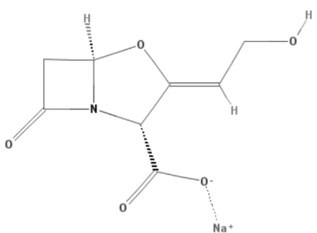
II.1. Amoxicillin in association with clavulanic acid ... 28
II.2. Metronidazole in association with amoxicillin ... 30
II.3. Clindamycin ... 32
II.4. Minocycline ... 34
III. Antiseptics ... 35
III.1. Chlorhexidine ... 36
12
IV. Nanoparticles as antimicrobial agents ... 39
IV.1. Biocompatibility ... 41
IV.2. Silver nanoparticles ... 43
IV.3. Gold nanoparticles ... 46
IV.4. Copper and copper oxide nanoparticles ... 48
IV.5. Zinc oxide nanoparticles ... 50
IV.6. Titanium dioxide nanoparticles ... 51
IV.7. Quaternary ammonium compounds nanoparticles ... 52
IV.8. Chlorhexidine nanoparticles ... 54
IV.9. Calcium-phosphate nanoparticles ... 55
Conclusions ... 57
List of figures
13
List of figures
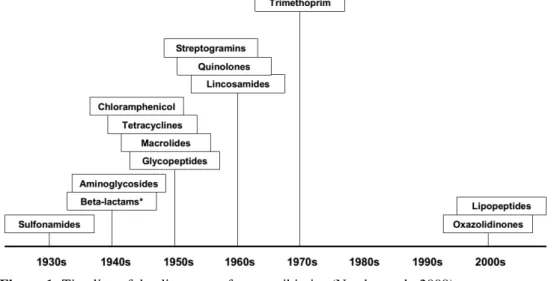
Figure 1: Timeline of the discovery of new antibiotics ... 19
Figure 2: The number of antibiotics approved has been decreasing throughout the years ... 20
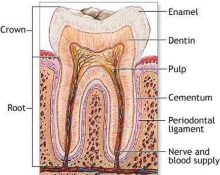
Figure 3: Structure of a healthy tooth. Cariogenic bacteria can damage enamel and dentin layers of the teeth ... 23
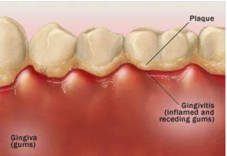
Figure 4:“Gingivitis can cause dusky red, swollen, tender gums that bleed easily” .... 25
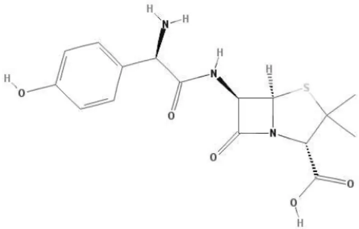
Figure 5: Chemical compostion of amoxicillin. ... 28
Figure 6: Chemical compostion of clavulanic acid . ... 29
Figure 7: Chemical composition of metronidazole. ... 30
Figure 8: Chemical composition of clindamycin. ... 32
Figure 9: Chemical composition of minocycline. ... 34
Figure 10: Chemical composition of chlorhexidine. ... 36
Figure 11: Chemical composition of triclosan. ... 37
Figure 12: Silver nanoparticles shaped as truncated triangles (A), spheres (B) and rods (C). ... 40
14
Figure 14: Some of the mechanisms of silver nanoparticles in the bacterial cell. ... 44
Figure 15: Electron microscopic image of AuNPs. ... 46
List of tables
15
List of tables
Table 1: Pathogenic bacteria found in oral infections. ... 21
Table 2: Spectrum of activity of clindamycin. ... 33
Table 3: Nanoparticle characteristics that influence the interactions with biological systems. ... 41
Table 4: Experimental effects of the use of nanoparticles and their possible outcome . 42
Table 5: Minimum inhibitory concentration and minimum bactericidal concentration values expressed in µg/mL of the tested bacterial species with AgNPs ... 44
Table 6: Minimum inhibitory concentration and minimum bactericidal concentration values expressed in µg/mL of the tested bacterial species CuONPs and AgNPs in combination with CuONPs. ... 49
Table 7: Minimum inhibitory concentration and minimum bactericidal concentration values expressed in µg/mL of the tested bacterial species Cu2ONPs 49
Table 8: Minimum inhibitory concentration and minimum bactericidal concentration expressed in µg/mL of the tested bacterial species with ZnONPs and AgNPs in combination with ZnONPs ... 50
Table 9: Minimum inhibitory concentration and minimum bactericidal concentration values expressed in µg/mL of the tested bacterial species with TiO2NPs ... 51
16
Table 11: Minimum bactericidal concentration values expressed in µg/mL of the tested bacterial species with DMAE-CB. ... 53
List of abbreviations
17
List of abbreviations
AgNPs Silver nanoparticles
ATP Adenosine triphosphate
CHX-HMP NPs Chlorhexidine-hexametaphosphate nanoparticles
CuONPs Copper oxide nanoparticles
Cu2ONPs Copper dioxide nanoparticles
DMAE-CB Methacryloxylethylcetyl dimethyl ammonium chloride
DNA Deoxyribonucleic acid
ENR Enoyl-acyl carrier protein reductase
MBC Minimum bactericidal concentration
MDBP 12-methacryloyloxydodecylpyridinium bromide
MIC Minimum inhibitory concentration
MRSA Methicillin-resistant Staphylococcus aureus
NAD+ Nicotinamide adenine dinucleotide
QPEI Quaternary ammonium polyethylenimine
STD Sexually transmitted disease
TiO2NPs Titanium dioxide nanoparticles
Introduction
19
Introduction
An antimicrobial agent is a compound that has a synthetic, semisynthetic or nat-ural source and can inhibit the growth or kill microorganisms such as bacteria, fungi, viruses and protozoa. Antibiotics and antiseptics are antimicrobials (Cole, 2014).
The use of antibiotics to treat infections has increased since the discovery of penicillin in 1928 by Alexander Fleming. He discovered that a culture he had inoculated with Staphylococcus aureus had been contaminated with mold, which would then be named Penicillium notatum. The culture appeared to have transparent halos without bacterial growth. After a few tests to this mold, Fleming discovered that it produced a compound, penicillin, which had inhibition markers and antimicrobial properties capa-ble of inhibiting and eliminate pathogenic bacteria. However, it was only after a few years that these results became a matter of interest to the scientific community (Ligon, 2004).
Since the discovery of penicillin and up until 1962, 20 new classes of antibiotics were developed and produced. The appearance of these new antibiotics made significant improvements to the health and life expectancy of the population. Without antibiotics, therapeutic interventions and treatments that are a routine nowadays (organ transplants, surgeries, for example) would not be possible (Coates, Halls, & Hu, 2011; Cole, 2014).
However, in the recent years, the research and development for new antibiotics has decayed probably due to the commercialization of analogues being more profitable to the pharmaceutical industry, to the absence of new metabolic targets, to the declining number of pharmaceutical companies involved in the development of new drugs or even
20
to the insufficient funding for the development of new drugs (Aminov, 2010; Coates et al., 2011; Cole, 2014).
Pathogenic bacteria in the oral cavity have been linked to antibiotic resistance (Laxminarayan, 2014). With the emergence of bacterial resistance to the currently used antibiotics, a new approach has been adopted to combat these resistances and to treat multi-drug resistant infections. This approach may consist in (Aminov, 2010; Cushnie, Cushnie, & Lamb, 2014; Nuñez-Anita et al., 2014):
o Modifying the antibiotics currently available in order to change their targets, to
improve their activity, to decrease their toxicity and decrease the bacterial re-sistances;
o Using peptides, alkaloids and other compounds with antimicrobial properties; o Incorporating metals, such as silver, titanium and zinc, in nanoparticles.
This dissertation will address the antibiotics and antiseptics that are currently used, in Portugal, to treat and to help prevent possible oral infections, such as dental caries, gingivitis and periodontitis. Since the use of antibiotics is being affected by re-sistant strains of bacteria, an investigation about possible new antimicrobials was con-ducted in order to evaluate their possible use in the dentistry field. For that, Google Scholar and PubMed databases were used to research the articles needed. The following keywords were used: “oral infection”; “gingivitis”; “periodontitis”; “caries”; “symp-toms”; “side effects”; “antibiotic”; “amoxicillin”; “clavulanic acid”; “metronidazole”; “clindamycin”; “minocycline”; “antiseptic”; “chlorhexidine”; “triclosan”; “spectrum of activity”; “nanoparticle”; “silver”; “copper”; “gold”; “zinc”; “titanium”; “quaternary ammonium compounds”; “calcium phosphate”.
I. Oral Infections
21
I. Oral Infections
Oral infections can be originated by bacteria, fungi and even viruses. They can be caused by external pathogens or by opportunist pathogens living in the oral cavity.
Bacterial infections in the oral cavity usually cause pain, fever and damage to the gingiva. The most common cause for these infections is bad maintenance of a proper oral hygiene since the patient may feel pain, see gingival bleeding or be clueless about oral health status (Lôbo & Martins, 2009). They are mostly caused by Gram-negative bacteria such as Escherichia coli and Pseudomonas spp.; and by oral colonizers such as
Streptococcus mutans, Streptococcus sanguis, Streptococcus mitis and Streptococcus oralis (Table 1).
Anaerobic bacteria Facultative anaerobic bacteria
Peptostreptococcus micros Streptococcus spp.
Actinomyces spp. β-hemolytic streptococci
Eubacterium spp. Streptococcus milleri
Propionibacterium spp. Lactobacillus spp.
Veilonella spp. Aggregatibacter actinomycetemcomitans
Prevotella nigrescens Capnocytophaga spp.
Prevotella intermedia Porphyromonas gingivalis Bacteroides forsythus Rothia dentocariosa Fusobacterium nucleatum Treponema denticola Treponema sokranskii
Table 1: Pathogenic bacteria found in oral infections, adapted fromBrook et al. (2005); Tancawan et al. (2015).
22
eliminate dental biofilm by correctly educating the patient on how to properly brush and floss the teeth. If these strategies are insufficient, the intervention of a specialist is need-ed to prevent further complications (Varoni, Tarce, Lodi, & Carrassi, 2012).
Oral infections can also be caused by fungi and are mostly by Candida albicans
and other species of Candida spp (Lôbo & Martins, 2009). They are common with pa-tients that have head and neck cancers; are submitted to radiotherapy; wear dentures; and with leukemia and transplant patients (Waltimo, Sirén, Torkko, Olsen, & Haapasalo, 1997; Brown et al., 2012).
Viruses can also cause infections in the oral cavity. They are mostly caused by
I. Oral Infections
23
I.1. Dental caries
Dental caries are a form of tooth decay that occurs when specific strains of bac-teria produce cariogenic acids that destroy and damage the various layers of the tooth (Takahashi & Nyvad, 2011). The oral cavity is home to these bacteria and they can ac-cumulate in the form of biofilms (also known as dental plaque) in certain areas: near the gum line; between the teeth; around dental fillings; on cracks, pits and rough surfaces; in areas with a low salivary flow (Moreau, Sun, Chow, & Xu, 2011).
Most dental caries are caused by Streptococcus mutans but other strains of bac-teria can also been found, such as Streptococcus sobrinus, Veilonella spp., Lactobacillus acidophilus, Eubacterium spp., Streptococcus salivarius and Actinomyces spp. (Aas et al., 2008). When in the presence of certain foods, such as carbohydrates, these bacteria can convert them into acids through fermentation. If these acids remain on the tooth, the acids will start to interact with the calcium and phosphate from the teeth (Melo, Guedes, Xu, & Rodrigues, 2013), therefore demineralizing it and beginning to form cavities on the surface of the teeth (Takahashi & Nyvad, 2011).
Saliva can neutralize the effect of the acids produced by cariogenic bacteria. However, it is also necessary to have a good oral hygiene to prevent the formation of cavities. One of the ways to do it is by brushing the teeth after meals and by flossing daily. Antibacterial mouth rinses can also be used since they are able to decrease the
24
levels of cariogenic bacteria. The use of toothpaste and mouth rinses containing fluo-ride, chlorhexidine and/or triclosan effectively reduces the formation of dental caries. Another way to prevent dental caries involves the use of fluoride to strengthen and re-mineralize the teeth and to inhibit the growth of cariogenic bacteria. Molar teeth can also be sealed in order to decrease or prevent their decay (Darby & Walsh, 2014).
Once the enamel is damaged, only a specialist can repair the tooth. Generally, the treatment involves the use of fillings, crowns or root canals. The specialist will start to remove the damaged parts with a drill or a laser (Park et al., 2014) and then replace it with restorative materials such as amalgam, acrylic resin, composites, gold or porcelain (Heymann, Swift Jr., & Ritter, 2014). If the tooth is extensively decayed, the use of fill-ings is not recommended as it can break the remainder of the tooth. In these cases, a crown made of porcelain or gold is usually used to protect the tooth (Powers & Wataha, 2014).
If the decay is too extensive and damages the nerve in the tooth, a root canal needs to be performed. The nerve, the pulp and the damaged parts of the tooth are re-moved and filled with restorative materials. A crown is then applied on top of the tooth in order to protect it against future infections (Powers & Wataha, 2014).
I. Oral Infections
25
I.2. Periodontal disease
Periodontal disease is an inflammation of the gingiva that can also affect the bones surrounding and supporting the teeth. This inflammation is caused by toxins pro-duced by bacteria in dental plaque, as described in chapter I.1.
Gingivitis is a mild form and the earliest stage of periodontal disease caused by the accumulation of dental plaque and tartar at the gum line. This accumulation can in-duce inflammation, irritation and redness and, if not treated properly, can evolve into a more severe form. The most common bacteria involved are Actinomyces spp., Prevotel-la intermedia, Aggregatibacter actinomycetemcomitans, Porphyromonas gingivalis,
Fusobacterium nucleatum and Tanerella forsythia (Tsaousoglou, Nietzsche, Cachovan, Sculean, & Eick, 2014; Yang, Zhang, Li, Yang, & Shi, 2014).
Some of the signs and symptoms of gingivitis include changes in the appearance of the gingiva as it may be swollen, tender, bleeding and with more accentuated red coloration (Figure 4). Other signs also include the formation of pockets with plaque in the gingiva, bad breath and receding gingiva (Cope & Cope, 2011).
The most common cause is a poor oral hygiene but the effects of gingivitis can be reversed if they are treated in the early stages. Maintenance of a good oral health is essential in order to remove the plaque and it can be achieved by brushing the teeth after meals, by flossing daily and by using mouth rinses (Powers & Wataha, 2014). If the plaque hardens (it becomes tartar), it can only be removed through a root planning
26
cess of smoothing a tooth’s root by removing the affected part of the dentin and ceme n-tum) or a scaling (a professional cleaning that uses hand and ultrasonic instruments) (Darby & Walsh, 2014; Heymann et al., 2014).
If gingivitis is not treated properly, the inflammation can reach the area around the tooth and advance into periodontitis, a more severe form of periodontal disease. Per-iodontitis damages the soft tissue and can even destroy the bone supporting the affected teeth. The gingival symptoms present in periodontitis are identical to gingivitis. Other signs may also include deep pockets in the gingiva, loose teeth, receding gingiva, bad breath, pus between the tooth and the gingiva, and malocclusion (Cope & Cope, 2011; Anwar, Amir, & Khan, 2014).
II. Antibiotics
27
II. Antibiotics
An antibiotic consists in an antibacterial agent produced by a microorganism that has the ability to kill or inhibit the growth of bacteria by disrupting their cellular functions (Norrby et al., 2009). They can be classified according to their spectrum of activity, their effect and their mode of action.
The mechanism of action of an antibiotic may consist in (Norrby et al., 2009):
o The disruption of cell membrane functions; o The inhibition of DNA replication;
o Or the inhibition of the synthesis of new proteins and/or cell wall materials.
The spectrum of activity of an antibacterial agent can be classified as narrow or broad. A broad spectrum antibiotic covers most of the gram-positive and gram-negative bacteria whether a narrow spectrum antibiotic will only cover specific strains of bacte-ria.
Antibiotics also have a bactericidal and/or a bacteriostatic effect on bacteria, depending on the dosage and duration of the treatment. A bactericidal antibiotic can eliminate the intended bacteria whether a bacteriostatic antibiotic will only inhibit and delay the growth of bacteria.
28
II.1. Amoxicillin in association with clavulanic acid
Amoxicillin is a semisynthetic beta-lactam antibiotic from the aminopenicillin family. Its mode of action consists in the inhibition of penicillin-binding proteins during the metabolic synthesis of the bacterial peptidoglycan. The inhibition of the peptidogcan synthesis leads to the weakening of the bacterial cell structure, followed by cell ly-sis and the death of the bacteria (Dörr, Davis, & Waldor, 2015).However, amoxicillin is susceptible to beta-lactamases produced by resistant bacteria.
Some of the most common side effects of amoxicillin include nausea, diarrhea and rash. Although renal side effects are rare, the dosage of amoxicillin needs to be ad-justed if the patient has renal insufficiency (Manuel José Guedes da Silva Lda & Guia de Saúde: Edição e Comunicação Audio-Visual, 2014).
Amoxicillin has a broad spectrum of action that includes Gram-positive coccus,
Haemophilus influenza, Escherichia coli, Proteus mirabillis, Salmonella, Fusobacte-rium spp. and Shigella. It is inactivated by staphylococci that produce beta-lactamases,
Enterobactereaceae, Bacteroides fragillis and Pseudomonas (MJGSLda & GS: ECAV, 2014).
Clavulanic acid is a semisynthetic compound obtained from the fermentation of
Streptomyces clavuligerus and normally used in combination with beta-lactam antibiot-ics in order to prevent their inactivation by beta-lactamases. Its mechanism of action consists in irreversibly binding with beta-lactamase enzymes so that susceptible antibi-otics, such as amoxicillin, can induce their activity on the bacteria (Jensen, 2012). When used in combination with amoxicillin, the treatment may need to be monitored in pa-tients with hepatic and renal insufficiency (MJGSLda & GS: ECAV, 2014).
II. Antibiotics
29
The combination of amoxicillin with clavulanic acid expands the spectrum of activity since clavulanic acid inhibits bacteria that produce beta-lactamases. They are the first choice antibiotics to use when treating oral infections (Direcção Geral da Saúde, 2014). There are many dosages of this association that are used depending on the degree of the infection and/or the age of the patient. For adults, the dosage, expressed in mg of amoxicillin, can vary between 250 mg and 875 mg every 12 to 6 hours (Infarmed, 2015). If it is administered via IV therapy, the recommended dosage is 1 g to 2 g of amoxicillin every 8 or 6 hours. The treatment should not exceed 14 days without further evaluations of the patient’s condition (MJGSLda & GS: ECAV, 2014).
However, if the patient being treated is allergic to amoxicillin, it can be replaced by clindamycin. Clindamycin can also be associated with clavulanic acid to show simi-lar results and effectiveness (Tancawan et al., 2015).
30
II.2. Metronidazole in association with amoxicillin
When antibiotic therapy is required to treat gum disease, metronidazole, alone or in combination with amoxicillin, can be an alternative to the amoxicillin-clavulanic acid association (Direcção Geral da Saúde, 2014). It is a compound derived from nitro-imidazole that can be used as an antibacterial or as an anti-parasitic agent (Soares et al., 2012). It is commonly used in the treatment of periodontitis, acne, rosacea, bacterial vaginosis, Trichomonas vaginalis infections, Clostridium difficile infections and amoe-bic dysentery (Finberg & Guharoy, 2012; Khodaeiani et al., 2012).
Metronidazole has a bactericidal effect on bacteria. Its mechanism of action con-sists in inhibiting nucleic acid synthesis. The molecule enters the bacterial cell by pas-sive diffusion and it is metabolized inside the cell. The metabolites bind to the DNA, preventing cellular replication and inhibiting enzymes responsible for energy production (Finberg & Guharoy, 2012; Soares et al., 2012). Metronidazole is active against some parasites, for instance Trichomonas vaginalis, Helicobacter pylori and Giardia lamblia. It is also active against anaerobic bacteria, such as Clostridium spp., Fusobacterium
spp., Prevotella spp. and other anaerobic cocci and bacilli. Certain bacterial strains pre-sent in the oral cavity, that include Actinomyces spp. and Propionibacterium spp., are resistant to metronidazole (Finberg & Guharoy, 2012).
Some of the most common side effects of metronidazole include nausea, head-aches, diarrhea, abdominal pain, metallic taste, rash, anorexia and candidiasis (Infarmed, 2015).
When used alone, the recommended dosage for metronidazole per os is 250 mg to 500 mg every 8 hours or 250 mg every 6 hours when treating infections caused by
II. Antibiotics
31
Helicobacter pylori. For IV therapy, it is recommended a dosage of 500 mg every 8 hours (Infarmed, 2015).
Metronidazole can be combined with amoxicillin to improve its spectrum of activity. The use of this antibiotic association to treat oral infections has shown positive results and improvements in treatments when combined with scaling and root planning (Cionca, Giannopoulou, Ugolotti, & Mombelli, 2009; Powell, 2013). Both antibiotics have also shown antibacterial activity against Aggregatibacter actinomycetemcomitans,
32
II.3. Clindamycin
Clindamycin is a semi synthetic macrolide antibiotic with a bacteriostatic and bactericidal (if used in higher concentrations) effects on bacteria. If a patient is allergic to beta-lactamic antibiotics, such as amoxicillin, clindamycin is prescribed to treat peri-odontal disease (Direcção Geral da Saúde, 2014). It can be used to treat other condi-tions, such as amygdalae pharyngitis, acute sinusitis, acne vulgaris, acute otitis media, scarlet fever, intra-abdominal infections, endometritis, lung abscess and osteomyelitis. Its mode of action consists in binding to the 50s ribosomal subunits to stop the for-mation of peptide bonds, inhibiting protein synthesis (Brook et al., 2005).
Some of the most common side effects of clindamycin include abdominal pain, diarrhea, nausea, rash, anorexia, vomiting and flatulence (Finberg & Guharoy, 2012). There are also other side effects that despite not being common need to be accounted for. These include eosinophilia, neutropenia, pseudomembranous colitis and thrombocy-topenia (Finberg & Guharoy, 2012; Tancawan et al., 2015).
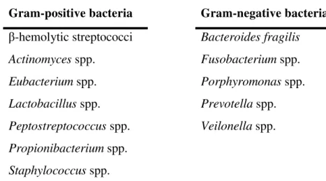
Clindamycin is a broad-spectrum antibiotic with activity against anaerobic, Gram-negative and Gram-positive bacteria (Table 2).
II. Antibiotics
33
Gram-positive bacteria Gram-negative bacteria
β-hemolytic streptococci Bacteroides fragilis
Actinomyces spp. Fusobacterium spp.
Eubacterium spp. Porphyromonas spp.
Lactobacillus spp. Prevotella spp.
Peptostreptococcus spp. Veilonella spp.
Propionibacterium spp.
Staphylococcus spp.
Table 2: Spectrum of activity of clindamycin, adapted from Brook et al. (2005).
34
II.4. Minocycline
Minocycline is a semi-synthetic antibiotic that belongs to the second generation of the tetracycline family. It is a bacteriostatic antibiotic that has a broad spectrum of activity against Gram-positive and Gram-negative bacteria. It is used in the treatment of respiratory tract infections, such as acute and chronic bronchitis, bronchiectasis, pulmo-nary abscesses and pneumonia; STD’s, such as gonorrhea, chlamydia, non-gonococcal urethritis and pelvic inflammatory disease; acne; periodontitis, gingivitis and dental abscesses (Garrido-Mesa, Zarzuelo, & Gálvez, 2013).
When in the presence of minocycline and other tetracyclines, the bacterial mem-brane uses an energy-dependent system that actively transports these antibiotics into the cytoplasm. Its mode of action consists in blocking protein synthesis by reversibly bind-ing to the 30s ribosomal subunit of bacteria in order to prevent the connection to tRNA and inhibit protein synthesis (Garrido-Mesa et al., 2013).
The most common side effects may include nausea, dizziness and vertigo during the administration. Patients taking minocycline need to be monitored since it is a hepa-totoxic antibiotic and it could lead to appearance of pigmentation and systemic lupus erythematosus (Infarmed, 2015).
Minocycline is mainly used to treat acne vulgaris and some STD’s. Due to its anti-apoptotic, anti-collagenase and anti-inflammatory properties, it is also used in the treatment of periodontitis (Garrido-Mesa et al., 2013). When taken in doses between 100 and 200 mg per day for 7 to 14 days, minocycline has shown to effectively control the progression of periodontitis and promote the healing of the affected area. Therefore, the suggested dosage for the treatment is 200 mg initially followed by 100 mg every 12 hours. In children aged 12 or older, the recommended initial dose is between 2 to 4 mg/kg followed by 1-2 mg/kg every 12 hours (Garrido-Mesa et al., 2013; Infarmed, 2015).
III. Antiseptics
35
III. Antiseptics
Oral antiseptics can help control the formation of bacterial biofilms in the oral cavity byreducing bacterial growth, delaying accumulation of dental plaque and elimi-nating bacteria. Antiseptics can be used on the skin and on the mucous membrane (Corbin, Pitts, Parker, & Stewart, 2011).
These antimicrobials have side effects that usually appear after long periods of usage. For example, the continuous usage of chlorhexidine can induce teeth staining, dysgeusia and xerostomia (Park et al., 2014).
36
III.1. Chlorhexidine
Chlorhexidine is an antiseptic agent with antimicrobial and antifungal properties that belongs to the bisbiguanide family (Parashar, 2015). This compound is extensively studied and it is applied in the medical and dentistry fields as, for example, in pre-surgical cleansing agents, antibacterial mouth rinses, gels and eye drops. Chlorhexidine can help reduce the frequency of periodontal disease, reduce gingival inflammation and bleeding, and reduce dental plaque formation (Varoni et al., 2012).
Its mode of action targets different areas of the bacterial cell, depending on the concentration used. A bacteriostatic effect is shown when used in lower concentrations and targets the osmotic balance of the cell. At higher concentrations, chlorhexidine acts as a bactericidal antiseptic and targets the cell membrane, resulting in its rupture and leakage of its intracellular components (Varoni et al., 2012).
Chlorhexidine usually has a better antimicrobial activity when pH is between 7 and 8. It has shown a wide antibacterial activity against negative and Gram-positive bacteria, yeasts, virus and fungi. With regard to the bacteria responsible for infections in the oral cavity, it was shown that Aggregatibacter actinomycetemcomitans,
Streptococcus mutans, Fusobacterium nucleatum, Porphyromonas gingivalis and En-terobacteria were susceptible to chlorhexidine (Varoni et al., 2012; Parashar, 2015).
There are various concentrations and formulations available. Chlorhexidine can be presented as a gel, mouth wash, spray and aerosol with concentrations that can range between 0,02% and 0,3% (Varoni et al., 2012).
The side effects that follow a treatment with chlorhexidine are usually reversible once the treatment ends. These include mucosa and teeth staining, dysgeusia, xerosto-mia and hypersensitivity reactions (Varoni et al., 2012; Lachenmeier, 2014).
III. Antiseptics
37
III.2. Triclosan
Triclosan (or 5-Chloro-2-(2,4-dichlorophenoxy) phenol) is a non-ionic antiseptic used in diverse consumer products in order to reduce and prevent contamination by bac-teria and fungus (Varoni et al., 2012; Dhillon et al., 2015). It is commonly used in di-verse products such as mouth washes, toothpastes, shampoos, deodorants, soaps, surgi-cal cleaning treatments, textiles, among others. As an ingredient in dentistry products such as toothpastes and mouth washes, it has shown to reduce tooth cavities, dental plaque and gingival related diseases (Yazdankhah et al., 2006; Haraszthy, Sreenivasan, & Zambon, 2014).
This compound acts as a biocide if used in higher concentrations and targets specific areas in the cytoplasm and membrane. If used in lower concentrations, such as in dentistry related consumer products, triclosan is essentially bacteriostatic. Its mecha-nism of action inhibits the bacterial synthesis of fatty acids. Triclosan binds to the ENR enzyme, creating a stable complex with NAD+. This complex will no longer participate in the fatty acid synthesis thus preventing the formation of cell membranes (Dann & Hontela, 2011; Dhillon et al., 2015). Triclosan can also inhibit the cyclooxygenase and lipoxygenase pathways by reducing the levels of leukotrienes and prostaglandins in-volved in the inflammatory process (Parashar, 2015).
The use of triclosan has been linked to side effects such as allergic sensitization to allergens, altered levels of thyroid hormones, rash and bacterial resistance to some antibiotics (Davison & Maillard, 2010; Li et al., 2013; Lachenmeier, 2014).
Despite its antibacterial activity, there has been some controversy about its use. In 2009, Denmark suggested that triclosan should be banned due to being linked to
38
IV. Nanoparticles as antimicrobial agents
39
IV. Nanoparticles as antimicrobial agents
Since the discovery of penicillin, antibiotics have been used and developed to treat bacterial infections. However, due to the abusive use and prescription, many bacte-ria are becoming resistant to these compounds, making it more difficult to treat some infections (Khan, Ahamed, Musarrat, & Al-Khedhairy, 2014). In spite of this resistance, scientists are trying to develop new antimicrobial and inorganic materials to mimic the same killing effect that antibiotics have.
Nanotechnology can be applied to the dental field in order to develop new mate-rials with better antimicrobial properties (Melo et al., 2013). Nanoparticles made of me-tallic elements, such as gold, mercury, silver, titanium and zinc, are being developed due to their antimicrobial properties and spectrum of activity. These metals have been used for many years in the dental field (Fernandes et al., 2015) and to treat infectious diseases (Rizzoto, 2012). One of the first uses of mercury can be traced back to China in the 2nd century BC (Sloane, 2012). Before the discovery of antibiotics, mercury was also used to treat syphilis and skin diseases (The Trustees of Indiana University, 2002). In the 20th century, it was commonly applied as an antiseptic and a disinfectant but, due to its toxicity, there has been a decline in its use. Zinc also has antibacterial and antifun-gal properties and can be found in ointments to treat skin diseases (Gupta, Mahajan, Mehta, & Chauhan, 2014) and in oral products to decrease the formation of dental plaque (Allaker, 2010).
nanopar-40
ticles shaped like spheres (figure 12B) and rods (figure 12C), due to the proportion of active facets in the particles (Pal, Tak, & Song, 2007).
Metal nanoparticles toxicity studies have brought new antimicrobial applica-tions. In a study, copper, copper oxide, silver, titanium dioxide and zinc oxide showed a significant activity against Porphyromonas gingivalis, Fusobacterium nucleatum,
Prevotella intermedia and Aggregibacter actinomycetemcomitans with minimum bacte-ricidal concentrations ranging 250 µg/mL and 2500 µg/mL. Activity against Pseudomo-nas aeruginosa and MRSA was also shown with minimum bactericidal concentrations between 100 µg/mL and 5 mg/mL (Vargas-Reus, Memarzadeh, Huang, Ren, & Allaker, 2012).
In this chapter, nanoparticles made of metallic elements, chlorhexidine and cal-cium-phosphate will be addressed. Despite calcal-cium-phosphate not having antibacterial properties, it has improved the activity of those properties when combined with other nanoparticles.
IV. Nanoparticles as antimicrobial agents
41
IV.1. Biocompatibility
There is a considerable interest in the development and application of nanotech-nology to the human body. Some nanoparticles could accumulate in certain organs, tis-sues and cells due to their small size, which is not verified by larger sized particles such as antibiotics (Oyar, 2014; B. Sutariya & Pathak, 2015). Although there are some sup-positions about the mode of action of some nanoparticles, it is not enough. It is neces-sary to have a thorough knowledge about their absorption, distribution, metabolism, excretion, physiochemical properties and genotoxic and immunogenic potential in the human body in order to build a safety profile with the desired biological effects and the minimal potential toxicity (B. Sutariya & Pathak, 2015).
To safely use nanotechnology and to design nanoparticles for biological applica-tions, it is necessary to know the interactions that occur between the human body and the nanoparticles, which are covered by the following: the surface/characteristics of the nanoparticle; the solid-liquid interface and the effects of its surroundings; and the con-tact zone with biological substrates (A. E. Nel et al., 2009). These components are in-fluenced by several characteristics as it is shown in Table 3.
Surface of the nanoparticle:
Chemical composition; Hydrophilicity;
Hydrophobicity; Number of sides; Porosity; Roughness; Shape; Surface crystallinity; Surface function; Size heterogeneity.
Solid-liquid interface and ef-fect of the surrounding media:
Ionic strength; pH;
Presence of organic molecules/detergents; Temperature.
Contact zone with biological substrates: Dispersion state; Hydration; Nanoparticle aggregation; Stability; Zeta charge.
42
However, data about the toxic effects of nanoparticles to humans are still limited to draw any conclusions in order to ensure a safe use (A. Nel, Xia, Mädler, & Li, 2006; Oyar, 2014). Some effects obtained in experiments can be found in Table 4. The re-search currently available is expensive and inconsistent in the matter of the preparation and dosimetry of the nanoparticles being used. Therefore, results may vary depending on the researcher and on the materials used to perform these experiments. There is a need to standardize these studies so that they can correctly evaluate the potential bene-fits and risks of existing and newly-developed nanoparticles (B. Sutariya & Pathak, 2015).
Experimental nanoparticle effects Possible outcomes
Reactive oxygen species generation Protein, DNA and membrane injury; Oxidative stress.
Oxidative stress
Phase II enzyme induction; Inflammation; Mitochondrial perturbation.
Mitochondrial perturbation
Inner membrane damage; Permeability transition; Pore opening; Energy failure;
Apoptosis; Apo-necrosis; Cytotoxicity.
Inflammation
Tissue infiltration with inflammatory cells; Fibrosis; Granulomas; Atherogenesis; Acute phase protein expression.
Uptake by reticulo-endothelial system
Asymptomatic sequestration and storage in liver, spleen and lymph nodes; Possible organ enlargement and dysfunction.
Protein denaturation/degradation
Loss of enzyme activity; Auto-antigenicity.
Nuclear uptake
DNA damage; Nucleoprotein clumping; Autoantigens.
Neuronal tissue uptake Brain and peripheral nervous system injury.
Perturbations in phagocytic function, particle overload and mediator release
Chronic inflammation; Fibrosis;
Granulomas; Interference in clearance of infectious agents.
Endothelial dysfunction and effects on blood clotting
Atherogenesis; Thrombosis; Stroke; Myocardial infarction.
Generation of neoantigens and break-down in immune tolerance
Autoimmunity; Adjuvant effects.
Altered cell cycle regulation Proliferation; Cell cycle arrest; Senescence.
DNA damage Mutagenesis; Metaplasia; Carcinogenesis.
IV. Nanoparticles as antimicrobial agents
43
IV.2. Silver nanoparticles
Silver has been known throughout history for its antimicrobial effects and can be used in different areas: in the treatment of burns, wounds and ulcers (Rai, Yadav, & Gade, 2009); making water potable (Chopra, 2007); and in dentistry, catheters, wound dressings and sutures, surgical instruments and prostheses (Kim et al., 2007; Corrêa et al., 2014). Silver also exhibits a good biocompatibility with the human cells, low bacte-rial resistance and a long-term antimicrobial action (Corrêa et al., 2014). It is also capa-ble of inactivating specific microorganisms such as the human immunodeficiency virus type 1 (Elechiguerra et al., 2005) and the hepatitis B virus (Lara, Garza-Treviño, Ixtepan-Turrent, & Singh, 2011).
The mechanism of action of silver nanoparticles is not well known but it is thought that it might involve multiple targets. This inconsistency may be caused by the different physicochemical properties of the silver nanoparticles that are used in these studies (Markowska, Grudniak, & Wolska, 2013). Some studies suggest that the posi-tive-charged nanoparticles establish an electrostatic connection with the negative-charged cell membrane of bacteria. The nanoparticles will then bind with proteins that contain either sulphur (thiol groups) or phosphorus and release silver ions into the mi-crobial cell. These ions can stop the replication of DNA and inhibit respiratory chain enzymes and cellular proteins, releasing reactive oxygen species and compromising the levels of ATP (Figures 13 and 14)(Kim et al., 2007; Lara et al., 2011). These mecha-nisms can help improve the reduction of the wound burden since silver nanoparticles can release more ions than molecular silver (Habiboallah et al., 2014).
Figure 13:“Some studies have reported that nano-silver causes oxidative damage, leading to the production
44
In a study conducted after surgery in the oral cavity of healthy rabbits, silver nanoparticles effectively reduced the inflammatory parameters during the four days af-ter the surgery when using a high concentration of nanoparticles in the periodontal dressing. However, when determining the “dose-dependent histopathological and bio-logical effects”, researchers found that using a higher concentration of nanoparticles could cause mild cytotoxicity (Habiboallah et al., 2014).
When compared to other antimicrobials currently used (such as chlorhexidine), silver nanoparticles have shown a better antimicrobial action against strains of Strepto-coccus mutans with a MIC of 50 µg/mL (Besinis, De Peralta, & Handy, 2014). Silver nanoparticles also show activity against Porphyromonas gingivalis, with a MIC of 250 µg/mL; Aggregatibacter actinomycetemcomitans and Fusobacterium nucleatum with a minimum inhibitory concentration of 100 µg/mL (Table 5).
MIC MBC
Aggregatibacter actinomycetemcomitans 100 100 Fusobaterium nucleatum 100 100 Prevotella intermedia 100 100 Porphyromonas gingivalis 250 250
Table 5: Minimum inhibitory concentration and minimum bactericidal concentration values ex-pressed in µg/mL of the tested bacterial species with AgNPs, adapted from Vargas-Reus et al. (2012).
IV. Nanoparticles as antimicrobial agents
45
In another study conducted by Lu et al. (2013), a lower MIC was obtained for
46
IV.3. Gold nanoparticles
Gold is one of the oldest elements known to mankind and its first uses can be traced back to ancient China and Egypt in 2500 BC (Thakor, Jokerst, Zavaleta, Massoud, & Gambhir, 2011; Dykman & Khlebtsov, 2012). It was used to treat fevers and syphilis between the 17th and the 19th century. Currently, gold is used in cavity fill-ing, prostheses, stents and ophthalmology. It can also treat some forms of arthritis such as rheumatoid arthritis and psoriatic arthritis. As a nanoparticle, it can be used in Raman Imaging, photothermal ablation, antitumor drug delivery, siRNA delivery, thermal im-aging and as contrasting agents (Thakor et al., 2011; Rieznichenko et al., 2012).
Although the mechanism of action of gold as a molecule is known, its mecha-nism as a nanoparticle is still unknown. Due to its small size, gold nanoparticles have a high surface area to volume ratio and may have their physical and chemical properties changed (Thakor et al., 2011). These factors can lead to unexpected reactions in terms of their toxicity and their interaction in the human body. According to a study by Riez-nichenko et al. (2012), the antimicrobial and biological activity of gold nanoparticles depends on the size of the nanoparticle itself. The size and shape of the nanoparticle can be manipulated in order to achieve biocompatibility in the human body (Park et al., 2014). In similarity to silver nanoparticles, it is believed that gold nanoparticles may also cause oxidative stress due to the release of reactive oxygen species (Thakor et al., 2011; Park et al., 2014).
IV. Nanoparticles as antimicrobial agents
47
48
IV.4. Copper and copper oxide nanoparticles
Copper is one of the essential metallic elements to the human health. It has been used for many centuries for the disinfection of biological tissues and liquids (Perelshtein et al., 2009; Ahmad et al., 2012). It has been reported that in 400 B.C. Hippocrates pre-scribed copper to purify water and to treat pulmonary diseases (Ingle, Duran, & Rai, 2014). Nowadays, it is mostly used to purify water, as an antibacterial, as a fungicide, as a nematocide and as an antifouling agent. In fact, copper is an essential metal required to the human health and can be found in many vitamins and amino and fatty acids re-quired for metabolic processes (Perelshtein et al., 2009).
Copper oxide is very stable in terms of its chemical and physical properties: electron correlation effects; high temperature superconductivity; photoconductivity; and spin dynamics (Ren et al., 2009). It is also cheaper than other metals, which can make it a better cost-effective option (Ahmad et al., 2012; Ingle et al., 2014). Copper oxide can be mixed with various polymers and can be prepared with multiple crystal morpholo-gies. Since the documentation about the antimicrobial effects of copper oxide nanoparti-cles is limited, these features can influence future studies and production of these nano-particles (Ren et al., 2009).
Despite the lack of information about the antimicrobial activity of copper and copper oxide nanoparticles, some studies report that they share a similar mode of action with silver nanoparticles (Ruparelia, Chatterjee, Duttagupta, & Mukherji, 2008). Copper based nanoparticles have a wide antibacterial range of action due to the nanoparticle binding with thiol groups and the production of reactive oxygen species. This weakens
IV. Nanoparticles as antimicrobial agents
49
the bacterial cell wall and enables the entrance of more nanoparticles into the cell. The nanoparticles also bind with DNA and some enzymes, leading to cell death (Kim et al., 2007; Ingle et al., 2014).
Copper nanoparticles have shown antibacterial properties against Streptococcus mutans, with a MBC and MIC of 100 µg/mL (Eshed, Lellouche, Matalon, Gedanken, & Banin, 2012).
Copper oxide nanoparticles have shown inhibitory effects against Porphyromo-nas gingivalis, with a MIC of 500 µg/mL and MBC of 2500 µg/mL; Aggregatibacter actinomycetemcomitans, Prevotella intermedia and Fusobacterium nucleatum with a MIC of 250 µg/mL (Table 6). However, when in combination with silver nanoparticles, the MIC needed to have an effect on Porphyromonas gingivalis and Prevotella interme-dia was inferior to 100 µg/mL and for Fusobacterium nucleatum was 500 µg/mL (Vargas-Reus et al., 2012). These nanoparticles also show biofilm inhibition when test-ed against Streptococcus mutans (Eshed et al., 2012).
CuO Ag + CuO
MIC MBC MIC MBC Aggregatibacter actinomycetemcomitans 250 250 250 250 Fusobaterium nucleatum 250 250 500 500 Prevotella intermedia 250 250 <100 <100 Porphyromonas gingivalis 500 2500 <100 <100
Table 6: Minimum inhibitory concentration and minimum bactericidal concentration values ex-pressed in µg/mL of the tested bacterial species CuONPs and AgNPs in combination with CuONPs, adapted from Vargas-Reus et al. (2012).
Copper dioxide nanoparticles also offer a good antimicrobial activity. It has shown a MBC that was less than 100 µg/mL against Fusobacterium nucleatum,
Prevotella intermedia and Porphryromonas gingivalis. Against Aggregatibacter acti-nomycetemcomitans, the results of the MBC obtained were slightly higher than the other bacteria tested, with a value of 1000 µg/mL (Table 7).
MIC MBC
Aggregatibacter actinomycetemcomitans 1000 1000 Fusobaterium nucleatum <100 <100 Prevotella intermedia <100 <100 Porphyromonas gingivalis <100 <100
50
IV.5. Zinc oxide nanoparticles
As it was previously mentioned, zinc has antibacterial and antifungal properties and can be found in ointments to treat skin diseases (Gupta et al., 2014) and in oral products to decrease the formation of dental plaque (Allaker, 2010). It can also be used in the formulation of paints, cosmetics and food products (Shukla et al., 2011).
Like many other nanoparticles being studied for potential use in the human body, the mechanism of action and potential toxicity is not entirely known. Some stud-ies indicate that zinc oxide nanoparticles have a selective toxicity against bacteria, with almost no effects in the human body (Allaker & Douglas, 2015). Other studies report that the nanoparticles may induce genotoxicity and damage the cell membrane by re-leasing reactive oxygen species (Shukla et al., 2011; Wang, Deng, Zhang, Chen, & Ding, 2014).
Zinc oxide nanoparticles inhibit the growth of Porphyromonas gingivalis, Ag-gregatibacter actinomycetemcomitans and Fusobacterium nucleatum with a MIC of 250 µg/mL; and Prevotella intermedia with a MIC of 1000 µg/mL (Table 8). If these bacte-ria are under anaerobic conditions, the MIC ranges from 250 µg/mL to 2500 µg/mL (Vargas-Reus et al., 2012). These nanoparticles also show antimicrobial activity against
Streptococcus mutans (with a MBC of 500 µg/mL) and Rothia dentocariosa (Eshed et al., 2012; Khan et al., 2014).
Silver nanoparticles can help improve the antimicrobial effect of zinc oxide na-noparticles. When combined, the MIC on Porphyromonas gingivalis and Aggregatibac-ter actinomycetemcomitans is 100 µg/mL and on Prevotella intermedia is 250 µg/mL (Vargas-Reus et al., 2012).
ZnO Ag + ZnO
MIC MBC MIC MBC Aggregatibacter actinomycetemcomitans 250 250 100 100 Fusobaterium nucleatum 250 500 1000 1000 Prevotella intermedia 1000 1000 250 500 Porphyromonas gingivalis 250 250 <100 500
IV. Nanoparticles as antimicrobial agents
51
IV.6. Titanium dioxide nanoparticles
Titanium dioxide is the most common form of titanium and, in bulk, is used as a whitener in toothpastes (Allaker, 2010); as a pigment in diverse areas such as tooth-pastes, paints, food industry and plastics; as an antimicrobial agent and as a water puri-fier (Weir, Westerhoff, Fabricius, & von Goetz, 2013). Titanium is also one of the main components of dental implants used nowadays since it can offer biocompatibility and the ability to be osseointegrated (Wood et al., 2015).
In order to have a considerable antimicrobial effect, titanium dioxide nanoparti-cles have to be in anatase form and in the presence of UV light, since photoactivation increases their antibacterial properties. These factors will lead to the formation of reac-tive oxygen species, causing damage in the cell membrane, in the DNA and in the res-piratory activity which will evidently lead to cell death. Its action depends on the con-centration employed, the intensity of the UV light and on the crystal structure (Hartmann et al., 2010; Foster, Ditta, Varghese, & Steele, 2011).
Titanium dioxide nanoparticles have shown antibacterial activity against Strep-tococcus mutans and Streptococcus sanguis, bacteria involved in the formation of dental caries (Ahrari, Eslami, Rajabi, Ghazvini, & Barati, 2015). The minimum inhibitory concentration needed against Aggregatibacter actinomycetemcomitans is 250 µg/mL;
Fusobacterium nucleatum and Prevotella intermedia is 1000 µg/mL; and Porphyromo-nas gingivalis is 2500 µg/mL (Table 9). Under anaerobic conditions, the MIC ranges between 250 µg/mL and 2500 µg/mL (Vargas-Reus et al., 2012).
MIC MBC Aggregatibacter actinomycetemcomitans 250 >2500 Fusobaterium nucleatum 1000 >2500 Prevotella intermedia 1000 >2500 Porphyromonas gingivalis 2500 >2500
Table 9: Minimum inhibitory concentration and minimum bactericidal concentration values ex-pressed in µg/mL of the tested bacterial species with TiO2NPs, adapted from Vargas-Reus et al.
52
IV.7. Quaternary ammonium compounds nanoparticles
The frequency of recurrent dental caries creates the necessity for new restorative materials with antibacterial effects (Antonucci et al., 2012; Imazato, Chen, Ma, Izutani, & Li, 2012). Common restorative composites may allow the colonization of bacteria, leading to tooth demineralization and, if not treated, dental caries (Antonucci et al., 2012). In order to prevent this situation, quaternary ammonium monomers have been developed due to their antimicrobial activity. They can be used in healthcare and textile products, food industry and water treatment (Kenawy, Worley, & Broughton, 2007). Compounds such as 12-methacryloyloxydodecylpyridinium bromide, methacryloxy-lethylcetyl dimethyl ammonium chloride, quaternary ammonium dimethacrylate and quaternary ammonium polyethylenimine (Ge et al., 2015) are polymers known for their bactericidal activity against a wide range of pathogenic bacteria found in the oral cavity (Tables 10 and 11). They also have remineralization properties, good mechanical prop-erties and can also promote biofilm growth inhibition (Ge et al., 2015). However, when using a higher concentration of these polymers, their properties and structures might change (Antonucci et al., 2012).
MIC MBC MIC MBC
Streptococcus mutans 15.6 62.5 Eubacterium alactolyticum 31.3 125
Enterococcus faecalis 31.25 62.5 Bifidobacterium bifidum 31.3 62.5
Fusobacterium nucleatum 31.25 62.5 Peptostreptococcus asaccharolyticus 31.3 31.3
Prevotella nigrescens 1.95 7.81 Lactobacillus plantarum 7.8 15.6
Streptococcus sobrinus 7.8 62.5 Lactobacillus salivarius ssp. Salivarius 7.8 62.5
Streptococcus oralis 16.7 31.3 Lactobacillus acidophilus 15.6 62.5
Streptococcus mitis 25 31.3 Lactobacillus paracasei spp. Paracasei 15.6 62.5
Streptococcus sanguis 16.7 31.3 Lactobacillus brevis 15.6 31.3
Streptococcus gordonii 16.7 31.3 Lactobacillus salivalius ssp. Salicinius 15.6 125
Streptococcus salicarius 15.6 31.3 Lactobacillus fermentum 15.6 15.6
Propionibacterium acnes 3.9 62.5
IV. Nanoparticles as antimicrobial agents
53
MBC
Streptococcus mutans 4.9
Streptococcus sobrinus 2.4
Streptococcus sanguinis 4.9
Lactobacillus acidophilus 2.4
Actinomyces viscosus 2.4
Table 11: Minimum bactericidal concentration values expressed in µg/mL of the tested bacterial species with DMAE-CB, adapted fromImazato et al. (2012).
54
IV.8. Chlorhexidine nanoparticles
As it was previously mentioned in chapter III.1., chlorhexidine is an antiseptic agent with antifungal and antimicrobial properties. It has a non-specific mode of action that can be applied in nanotechnology, improving the antimicrobial properties of current dental implants and the treatment of peri-implant infections (Varoni et al., 2012; Wood et al., 2015). Chlorhexidine is effective against a wide range of bacteria. When applied as a nanoparticle to filling materials, such as cements and ceramics, it can provide these materials with its antimicrobial and antifungal properties (Hook et al., 2014).
Chlorhexidine-hexametaphosphate nanoparticles have been developed and they can provide a continuous and slow release of chlorhexidine. In a study conducted by Wood, N.J. et al. (Wood et al., 2015), CHX-HMP NPs were applied to titanium dental implants as a coating. The purpose of the study was to evaluate the antimicrobial activi-ty of chlorhexidine as a nanoparticle. The dental implants were coated with CHX-HMP NPs and tested against Streptococcus gordonii in the presence and in the absence of human saliva. Results have shown that, after 24 hours, there was significant decrease in
the CFU’s on the implants coated with CHX-HMP NPs. Differences were observed in
the presence of saliva: in its presence, the nanoparticles had a bactericidal effect on the colonies; in its absence, the nanoparticles had a bacteriostatic effect (Wood et al., 2015). Chlorhexidine nanoparticles have also shown positive results against biofilms. A MIC of 19,5 µg/mL and a MBC of 312,5 µg/mL were observed when these nanoparti-cles were used against Streptococcus mutans. The nanoparticles were also tested in the form of a 24 and a 48-hour treatment against mono-species biofilms and were effective against Acinetobacter actinomycetemcomitans, Fusobacterium nucleatum and Strepto-coccus mutans with a MIC with a range of 100 µg/mL. It was also tested against Strep-tococcus sobrinus and MIC of 200 µg/mL was obtained (Seneviratne et al., 2014). When using chlorhexidine nanoparticles against mixed-species biofilms, the results shown in Table 12 were obtained.
24 h 48 h 72h
S. mutans, F. nucleatum and P. gingivalis 12,5 50 50
Streptococcus sobrinus, F. nucleatum and P. gingivalis 25 50 100
A. actinomycetemcomitans, S. mutans, F. nucleatum and P. gingivalis 25 50 50
IV. Nanoparticles as antimicrobial agents
55
IV.9. Calcium-phosphate nanoparticles
Calcium and phosphate compounds belong to the amorphous calcium-phosphates and have the potential to remineralize the damaged enamel by providing the needed ions to repair it. These compounds are soluble and react with saliva in order to release calcium and phosphate ions, which are then transformed into apatite, a major component of the teeth’s enamel (Moreau et al., 2011). They are also compatible with the bone since they are identical at a biochemical level.
In a study conducted by Kovtun et al., (2012), calcium-phosphate nanoparticles were loaded with chlorhexidine in order to test their mineralizing and antibacterial properties when combined. Extracted human teeth without caries were used in this ex-periment to test the remineralizing effects. Lactobacillus casei and Escherichia coli
Conclusions
57
Conclusions
The evidence of the connection of bacteria with the development of antibiotic resistance is growing (Laxminarayan, 2014). However, there have been a few scientific advancements that will improve and reduce the dependence of antibiotic therapy to treat bacterial infections.
Studies about nanoparticles are being developed in order to use them in the hu-man body. Gold, titanium dioxide and zinc oxide nanoparticles are already used in areas such as cosmetics and the food industry. The next step would be to apply them to treat specific infections, specifically those in the oral cavity since these nanoparticles already exhibit antimicrobial properties against pathogenic bacteria in this area. Nevertheless, most studies focus only on the antimicrobial aspects of the nanoparticle and neglect their potential toxic effect in the human body. Also, nanoparticles are smaller than the antibiotic molecules, making them susceptible to accumulate in organs, tissues and cells that antibiotics would not. For example, silver nanoparticles have shown good antimi-crobial properties but the insufficient studies about their toxic effects may affect their development for future use in the dental area. In order to change this outcome, more studies about the mechanisms of action and toxic effects of metal or other potential na-noparticles are needed to understand their impact on bacteria and oral infections.
With this dissertation, it can be concluded that the choice of antibiotics to treat oral infections has not changed and that the current guidelines for Portugal are up-to-date. However, development of new antibiotics has stagnated along the years. Since chronic diseases are more profitable, fewer pharmaceutical companies are interested in developing new antibiotics.
References
59
References
Aas, J. a., Griffen, A. L., Dardis, S. R., Lee, A. M., Olsen, I., Dewhirst, F. E., … Paster, B. J. (2008). Bacteria of dental caries in primary and permanent teeth in children and young adults. Journal of Clinical Microbiology, 46(4), 1407–1417.
http://doi.org/10.1128/JCM.01410-07
Ahmad, Z., Vargas-Reus, M. a., Bakhshi, R., Ryan, F., Ren, G. G., Oktar, F., & Allaker, R. P. (2012). Antimicrobial properties of electrically formed elastomeric polyurethane-copper oxide nanocomposites for medical and dental applications.
Methods in Enzymology, 509, 87–99. http://doi.org/10.1016/B978-0-12-391858-1.00005-8
Ahrari, F., Eslami, N., Rajabi, O., Ghazvini, K., & Barati, S. (2015). The antimicrobial sensitivity of Streptococcus mutans and Streptococcus sangius to colloidal solutions of different nanoparticles applied as mouthwashes. Dental Research Journal, 12(1), 44–49.
Allaker, R. P. (2010). The use of nanoparticles to control oral biofilm formation.
Journal of Dental Research, 89(11), 1175–1186.
http://doi.org/10.1177/0022034510377794
Allaker, R. P., & Douglas, C. W. I. (2015). Non-conventional therapeutics for oral
infections. Virulence, 6(3), 196–207.
http://doi.org/10.4161/21505594.2014.983783
Aminov, R. I. (2010). A Brief History of the Antibiotic Era: Lessons Learned and Challenges for the Future. Frontiers in Microbiology, 1(December), 1–7.
http://doi.org/10.3389/fmicb.2010.00134
Antonucci, J. M., Zeiger, D. N., Tang, K., Lin-gibson, S., Fowler, B. O., & Lin, N. J. (2012). Synthesis and characterization of dimethacrylates containing quaternary ammonium funtionalities for dental applications. Dent Mater, 28(2), 219–228. http://doi.org/10.1016/j.dental.2011.10.004.Synthesis
Anwar, A., Amir, Q., & Khan, M. W. (2014). Chronic Periodontitis , A Silent Hazardous Disease. Biomedica, 30(1), 34–39.
B. Sutariya, V., & Pathak, Y. (2015). Biointeractions of Nanomaterials. CRC Press. Barros, J., Silva, M. G., Rôças, I. N., Gonçalves, L. S., Alves, F. F., Lopes, M. A.,
60
quaternary ammonium polyethylenimine nanoparticles. Journal of Endodontics,
40(8), 1167–1171. http://doi.org/10.1016/j.joen.2013.12.021
Berglundh, T., Krok, L., Liljenberg, B., Westfelt, E., Serino, G., & Lindhe, J. (1998). The use of metronidazole and amoxicillin in the treatment of advanced periodontal disease. A prospective, controlled clinical trial. Journal of Clinical Periodontology,
25(5), 354–362. http://doi.org/10.1111/j.1600-051X.1998.tb02455.x
Besinis, A., De Peralta, T., & Handy, R. D. (2014). The antibacterial effects of Ag, TiO2 and SiO2 nanoparticles compared to the Dental Disinfectant Chlorhexidine on Streptococcus mutans Using a Suite of Bioassays. Nanotoxicology, 1(8), 1–16.
http://doi.org/10.3109/17435390.2012.742935
Beyth, N., Houri-Haddad, Y., Baraness-Hadar, L., Yudovin-Farber, I., Domb, A. J., & Weiss, E. I. (2008). Surface antimicrobial activity and biocompatibility of incorporated polyethylenimine nanoparticles. Biomaterials, 29(31), 4157–4163. http://doi.org/10.1016/j.biomaterials.2008.07.003
Beyth, N., Yudovin-Farber, I., Perez-Davidi, M., Domb, A. J., & Weiss, E. I. (2010). Polyethyleneimine nanoparticles incorporated into resin composite cause cell death and trigger biofilm stress in vivo. Proceedings of the National Academy of Sciences of the United States of America, 107(51), 22038–22043. http://doi.org/10.1073/pnas.1010341107
Brook, I., Lewis, M. a O., Sándor, G. K. B., Jeffcoat, M., Samaranayake, L. P., & Rojas, J. V. (2005). Clindamycin in dentistry: More than just effective prophylaxis for endocarditis? Oral Surgery, Oral Medicine, Oral Pathology, Oral Radiology and Endodontology, 100(5), 550–558. http://doi.org/10.1016/j.tripleo.2005.02.086 Brown, G. D., Denning, D. W., Gow, N. a. R., Levitz, S. M., Netea, M. G., & White, T.
C. (2012). Hidden Killers: Human Fungal Infections. Science Translational Medicine, 4(165), 165rv13. http://doi.org/10.1126/scitranslmed.3004404
Chopra, I. (2007). The increasing use of silver-based products as antimicrobial agents: A useful development or a cause for concern? Journal of Antimicrobial Chemotherapy, 59(4), 587–590. http://doi.org/10.1093/jac/dkm006