(1) Centro de apoio à pesquisa de análise de movimento, Universidade Nove de Julho - UNINOVE, São Paulo, São Paulo, Brasil.
Conflict of interests: Nonexistent
Evaluation of pain threshold upon palpation
of the masticatory muscles in women with
temporomandibular disorder according
to the Research Diagnostic Criteria of
Temporomandibular Disorders
Carolina Marciela Herpich(1) Cid Andre Fidelis de Paula Gomes(1) Igor Phillip dos Santos Gloria(1) Ana Paula Amaral(1) Maitê de Freitas Rocha de Souza Amaral(1) Graciela Herpich(1) Sandra Bussadori Kalil(1) Tabajara de Oliveira Gonzalez(1) Fabiano Politti(1) Daniela Aparecida Biasotto-Gonzalez(1)
Received on: August 1, 2016 Accepted on: January 25, 2018
Mailing address:
Carolina Marciela Herpich
Rua Prof. José Maria Barone Fernandes, 300, Vila Maria
CEP: 02117-020 - São Paulo, São Paulo, Brasil
E-mail: carolinaherpich12@hotmail.com
ABSTRACT
Purpose: to evaluate the pain threshold upon palpation of the masticatory muscles in
women with temporomandibular disorder (TMD) according to the Research Diagnostic Criteria of Temporomandibular Disorders (RDC/TMD).
Methods: a cross-sectional study was conducted involving the evaluation of pain
threshold upon palpation of the extraoral muscles (temporal, masseter, posterior man
-dibular region, subman-dibular region) and intraoral muscles (lateral pterygoid area and temporal tendon) in women using the RDC/TMD clinical examination.
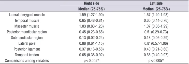
Results: 60 women were evaluated. Statistically significant differences were found
among the muscles evaluated regarding the pain threshold. The lateral pterygoid area, bilaterally, had the lowest pain threshold, followed by the masseter and temporal muscles.
Conclusion: this study suggests that the lateral pterygoid muscle, bilaterally, has the
lowest pain threshold upon palpation among the masticatory muscles, followed by masseter and temporal muscles, in women with TMD, according to the RDC/TMD
evaluation.
Keywords: Temporomandibular Joint Disorders; Physical Therapy Modalities;
INTRODUCTION
Temporomandibular disorder (TMD) is characterized as a set of disorders that mainly affect the masticatory muscles and temporomandibular joints1. The main symptom is referred pain, especially upon palpation2,3, accompanied by a reduction in range of motion. The multifactor etiology4,5 includes functional, anatomic and psychosocial elements6-9, illustrating a complex inter-action among these mechanisms that can reduce the quality of life of affected individuals10.
For the diagnosis of TMD, studies and clinicians have used indexes and questionnaires that assist in
the classification of the disorder, the evaluation of the
severity and the determination of the effectiveness of the most diverse interventions11,12. The Research Diagnostic Criteria of Temporomandibular Disorders (RDC/TMD) is among the most widely used for this purpose and is characterized as a complete assessment tool for the diagnosis of TMD with muscle and/or joint involvement as well as the determination of both physical and psychological aspects related to the disorder13,14. The RDC/TMD also enables a discerning evaluation of pain symptoms in the masticatory muscles and temporo-mandibular joints through palpation. Characterized as a widely employed evaluation in clinical practice, palpation offers the examiner the ability to investigate the harmony among the actions of the masticatory muscles and the functioning of the temporomandibular joint15.
Besides the evaluation of affected structures, several authors report different clinical responses to pain between genders16. The higher prevalence rates of pain states in the female gender has been attributed to a possible link to changes in the hormonal system17, which may explain proportions that range from two to six women for each man diagnosed with TMD18.
Understanding these characteristics and that TMD is a complex clinical entity, studies are needed to compare
symptoms using specific methods and recruiting the
population that is truly more affected. Such studies can provide knowledge for the choice of the best thera-peutic approach as well as improve the chances of the resolution of the signs and symptoms and, conse-quently, the rehabilitation of affected individuals.
The aim of the present study was to evaluate the pain threshold upon palpation of the masticatory muscles in women with TMD according to the RDC/ TMD. The hypothesis is that differences in the pain threshold are found among the different masticatory muscles involved in TMD when submitted to palpation,
with the lowest threshold expected for the lateral pterygoid muscle.
METHODS
This study received approval from the Human Research Ethics Committee of Nove de Julho University
(certificate number: 18032013.4.0000.5511). All volun -teers signed a statement of informed consent, which was drafted in compliance with Resolution 466/12 from 2012 of the Brazilian National Board of Health.
A cross-sectional study was conducted at a physical therapy clinic in the city of São Paulo, Brazil. One physiotherapist was in charge of recruiting the patients
and determining the fulfillment of the eligibility criteria.
A second physiotherapist administered the RDC/TMD and determined the diagnosis. A third physiotherapist performed the data processing and analysis. All physiotherapists had an average of 10-year experience involving patients with TMD and received three months of training with the tools employed in the study.
Volunteers were recruited through verbal
invita-tions and flyers placed in dental and physical therapy
clinics in the city of São Paulo between February and November 2014. Female volunteers (due to the high prevalence of TMD in this gender19) with a diagnosis of TMD using the RDC/TMD were included. The exclusion criteria were age under 18 and above 40 years, currently undergoing orthodontic, physiotherapeutic
or medicinal (analgesic, anti-inflammatory or muscle
relaxant) therapy, missing teeth and/or the use of partial or complete dentures, bite plate usage, a history of trauma to the face or temporomandibular joint and a diagnosis of osteoarthritis (IIIb) or osteoarthrosis (IIIc), using the RDC/TMD.
Table 1. Distribution of diagnoses of temporamandibular disorders according to Research Diagnostic Criteria of Temporomandibular Disorders (RDC/TMD)
Diagnosis n %
Only group I (muscle) Only group II (disc displacement)
Only group III (arthralgia) Groups I and II Groups I and III Groups II and III Groups I. II and III
24 4 2 16
5 2 7
40 6.6 3.3 26.6
8.3 3.3 11.6
Total 60 100
Outcomes
Pain threshold upon palpation
Items 8 and 10 of the RDC/TMD clinical exam were selected for the evaluation of the pain threshold upon palpation. These items rate pain intensity upon palpation of the masticatory muscles bilaterally, as follows: no pain = 0, mild pain = 1, moderate pain = 2, and severe pain = 3. The muscles were evaluated always from right to left in accordance with the experience of the examiner. The following muscles were evaluated:
- Extraoral muscle palpation
A – Posterior temporal – behind the temple (behind and immediately above the ears).
B – Mid temporal – mid region of temple (4 to 5 cm to the side of the lateral margin of the eyebrows). C – Anterior temporal – anterior part of temple (superior
to the infratemporal fossa and immediately above the zygomatic process).
D – Superior masseter – cheek below zygoma (1 cm in front of the temporomandibular joint and immediately below the zygomatic arch).
E – Mid masseter – cheek, side of face (anterior margin descending to the angle of the mandible).
F – Inferior masseter – cheek, line of mandible (1 cm above and anterior to the angle of the mandible). G – Posterior mandibular region (stylohyoid) –
posterior region of digastric muscle (mandible, throat region – area between the insertion of the sternocleidomastoid muscle and posterior edge of the mandible).
H - Submandibular region – mid pterygoid/supra--hyoid/anterior region of digastric (below the
mandible, 2 cm in front of the angle of the mandible).
The examiner performed single manual palpation of the muscles with 1 Kg of pressure for muscles A to F and 0.5 Kg of pressure for muscles G and H.
- Joint palpation
A – Lateral pole (anterior to tragus and on temporo-mandibular joint)
B – Posterior ligament (inside ear)
- Intraoral muscle palpation
A – Lateral pterygoid area – anterior portion of maxillary molars.
B - Temporal tendon – anterior edge of coronoid process.
Single manual palpation of these muscles was performed with 0.5 Kg of pressure.
For the definition of the pain threshold upon
palpation of the temporal and masseter muscles, the mean corresponding to the score of each of the portions of these muscles was calculated.
Data analysis
The Shapiro-Wilk test was used to determine the normality of the data. As the data exhibited asymmet-rical distribution, the Kruskal-Wallis test with Dunn’s post hoc test for multiple comparisons was used to measure differences in the pain threshold upon palpation. The SPSS 16.0 program (SPSS Inc., Chicago, USA) was used for the statistical analyses, with a p-value < 0.05
significant differences among the structures investi -gated. Table 3 displays the results of Dunn’s post hoc
test, demonstrating significant differences in the pain
threshold among the structures investigated.
RESULTS
Table 1 displays the distribution of the diagnoses of TMD of the 60 volunteers according to the RDC/TMD. Table 2 displays the pain thresholds of the regions palpated indicated on the RDC/TMD, demonstrating
Table 2. Median and interquartile range (25-75%) of pain threshold in palpated structures indicated by Research Diagnostic Criteria of Temporomandibular Disorders (RDC/TMD) (0 = no pain, 1 = mild pain, 2 = moderate pain, and 3 = severe pain)
Right side Left side
Median (25-75%) Median (25-75%)
Lateral pterygoid muscle 1.59 (1.27-1.90) 1.67 (1.40-1.93)
Temporal muscle 0.65 (0.48-0.81) 0.60 (0.44-0.76)
Masseter muscle 1.03 (0.83-1.23) 1.07 (0.86-1.29)
Posterior mandibular region 0.45 (0.23-0.68) 0.51(0.29-0.73)
Submandibular region 0.13 (0.02-0.24) 0.18 (0.06-0.29)
Lateral pole 0.88 (0.61-1.15) 0.81(0.57-1.06)
Posterior ligament 0.37 (0.16-0.58) 0.40 (0.21-0.60)
Temporal tendon 0.65 (0.38-0.92) 0.68 (0.40-0.97)
Comparisons among variables p<0.005* p<0.005*
* Denotes significant differences among structures (Kruskall-Wallis ANOVA).
Table 3. Comparison of pain upon palpation among structures described on Research Diagnostic Criteria of Temporomandibular Disorders (RDC/TMD) (Dunn’s post hoc text)
MT MM PMR SR LP PL TT
R L R L R L R L R L R L R L
LPA <0.05 <0.001 ns ns <0.001 <0.001 <0.001 <0.001 <0.001 <0.001 <0.001 <0.001 <0.001 <0.001
MT ns ns ns ns <0.001 <0.001 ns ns <0.05 ns ns ns
MM <0.001 <0.001 <0.001 <0.001 ns ns <0.001 ns <0.01 <0.01
PMR ns ns ns ns ns ns ns ns
SR <0.001 <0.001 <0.001 ns <0.001 ns
LP <0.001 ns <0.001 <0.001
PL ns ns
R: right; L: left; LPA: lateral pterygoid area; MT: mid temporal; MS: mid masseter; PMR: posterior mandibular region; SR: submandibular region; TT: temporal tendon; LP: lateral pole; PL: posterior ligament
DISCUSSION
The aim of the present study was to evaluate the pain threshold upon palpation of the masticatory muscles in women with TMD according to the RDC/TMD. The main
finding was the lower pain threshold found in the area of
the lateral pterygoid muscle (LPM) bilaterally, followed by the masseter and temporal muscles. Besides the high degree of representativeness of the population with TMD in clinical practice, which is characterized by the female gender16, this study differs from previous
investigations by using a musculoskeletal pain threshold assessment method that is widely employed in clinical practice and considered one of the founda-tions for the diagnosis of TMD using the RDC/TMD21.
Despite the wide use in the clinical setting, divergent opinions are found regarding the use of palpation for the evaluation and diagnosis of individuals with TMD. Gomes et al. (2008)22 report acceptable
speci-ficity values (higher than 0.90), whereas Chaves et al.
volunteers in the present study exhibited myofascial pain associated with disc displacement and 8.3% had myofascial pain associated with arthralgia.
Further demonstrating the clinical importance of the LPM, Stelzenmueller et al. (2016)33 state that manual palpation is essential to the clinical evaluation of this muscle. The basic requirement for successfully palpating the LPM is precise knowledge of the muscle topography and the proper route for intraoral palpation. These requirements were met by the physiotherapist in charge of the evaluations in the present study, who had 10 years of experience in the evaluation and treatment of patients with TMD.
The present study has limitations that should be addressed. There was no group without TMD to serve as a baseline in the follow up of the volunteers. Moreover, there was no establishment of different groups based on the type of TMD and there were no imaging exams to complement the diagnosis and enable a better analysis of the LPM area. Thus, further studies are needed to overcome these limitations.
CONCLUSION
The present findings suggest that the lateral
pterygoid muscle, bilaterally, has the lowest pain threshold upon palpation among the masticatory muscles, followed by masseter and temporal muscles in women with TMD, according to the RDC/TMD evaluation.
REFERENCES
1. American Society of Temporomandibular
Joint Surgeons (ASTJS). Guidelines for diagnosis and management of disorders involving the temporomandibular joint and related musculoskeletal structures. Cranio. 2003;21(1):68-76.
2. Schiffman E, Ohrbach R, Truelove E, Look J, Anderson G, Goulet JP et al. Diagnostic Criteria for Temporomandibular Disorders (DC/ TMD) for Clinical and Research Applications: recommendations of the International RDC/TMD Consortium Network* and Orofacial Pain Special Interest Groupdagger. J Oral Facial Pain Headache. 2014;28(1):6-27.
3. Peck CC, Goulet JP, Lobbezoo F, Schiffman EL, Alstergren P, Anderson GC et al. Expanding the taxonomy of the diagnostic criteria with the aid of an algometer has greater reliability than
manual palpation. However, unlike the present study, Chaves et al. (2010)23 had a sample of only children, the majority of whom was male, which are character-istics that do not represent the main population affected
by TMD. Thus, the practice of palpation is justified in
the clinical setting due to the fact that it enables the determination of other clinical factors beyond pain alone, such as tissue quality and the state of muscle
fibers, which are essential factors for the evaluation and
diagnosis of musculoskeletal disorders23.
The present findings demonstrate the importance
of investigating the lateral pterygoid, masseter and temporal muscles bilaterally. These muscles have considerable representativeness with regard to the characteristic pain in individuals with a diagnosis of TMD9. Santos et al. (2005)24 report that the masseter muscle is the most sensitive site in cases of TMD, which may involve functional overload of the joint, occlusal factors and consequent overactivity of the LPM, although the evidence is limited25,26. Differently from the study conducted by Santos et al. (2005)24,however, the
present findings show the greater representativeness
of the LPM regarding pain upon palpation in cases of TMD, demonstrating that this muscle may have funda-mental importance in the evaluation of patients with a diagnosis of this disorder. Bonjardim et al. (2005)27 also point out the importance of the LPM, although the sample of individuals with a diagnosis of TMD was in a different age range (12 to 18 years). Moreover, the authors found greater pain sensitivity in this muscle in the female gender.
The LPM is directly involved in lateral functional movements in unilateral action and protrusion of the mandible in bilateral action, playing an important modulating role in the joint movements28,29. Thus, changes in the LPM could facilitate joint derangement, exerting a negative impact on the biomechanical
harmony of the temporomandibular joint, which justifies
the use of resources directed at this muscle30,31.The
present findings underscore the clinical importance of
a detailed evaluation and the application of therapies directed at the LPM. Lopes (2015)32 found that individuals with simultaneous diagnoses of TMD and migraine tend to exhibit hypertrophy of the LPM and have greater pain symptoms during palpation of this muscle. The authors also found strong associations between myofascial pain and both disc displacement and arthralgia in the diagnosis using the RDC/
for temporomandibular disorders. J Oral Rehabil.2014;41(1):2-23.
4. Oral K, Bal Küçük B, Ebeoğlu B, Dinçer S. Etiology
of temporomandibular disorder pain. Agri. 2009;21(3):89-94.
5. Munhoz WC, Marques AP, Siqueira JTT. Evaluation of body posture in individuals with internal temporomandibular joint derangement. J. of Craniomandibular Practice. 2005;23(4):269-77. 6. De Leeuw R. American academy of orafacial
pain: guidelines for assessment diagnosis and managements. 4th Ed. Quintenssence Publishing, Chigago, 2008. p. 131-41.
7. Kafas P, Leeson R. Assessment of pain in temporomandibular disorders: the biopsychosocial complexity. Int J Oral Maxilofacial Surg. 2006;35(2):145-9.
8. Nassif NJ, Al-Salleeh F, Al-Admawi M. The prevalence and treatment needs of symptoms and signs of temporomandibular disorders among young adult males. J Oral Rehabil. 2003;30(9):944-50.
9. Magnusson T, Egermark I, Carlsson GE. A longitudinal epidemiologic study of signs and symptoms of temporomandibular joint disorders from 15 to 35 years of age. J Orofac Pain. 2000;14(4):310-9.
10. Biasotto-Gonzalez DA. Abordagem interdisciplinar das disfunções temporomandibulares. Barueri - SP: Ed. Manole, 2005
11. De Lucena LBS, Kosminsky M, DA Costa LJ, Góes PSA. Validation of the Portuguese version of the RDC/TMD Axis II questionnaire. Brazilian Oral Research. 2006;20(4):312-7.
12. Pereira-Junior FJ, Favilla EE, Dworkin SF, Huggins K. Critérios de diagnóstico para pesquisa das disfunções temporomandibulares (RDC/TMD).
Tradução oficial para a língua portuguesa. Jornal Brasileiro de Clínica Odontológica Integrada.
2004;8(47):384395.
13. Dworkin S F, Leresche L. Research diagnostic criteria for temporomandibular disorders: review,
criteria, examinations and specifications, critique. J
Craniomandib Disord.1992;6(4):301-55.
14. Look JO, Schiffman EL, Truelove EL, Ahmad M. Reliability and validity of Axis I of the Research Diagnostic Criteria for Temporomandibular Disorders (RDC/TMD) with proposed revisions. J Oral Rehabil. 2010;37(10):744-59. doi:
10.1111/j.1365-2842.2010.02121.x. Epub 2010 Jul 20.
15. Okeson JP: History and examination for temporomandibular disorders. In: Okeson JP (ed). Management of temporomandibular disorders and occlusion. 4th ed. St. Louis: Mosby, 1998. p.234-309.
16. Fillingim RB, King CD, Ribeiro-Dasilva MC, Rahim-Williams B, Riley JL 3rd. Sex, gender, and pain: a
review of recent clinical and experimental findings.
J Pain. 2009;10(5):447-85.
17. Abubaker AO, Hebda PC, Gunsolley JN. Effects of sex hormones on protein and collagen con- tent of the temporomandibular joint disc of the rat. J Oral Maxillofac Surg 1996;54(6):721-7.
18. Schmid-Schwap M, Bristela M, Kundi M,
Piehslinger E. Sex-specific differences in patients
with temporomandibular disorders. J Orofac Pain. 2013;27(1):42-50.
19. Manfredini D, Cocilovo F, Favero L, Ferronato G, Tonello S, Guarda-Nardini L. Surface electromyography of jaw muscles and kinesiographic recordings: diagnostic accuracy for myofascial pain. J. Oral Rehabil. 2011 Nov;38(11):791-9.
20. Rodrigues-Bigaton D, Dibai-Filho AV, Costa AC, Packer AC, Costa CS, de Castro EM. Accuracy of two forms of infrared image analysis of the masticatory muscles in the diagnosis of myogenous temporo-mandibular disorder. J. Bodyw. Mov. Ther. 2014;18(1):49-55.
21. Isselèe H, De Laat A, Lesaffre E, Lysens R. Short-term reproducibility of pressure pain thresholds in masseter and temporalis muscles of symptom-free subjects. Eur J Oral Sci. 1997;105(6):583e7.
22. Gomes MB, Guimarães JP, Guimarães FC, Neves AC. Palpation and pressure pain threshold reliability and validity in patients with temporomandibular disorders. Cranio. 2008;26(3):202e10
23. Chaves TC, Nagamine HM, Sousa LM, Oliveira AS, Grossi DB. Comparison between the reliability levels of manual palpation and pressure pain threshold in children who reported orofacial pain. Manual Therapy. 2010;15(5):508-12.
25. Manfredini D, Landi N, Romagnoli M, Cantini E, Bosco M. Etiopathogenesis of parafunctional habits of the stomatognathic system. Minerva Stomatol. 2003;52(7-8):339-45.
26. Wanman A, Agerberg G. Etiology of
craniomandibular disorders: evaluation of some occlusal and psychosocial factors in 19-year-olds. J Craniomandib Disord.1990;5(1):35-44.
27. Bonjardim LR, Gavião MBD, Pereira LJ, Castelo PM, Garcia RCMR. Signs and symptoms of temporomandibular disorders in adolescents. Braz Oral Res. 2005;19(2):93-8.
28. Taskaya-Yilmaz N, Ceylan G, Incesu L, Muglali M. A possible etiology of the internal derangement of the temporomandibular joint based on the MRI observations of the lateral pterygoid muscle. Surg Radiol Anat. 2005;27(1):19-24.
29. Mazza D, Marini M, Impara L, Cassetta M, Scarpato P, Barchetti F et al. Anatomic examination of the upper head of the lateral pterygoid muscle using magnetic resonance imaging and clinical data. Journal of Craniofacial Surgery. 2009;20(5):1508-11.
30. Gonzalez-Perez LM, Infante-Cossio P,
Granados-Nuñez M, Lopez FJU. Treatment of temporomandibular myofascial pain with deep dry needling. Med Oral Patol Oral Cir Bucal. 2012;17(5):e781-5.doi:10.4317/medoral.17822 http://dx.doi.org/doi:10.4317/medoral.17822
31. Gonzalez-Perez LM, Infante-Cossio P, Granados-Nuñez M, Lopez FJU, Lopez- Martos R, Ruiz-Canela-Mendez P. Deep dry needling of trigger points
located in the lateral pterygoid muscle: efficacy and
safety of treatment for management of myofascial pain and temporomandibular dysfunction. Med Oral Patol Oral Cir Bucal. 2015;20(3):326-33.doi: http://dx.doi.org/doi:10.4317/medoral.20384.
32. Lopes SLPC, Costa ALF, Gamba TO, Flores IL, Cruz AD, Min LL. Lateral pterygoid muscle volume and migraine in patients with temporomandibular disorders. Imaging Science in Dentistry. 2015;45(1):1-5.