Original article (short paper)
The effects of a global postural exercise program
on temporomandibular disorder
Alexandre Fiorelli Eduardo Aguilar Arca Carolina Menezes Fiorelli Alitéia Aparecida Souza Rodrigues
Ana Carla Furcin Alberto De Vitta Cesar Becalel Waisberg
Débora de Melo Trize Paulo Henrique Weckwerth
Universidade do Sagrado Coração, Bauru, SP, Brasil
Abstract — Changes in the suboccipital muscles and the hamstrings may interfere with head posture and the biomechanics of the temporomandibular joint, both of which contribute to the severity of temporomandibular disorders (TMD). The objective of this investigation was to evaluate the effects of a global postural exercise program (GPEP) on pain intensity and mouth-opening range of motion (ROM) in women with TMD. The participants were comprised of 30 women with TMD who were divided into two groups: an experimental group (EG) and a control group (CG). A pressure algometer was used for pain assessment and a paquimeter was used to measure ROM. The duration of the GPEP was six weeks. In the EG, there was a reduction in pain intensity and an increase in mouth-opening ROM compared to the CG. Therefore, we concluded that the GPEP was effective in relieving pain in all of the evaluated muscles and regions, and in increasing mouth-opening ROM in women with TMD.
Keywords: temporomandibular joint; facial pain; range of motion
Introduction
Temporomandibular disorders (TMD) are a group of disorders with signs and symptoms that include joint noises, such as click-ing and crepitus (gratclick-ing), pain in the masticatory muscles, limited jaw movements, craniofacial pain, and temporomandibular joint (TMJ) pain1.
TMD may involve alterations in the cervical roots, leading to pain in the cervical spine, craniofacial pain, and decreased mouth-opening range of motion (ROM) 2-4. In addition, TMD are the major cause of non-dental orofacial pain5-7.
Epidemiological investigations have shown that 40% - 75% of the population present with at least one TMD sign, such as joint noise, and 33% present with at least one TMD symptom, such as facial or joint pain8-10.
Some researchers have investigated the relationship between TMJ and several changes in different parts of the human body, e.g., the suboccipital muscles and the hamstrings11,12.
Myofascial chain tightness in any of the aforementioned parts of the body may interfere with head posture and TMJ biomechanics, which may in turn contribute to the severity of TMD11,13.
Global postural exercises have been shown to contribute to pain relief and increase muscular strength, muscle chain stretching, and balance in young, sedentary women14. Therefore, the aim of this study was to assess the effects of a global postural exercise program on pain intensity and mouth-opening ROM in women with TMD.
Method
Design and ethical aspects
This study was a randomized controlled clinical trial, which was conducted at the Manual Therapy and Research Lab at the Universidade do Sagrado Coração (USC), in the city of Bauru/SP/ Brazil. The research project was approved by the USC Research Ethics Committee (protocol number 541.201).
Participants
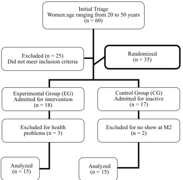
Sixty women who were diagnosed with TMD from the Dentistry and Physical Therapy School at USC were selected according to the inclusion and exclusion criteria.
The inclusion criteria were a diagnosis of TMD, according to the Research Diagnostic Criteria for Temporomandibular Disorders (RDC/TMD)15, and the absence of any physical functional impairment that could prevent participation in the exercise program.
The exclusion criteria were participation in another exercise
program, and treatment with painkillers or anti-inlammatory
the exercise-experimental group (EG) and 17 into the control-inactive group (CG). During the course of the study, three subjects left the EG and two subjects left the CG, as shown in Figure 1.
Procedure
The participants were individually assessed at the Physical Therapy Research Lab at USC. Data on age, life habits, and health conditions were gathered. To measure pain, a mechani-cal pressure algometer was used (Palpeter®).
This consisted of a pressure gauge with a probe area, 1cm in circumference, through which a constant pressure of 1.0 kg in seven different locations was applied. These locations were bilaterally located on the temporalis muscle (anterior, middle,
and posterior ibers), the supericial masseter (origin, body, and
insertion), and the upper trapezius (middle part).
A pressure of 0.5 Kg was applied in three locations bilat-erally, the submandibular region, the digastric muscle, and the lateral pole of the TMJ. Each procedure was repeated a maximum of three times in each location.
The pain intensity at each location was reported by the subjects as follows: 0 = no pain, only pressure; 1 = mild; 2 = moderate; and 3 = severe15. A paquimeter (JOMARCA®) was used to measure mouth-opening ROM. The subjects were seated, and the head was aligned with maximal active mouth opening. The paquimeter was positioned on the upper and lower central incisors (11, 21, 31 e 41) in accordance with a previous study2.
The subjects in the EG were then submitted to a global postural exercise program (GPEP).The GPEP lasted for six weeks. Exercises were performed twice weekly for six weeks and each session had a duration of 45 min.
The exercises, which consisted of stretching of the verte-bral column posterior muscle chain and the lower limbs, were associated with prolonged expiration techniques and self-stretching. In each session, a series of eight types of exercises were performed as follows: Exercise 1–supine position, hip
lexion of one lower limb and hip extension of the other with dorsilexion of the ankle, a total of three sets of 15 s each.
Exercise 2–supine position, bilateral extension of the lower
segments against the wall with ankle dorsilexion, holding the
position for 5 min. Exercise 3–supine position, one lower limb
with foot dorsilexion leaning on the wall and the contralateral
segment resting, a total of three alternate sets of 15 s. Exercise 4–supine position, bilateral hip external rotation with knees
lexed, feet leaning on the wall, holding the position for 5 min.
Exercise 5–seated with vertebral spine against the wall, one
lexed knee and the other extended, a total of three alternate
sets of 15 s each. Exercise 6–seated with vertebral spine against
the wall, knees extended, and feet in dorsilexion, holding the
position for 5 min. Exercise 7–seated with no support of the
torso, and one knee lexed and the other extended, a total of
three alternate sets of 15 s each. Exercise 8–seated position, with no support of the torso, and the knees extended with foot
dorsilexion, holding the position for 5 min.
At the end of the postural exercise program, a inal assess -ment of the participants (M2), with the same procedures as the initial assessment (M1), was performed.
Data collection and analysis
The data obtained in the study were expressed as mean and standard deviations. The Mann-Whitney U-test was used to compare the groups and the Wilcoxon rank test was used to
compare M1 and M2. A signiicance level of 5% was considered to be statistically signiicant.
Results
The casuistry consisted of 30 women (mean age: 36.2 ± 9.8 years) who were apparently healthy, sedentary, and not taking
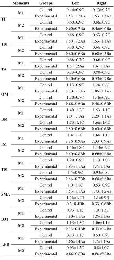
painkillers or anti-inlammatory medications. Table 1 shows
the mean and standard deviation values, the respective results of statistical testing on muscle pain intensity [temporalis (posterior, middle, and anterior portions), masseter, trapezium, submandibular area, digastric, and the lateral pole of the TMJ], and comparisons between the EG and CG groups.
The EG moments showed statistically signiicant differ -ences (p< 0.05) in the pain intensity of all variables studied.
The CG moments did not show statistically signiicant differ -ences in pain intensity, apart from the submandibular area. On
comparison of the groups, there were no statistically signiicant
differences in the superior portion of the left masseter, the inferior portion of the right masseter, the right trapezius, the temporalis muscle, the right digastric muscle, or the lateral pole of the TMJ.
Initial Triage
Women:age ranging from 20 to 50 years (n = 60)
Randomized (n = 35) Excluded (n = 25)
Did not meet inclusion criteria
Experimental Group (EG) Admitted for intervention
(n = 18)
Excluded for health problems (n = 3)
Control Group (CG) Admitted for inactive
(n = 17)
Excluded for no show at M2 (n = 2)
Analyzed
(n = 15) Analyzed(n = 15)
Table 1. Descriptive values for assessment of pain intensity in tempora-lis, masseter, trapezius, the submandibular area, digastric, and the later-al pole of the TMJ in the respective groups and the assessed moments.
Moments Groups Left Right
TP
M1 Control 0.46±0.9C 0.53±0.7C
Experimental 1.53±1.2Aa 1.53±1.3Aa
M2 Control 0.60±0.9C 0.66±0.9C
Experimental 0.60±0.7Ba 0.46±0.6Ba
TM
M1 Control 0.86±0.9C 0.53±0.7C
Experimental 1.60±1.2Aa 1.53±1.1Aa
M2 Control 0.80±0.9C 0.66±0.9C
Experimental 0.60±0.6Ba 0.60±0.5Ba
TA
M1 Control 0.66±0.7C 0.66±0.9C
Experimental 1.5±1.2Aa 1.6±1.1Aa
M2 Control 0.73±0.9C 0.80±0.9C
Experimental 0.40±0.6Ba 0.53±0.7Ba
OM
M1 Control 1.13±0.9C 1.20±0.6C
Experimental 0.20±1.1Aa 1.86±1.1Aa
M2 Control 1.20±0.7C 1.46±0.9C
Experimental 0.66±0.6Ba 0.46±0.6Bb
BM
M1 Control 1.40±1.2C 1.53±1.1C
Experimental 2.0±1.1Aa 2.20±1.1Aa
M2 Control 1.73±1.1C 1.66±1.0C
Experimental 0.80±0.6Bb 0.60±0.6Bb
IM
M1 Control 1.4±1.1C 1.60±1.1C
Experimental 2.26±0.9Aa 2.33±0.9Aa
M2 Control 1.46±1.8C 1.33±0.9C
Experimental 0.60±0.8Bb 0.66±0.6Ba
TM
M1 Control 1.20±0.9C 1.13±1.0C
Experimental 1.93±1.1Aa 1.7±1.1Aa
M2 Control 1.4±0.9C 0.93±0.8C
Experimental 0.46±0.7Bb 0.60±0.6Ba
SMA
M1 Control 1.0±1.1C 0.53±0.9C
Experimental 1.53±1.1Aa 1.73±1.2Aa
M2 Control 1.46±1.1D 1.1±0.9D
Experimental 0.3±0.4Bb 0.33±0.6Bb
DM
M1 Control 0.93±1.1C 1.0±1.3C
Experimental 1.80±1.1Aa 1.8±1.1Aa
M2 Control 1.13±1.5C 1.06±1.1C
Experimental 0.33±0.4Bb 0.33±0.4Ba
LPR
M1 Control 0.73±1.1C 0.53±0.9C
Experimental 1.66±1.4Aa 1.7±1.4Aa
M2 Control 0.93±1.2C 0.8±1.0C
Experimental 0.66±0.8Ba 0.80±0.8Ba M1: initial assessment; M2: inal assessment; TMPF: Temporalis Posterior;
TMMF: Temporalis Middle; TMAF: Temporalis Anterior; OM: Origin of Masseter; BM: Body of Masseter; IM: Insertion of Masseter; TM: Trapezius Muscle; SMA: Submandibular Area; DM: Digastric Muscle; RAPL: Lateral
Pole Region. Different upper case letters indicate a statistically signiicant
difference when comparing groups concerning M1 and M2 (Wilcoxon Test).
Different lower case letters indicate a statistically signiicant difference when
comparing M1 and M2 concerning groups (Mann–Whitney test).
Table 2. Descriptive values on maximal mouth-opening range of mo-tion in the respective groups and the assessed moments.
Moment Groups Mouth ROM (cm)
M1 Control 43.80±8.1C
Experimental 36.27±10.9Aa
M2 Control 43.20±7.3C
Experimental 41.87±7.8Bb M1: initial assessment; M2: inal assessment; cm: centimeters; ROM: range of motion. Different upper case letters indicate a statistically signiicant
difference when comparing groups concerning M1 and M2 (Wilcoxon Test).
Different lower case letters indicate a statistically signiicant difference when
comparing M1 and M2 concerning groups (Mann–Whitney test).
Table 2 shows the mean and standard deviation values, the respective results of statistical testing for maximal mouth-opening ROM, and comparison between the EG and the CG.
When comparing GE moments, there was a statistically
signiicant difference (p< 0.05) in maximal mouth-opening
ROM. The control group did not show a statistically signiicant
difference. When comparing both groups, there was a statistically
signiicant difference between the EG and the CG.
Discussion
The results of this study suggested that the GPEP signiicantly
contributed to relief in pain intensity of the assessed muscles and increase in maximal mouth-opening ROM in women with TMD, due to the intervention program, which consisted of pos-tural exercises based on a global approach focusing on posterior muscle chain balance.
The craniomandibular system is part of the upper quadrant, which involves the head, neck, and shoulder girdle. Therefore, muscles, ligaments, and TMJ fascia are closely related11. Any disorder, occlusal disturbance, postural change, or upper quad-rant trauma may lead to problems in those structures or their adjacent components16.
There is a functional relationship between the masticatory muscles, the paravertebral muscles, the hamstrings, soleus, and gastrocnemius, in addition to ascendant inhibitory coactivation
as a result of the stretch relex11. In light of this, the exercise pro-gram that was used in this study contributed to anti-gravitational postural adaptation because it used exercises in both the lying and sitting positions. A previous study compared subjects who received postural training and subjects with just awareness.
The postural training group showed signiicant improvements
with decreased TMD severity, relief of TMJ and neck pain, maxi-mal mouth opening without pain, and fewer TMD symptoms17. Another study assessed pain and quality of life in TMD individuals, using global postural re-education (GPR) and static stretching.
It was concluded that both techniques were equally eficient in
reducing symptom intensity, increasing pain thresholds in the evaluated muscles, and improving quality of life18.
In addition, another study that examined the effects of inhibi-tory techniques on suboccipital muscles in individuals with tight
hamstrings found a signiicant difference in the intervention group
Our indings showed a statistically signiicant difference
between the groups, and a relationship between the TMJ and lower limbs through myofascial chains. Postural adaptations
through the myofascial chains seem to explain the indings in
the present study, and indicate a functional relationship between the masticatory muscles and other structures, such as the para-vertebral muscles and the hamstrings20.
All of these associations contributed to better positioning of the head and neck, and better structuring of the relationship between the agonist and antagonist muscles, thereby, decreasing pain intensity and improving mouth-opening ROM of the participants.
Strengths and limitations
The strengths of the present study were the methods and assessment tools that were used to measure the variables, and the intervention program. It is known that manual palpation has obvious limitations because it is extremely subjective, and is hard
to quantify and standardize, thereby, compromising conidence
in the data and research reproducibility.
Therefore, the authors of the present study used a validated
pressure algometer to increase conidence in the results of vari -able pain21. In relation to the intervention program, we have highlighted its use in clinical practice. It can be used by several health care professionals, and the detailed description of the exercises makes their reproduction in future research easier.
However, the study has some limitations. Only female subjects participated in the study, which diminishes external validation of this study because the results may not be appli-cable to men. However, the internal validation for women was increased, which is important when considering that TMD has a greater incidence in women 20-45 years old, approximately 5
times compared to men. Therefore, this justiies the importance
of this study, which was only directed toward women.
Conclusion
Based on the results of this study, we concluded that global pos-tural exercises were effective on pain relief in all of the assessed muscles and areas. The exercise program was also effective on mouth-opening ROM in women with TMD. In future study, we recommend reassessing the patients after a period without the exercise program; analyzing other symptoms, such as crepitus, noises, and muscle strength; and comparison with other methods and physiotherapeutic techniques, the use of medication, and dental intervention.
References
1. Pimentel MJ, Gui MS, Martins de Aquino LM, Rizzatti-Barbosa CM. Features of temporomandibular disorders in ibromyal -gia syndrome. Cranio: J Craniomandibular Practice. 2013; 31 (1):40-45.
2. Ferragud PM, Gandia JJB. Efecto de lamanipulación de lacha-rnelaoccipito-atlo-axoideaen La apertura de La boca. Osteopatia Cientíica. 2008; 3 (2):45-51.
3. Biasotto-Gonzalez DA, Andrade DV, Gonzalez TO, Martins MD, Fernandes KPS, Corrêa JCF, et al. Correlação entre disfunção temporomandibular, postura e qualidade de vida. Rev bras cresci-mento desenvolvicresci-mento Hum. 2008; 18 (1):79-86.
4. Reiter S, Goldsmith C, Emodi-Perlman A, Friedman-Rubin P, Winocur E. Masticatory muscle disorders diagnostic criteria: the American Academy of Orofacial Pain versus the research diagnostic criteria ⁄temporomandibular disorders (RDC⁄TMD). J Oral Rehab. 2012; 39:941-947.
5. Kuroiwa DN, Marinelli JG, Rampani MS, Oliveira W, Nicodermo D. Temporomandibular disorders and orofacial pain: study of qual-ity of life measured by the Medical Outcomes Study 36 – Item Short Form Health Survey. Rev Dor Pesquisa, Clínica Terap. 2011; 12 (2):93-98.
6. Carrara SV, Conti PCR, Barbosa JS. Termo do 1º Consenso em Disfunção Temporomandibular e Dor Orofacial. Dental Press J Orthodontics. 2010; 15 (3):114-120.
7. Schiffman EL, Truelove EL, Ohrbach R, Anderson GC, John MT, List T, et al. The Research Diagnostic Criteria for Temporomandibular Disorders. I: overview and methodology for assessment of validity. J Oral Rehab. 2010; 24:7-24.
8. Al-Jundi MA, John MT, Setz JM, Szentpétery A, Kuss O. Meta-analysis of treatment need for temporomandibular disorders in adult nonpatients. J Orofacial Pain. 2008; 22 (2):97-107. 9. Barros VM, Seraidarian PI, Côrtes MIS, de Paula LV. The impact
of orofacial pain on the quality of life of patients with temporo-mandibular disorder. J Orofacial Pain. 2009; 23 (1):28-37. 10. Iodice G, Danzi G, Cimino R, Paduano S, Michelotti A.
Association between posterior crossbite, masticatory muscle pain, and disc displacement: a systematic review. Eur J Orthodontics. 2013; 35 (6):737-744.
11. Retischwerdt C, Rivas L, Palomeque L, Alburquerque F. Efectos inmediatos del estiramiento de los músculos isquiosurales em el sistema estomatognático en La cervicalgia mecânica. Osteopatiacientíica. 2009; 4 (2):39-46.
12. Packer AC, Pires PF, Dibai-Filho AV, Rodrigues-Bigaton D. Effects of upper thoracic manipulation on pressure pain sensitiv-ity in women with temporomandibular disorder: a randomized, double blind, clinical trial. Am J Physical Med Rehab. 2014; 93 (2):160-168.
13. Quintana E, Alburquerque F, Borallo L, Rodriguez-Blanco C. Inmediate effects of suboccipital muscles inhibition technique in subjects with short hamstring syndrome. J Manipulative Physiol Ther. 2009; 32(4):252-261.
14. Mann L, Kleinpaul JF, Weber P, Mota CB, Carpes FP. Efeito do treinamento de Isostretching sobre a dor lombar crônica: um estudo de casos. Motriz. 2009; 15 (1):50-60.
15. Dworkin SF, LeResche L. Research diagnostic criteria for tem-poromandibular disorders: review, criteria, examinations and speciications, critique. J Craniomandibular Disord. 1992; 6 (4):301-355.
di-agnósticos; uma contribuição para a prática clínica e de pesquisa. Fisioter. Pesqui. 2008; 15 (1):92-100.
17. Wright EF, Domenech MA, Fischer JR. Usefulness of posture training for patients with temporomandibular disorders. JADA. 2000; 131:202-210.
18. Maluf SA. Efeito da reeducação postural global e do alongamento estático em portadoras de disfunção temporomandibular: um estudo comparativo. [Tese Doutorado]. São Paulo: Faculdade de Medicina, Universidade de São Paulo; 2006.
19. Figueiredo GS, Oliveira LK. O efeito imediato da técnica de inibição dos músculos subocciptais em indivíduos com encur-tamento de isquiotibiais.[Trabalho de Conclusão de Curso]. Brasilia: Faculdade de Ciências da Educação e da Saúde, Curso de Fisioterapia, Centro Universitário de Brasília, 2011.
20. Fernández-de-las-Peñas C, Carratalá-Tejada M, Luna-Oliva L, Mian-golarra-Paje J. The immediate effect of hamstring muscle streching in subjects trigger points in the masseter muscle. J Musculoskeletal Pain. 2006; 14:27-35.
21. Futarmal S, Kothari M, Ayesh E, Baad-Hansen L, Svensson P. New palpometer with impliations for assessment of deep pain sensitvity. J Dental Res. 2011; 90 (7):918-922.
Corresponding author
Paulo Weckwerth
Pró-Reitoria de Pesquisa e Pós-Graduação, Universidade do Sagrado Coração Rua Irmã Arminda, 10-50, Jardim Brasil, Bauru-SP
Email: [email protected]
Manuscript received on March 09, 2016 Manuscript accepted on April 18, 2016