Carlos André Ramalho Simões
Construction of an innocuous
Salmonella
phage
Carlos André Ramalho Simões
Construction of an innocuous
Salmonella
phage
Dissertação de Mestrado
Mestrado em Bioengenharia
Trabalho efetuado sob a orientação da
Doutora Sanna Maria Sillankorva
DECLARAÇÃO
Nome: Carlos André Ramalho Simões
Endereço eletrónico: [email protected] Telefone:926814219
Bilhete de Identidade/Cartão do Cidadão: 14161592
Título da dissertação: Construction of an innocuous Salmonella phage
Orientadora
Professora Doutora Sanna Maria Silankorva
Ano de conclusão: 2015
Mestrado em Bioengenharia
DE ACORDO COM A LEGISLAÇÃO EM VIGOR, NÃO É PERMITIDA A REPRODUÇÃO DE QUALQUER
PARTE DESTA TESE/TRABALHO.
Universidade do Minho, _____/_____/_________
Desde já expresso o meu profundo agradecimento a todas as pessoas que me
ajudaram neste último ano e que me conseguiram ajudar a alcançar este patamar de
satisfação.
Em primeiro lugar, à Doutora Sanna Sillankorva pelo apoio dado, pelas sugestões e críticas,
pelo à vontade com que me pôs sempre, mas acima de tudo por me ter dado a oportunidade de
trabalhar com ela.
À Catarina por estar sempre presente para qualquer dúvida que precisasse no laboratório,
pelo à vontade que me pôs desde o primeiro dia em laboratório e pela grande ajuda que me deu na
escrita da tese. Ao Luís e à Graça por serem duas pessoas sempre dispostas a ajudar e a explicar
qualquer dúvida (ao Luís principalmente pela ajuda que me deu no estudo da Bioinformática e claro
aos grandes debates que tivemos). Ao Sílvio e à Ana pela grande e boa disposição que a presença
deles trazia ao trabalhar no laboratório.
Aos meus amigos Joana e Macedo, pois este ano de trabalho não seria a mesma coisa sem a
boa companhia e boa disposição deles. “Aturar” a Joana sem a ajuda do Macedo seria difícil.
Aos meus amigos Andreia, Rita, Sara e Pedro pelo apoio dado e pela “dor” partilhada ao
longo do último ano (bem ditos foram os intervalos que fazíamos nas máquinas) foi trabalhoso, mas
todos conseguimos no final.
Aos meus amigos Diana, Marco, Humberto e Ezequiel por todo o apoio dado e claro pela
paciência por me aturarem, uma irmandade que nunca se vai separar. Agradeço profundamente
pela grande ajuda que me deram e claro que espero retribuir já no próximo ano.
À minha família, principalmente, à minha irmã, mãe e pai por permitir que tudo isto fosse
possível, por me aguentarem nas alturas de maior pressão, mas acima de tudo por todo o apoio
dado durante esta fase e pelo exemplo que são para mim.
A
Salmonela é um dos principais agentes patogénicos alimentares responsáveis por
doenças e hospitalizações, sendo responsável por 38% das 3000 mortes reportadas anualmente,
devido a intoxicações alimentares nos EUA (Centro de Controlo de Doenças dos EUA). A Salmonela,
presente no trato gastrointestinal das aves domésticas, pode ser passada de ave para ave através
de comida, água contaminada, fontes/vetores ambientais. Os Bacteriófagos (fagos) são vírus
bacterianos e por isso são considerados os predadores naturais das bactérias. Os fagos são
inofensivos para os humanos e animais e têm vindo a ser utilizados extensivamente para a
prevenção e controlo de patogéneos bacterianos. Experiências in vitro têm vindo a demonstrar que
os fagos quando estão sozinhos ou misturados com outros fagos (cocktail), conseguem diminuir
significativamente os níveis de
Salmonela em alimentos contaminados. No entanto, o futuro dos
fagos na segurança alimentar encontra-se mais dependente das agências reguladoras, que ainda
mostram algum desconforto aquando da utilização dos fagos. Isto deve-se principalmente ao facto
de haver uma escassez de fortes evidências científicas, a uma emergência de fenótipos bacterianos
resistentes a fagos e devido à alta percentagem de genes fágicos com funções desconhecidas, que
podem ser fatores de virulência ou genes de toxinas.
O principal objetivo deste trabalho foi o de abordar o problema de genes com função
desconhecida no genoma de um fago. Para se conseguir isso a sequência do genoma deste fago foi
analisada através do uso de ferramentas bioinformáticas, todos os genes desconhecidos foram
identificados e mais tarde todo o seu genoma será introduzido num cromossoma artificial
bacteriano de maneira a ser geneticamente manipulado.
O fago vB_SenS_Φ38, que ataca Salmonella enterica serovar Enteritidis, foi geneticamente
caracterizado e apresentou um genoma com um tamanho de 42439 pb, DNA de cadeia dupla,
possuindo 60 open reading frames (ORFs). O estudo foi mais focado nas 28 proteínas hipotéticas
que existem no genoma do fago, as quais todas foram clonadas e tiveram a sua atividade
antimicrobiana avaliada. Estas 28 proteínas hipotéticas não demonstraram funções
antimicrobianas, quer quando expressas em meio líquido ou em meio sólido.
Salmonella is one of the leading foodborne pathogens responsible for illnesses and
hospitalizations, and responsible for 38% of the 3000 deaths reported annually (US Center for
Disease and Control). Salmonella, present in the gastrointestinal tract of poultry, is passed from bird
to bird for instance through contaminated feed and water, environmental sources/vectors, etc.
Bacteriophages are bacterial viruses and therefore natural predators of bacteria. Bacteriophages
(phages) are harmless to humans and animals, and have been extensively used for prevention and
control of bacterial pathogens.
In vitro experiments show that phages, alone and in cocktail, can
decrease significantly
Salmonella levels in contaminated foods; however, the future of phages in
food safety is further dependent on the regulatory agencies that still display uneasiness when it
comes to the use of phages. This is mostly due to a scarcity of strong scientific evidence, emergence
of phage resistant bacterial phenotypes, and the high percentage of genes in phages with unknown
function, which can be virulence factors or toxin genes.
The main objective of this work was to address the problem of genes of unknown function in
a phage genome. To achieve this, the genome sequence of a phage was analysed using
bioinformatics tools, all unknown genes were identified and later the whole genome of the phage will
be introduced in a bacterial artificial chromosome so that it could be genetically engineered.
Phage vB_SenS_Φ38, targeting Salmonella enterica serovar Enteritidis, was genetically
characterized and was shown to be 42439 bp in size, dsDNA and to contain 60 open reading
frames (ORFs). The study further focused in the 28 hypothetical proteins that exist in the phage
genome which were effectively cloned and their antimicrobial activity evaluated. These 28
hypothetical proteins have not shown any antibacterial function, either when expressed in liquid or in
solid medium.
This thesis is structured in six chapters.
The thesis starts with a thorough introduction to
Salmonella and the associated problems,
phages and their applications (Chapter I). The following chapter enumerates and describes all
materials and methods used during the work performed. Chapter III focuses not only in the
bioinformatics tools used and the consequent results, but also in results obtained in the expression
of phage proteins. Chapter IV describes the main conclusions and also reports some possible future
experiences that can be done.
Finally, includes a list of the references cited in this thesis and presents some important
attachments.
C
ONTENTS
Acknowledgements ... v
Resumo ... vii
Abstract ... ix
Outline of thesis ... xi
List of Tables ... xvii
List of Abbreviations ... xix
Chapter I ... 3
Introduction ... 3
1. Food contaminations ... 3
1.1
Foodborne Pathogens ... 4
1.1.1
Foodborne outbreaks in EU ... 5
1.2
Salmonella
Problems ... 5
1.2.1
Salmonella
spp. ... 6
2. Bacteriophages ... 7
2.1
Bacteriophages Discovery ... 7
2.2
Bacteriophages Classification ... 8
2.3
Bacteriophages Infection Cycles ... 8
2.4
Bacteriophages and their applications ... 9
2.4.1
Bacteriophage as biocontrol agents in food ... 10
2.4.2
Bacteriophage as therapy agents ... 11
2.4.3
Bacteriophage as biosanitizers of food contact surfaces and biopreservation agents ... 12
2.5
Bacteriophage infection of
Salmonella
... 12
3. Objectives ... 15
Chapter II ... 17
Materials and Methods ... 17
1. Bacterial strains and plasmids ... 19
2. DNA isolation and Genome sequencing ... 20
3. Primers construction for cloning ... 20
4. Bioinformatics ... 22
4.2
Compartive Genomics ... 23
4.3
Protein Analysis ... 23
4.4
Deletion of Hypothetical Proteins ... 23
5. Transformation ... 24
5.1
Electrocomptent cells... 24
5.2
Electroporation ... 24
6. DNA phage extraction ... 25
7. Cloning ... 25
8. Expression of Phage proteins... 27
8.1
Solid Medium ... 27
8.2
Liquid Medium ... 28
Chapter III ... 29
Results and Discussion ... 29
1. Genomic properties ... 31
2. Comparative genomics ... 32
3. Deletion of hypothetical proteins ... 36
4. Expression of phage proteins ... 37
Chapter IV ... 39
Conclusions ... 39
and Future approaches ... 39
References ... 43
Attachments ... 51
Attachment I – “Fred” file result obtained with myRast ... 53
Attachment II – Results obtained with Glimmer ... 74
Attachment III – Phylogenetic tree obtained by ClustalW2 already with distance corrections ... 76
Attachment IV – Circular map of the
Salmonella
phage vB_SenS_Φ38 genome prepared using CGView ... 77
L
IST OF
F
IGURES
Chapter 1
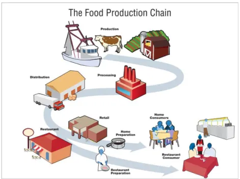
Figure 1 | Food Production Chain shows that food products contamination may happen at any
time since the moment they are produced until they are finally consumed (The Food Production
Chain, 2013)
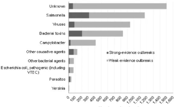
Figure 2 | Distribution of all foodborne outbreaks per causative agent in the EU, 2013. Adapted
from “The European Union summary report on trends and sources of zoonoses, zoonotic agents and
food-borne outbreaks in 2013”.
Figure 3 | Image of a typical bacteriophage. It can be seen a DNA molecule surrounded by a
protein coat (Microbial Food Webs, 2010)
Figure 4 | The general pattern of infection of a bacterial cell by a bacteriophage (Brown, 2010)
Chapter 2
Figure 1 | Representation scheme of pZE11G cloning vector used to clone each of unknown gene
sequences of Salmonella phage vB_SenS_Φ38
Figure 2 | Petri dish scheme used to analyse expression of phage proteins (Simões, 2015)
Chapter 3
Figure 1 | Weblogo multiple sequence alignment of consensus sequence
Figure 2 | Mauve results showing genome sequence comparison
Figure 3 | Phage genome sequence with all ORFs highlighted. The arrows represent the ORFs and
point in the direction of transcription
Figure 4 | Phage genome sequence with ORFs and without hypothetical proteins. The arrows
L
IST OF
T
ABLES
Chapter 1
Table 1 | Reported cases and notification rates per 100,000 of human salmonellosis in the
EU/EEA, 2009-2013. Adapted from “The European Union summary report on trends and sources of
zoonoses, zoonotic agents and food-borne outbreaks in 2013”
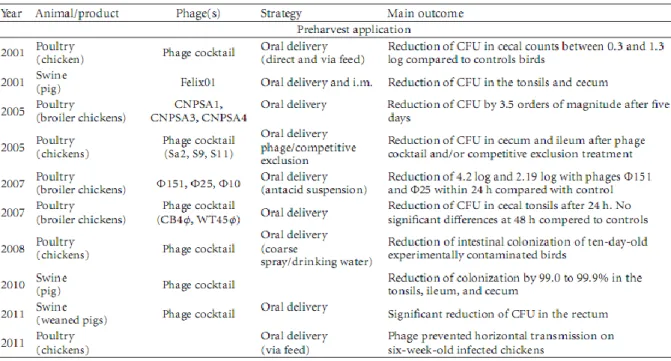
Table 2 | Preharvest
Salmonella phages application (S. M. Sillankorva, Oliveira, & Azeredo, 2012)
Table 3 | Postharvest
Salmonella phages application (S. M. Sillankorva et al., 2012)
Chapter 2
Table 1 | Microorganisms and vectors used in work development
Table 2 | Sequences of forward and reverse primers with restriction enzymes used. Where red
nucleotides are HindIII restriction sequence and purple nucleotides are BamHI restriction sequence
Table 3 | PCR components for amplification of each fragment. The volumes were calculated for 50
µl of final reaction volume
Table 4 | PCR Program Phusion conditions for amplification of each fragment using Phusion DNA
Polymerase
Table 5 | Colony PCR Components used
Table 6 | PCR Program Steps
Chapter 3
Table 1 | NCBI sequences of phages with higher homology with
Salmonella phage vB_SenS_Φ38
Table 2 | Alignment results of Emboss Stretcher with sequences with higher homology to
Salmonella phage vB_SenS_Φ38
Table 3 | Score values comparison between different phage sequences. 90-100% dark blue,
L
IST OF
A
BBREVIATIONS
HACCP - Hazard Analysis Critical Control Point
EU – European Union
EFSA - European Food Safety Authority
ECDC - European Centre for Disease Prevention and Control
EU/EEA - European Union/ European Economic Area
Spp. – species
DNA – Desoxyribonucleic Acid
RNA – Ribonucleic Acid
DEB - Department of Biological Engineering
LB - Lysogeny Broth
MCS - Multiple Cloning Site
GFP - Green Fluorescent Protein
IPTG - Isopropyl β-D-1-thiogalactopyranoside
ORF – Open Reading Frame
tRNA - transfer RNA
RNase – Ribonuclease
EDTA - Ethylenediaminetetraacetic acid
SDS - Sodium dodecyl sulfate
RT – Room Temperature
PCR – Polymerase Chain Reaction
T
mºC – Melting temperature
TAE - Tris-Acetate-EDTA
Amp – Ampicillin
C
HAPTER
I
1. F
OOD CONTAMINATIONS
Every day the quality and the food safety are put at risk and despite all the improvements that
have been made, such as the application of new technologies, good manufacturing practices,
guarantees of quality and hygiene. Food safety is continually put into question due changes in
lifestyle and requirements customers and also because the number of international exchanges
between different countries nowadays are bigger than ever (Rocourt, Moy, Vierk, & Schlundt, 2003).
World population is continually rising. It is therefore expected that food consumption,
particularly of ready-to-eat foods, will also rise. From such increment arises the need of creating
security measures that control food quality (Westrell, Ciampa, Boelaert, Helwigh, Korsgaard, Chríel,
Ammon, 2009).
Contaminations are still an issue and according to the current legislation no kind of food
should contain microorganisms or toxins that present a danger to public health (Westrell, Ciampa,
Boelaert, Helwigh, Korsgaard, Chríel, Ammon, 2009). It is important to note that food products
contamination may occur at any moment between production and consumption of food items
(Guntupalli et al., 2007) (see Food Production Chain, Figure 1). In order to avoid microbial growth in
a determined environment, good hygiene, biocides, and disinfectants should be used (Maukonen et
al., 2003). However, even applying these security measures bacteria continue to be found in food
(Holah, Taylor, Dawson, & Hall, 2002). Contamination problems may be further aggravated due to
cross contamination, i.e., when there is transference of pathogens from raw items to the surface of
others (this kind of contamination is responsible for about 40% of food related disease outbreaks)
(Oliveira, Oliveira, Teixeira, Azeredo, 2007; Teixeira, Silva, Araújo, Azeredo, & Oliveira, 2007).
Despite all the efforts made to fight these microorganisms, they possess defence mechanisms
which, in the end, allow them to surpass the measures put in place to prevent their growth
(Maukonen et al., 2003) in such a way it is still possible to find them in food and on food processing
surfaces (Holah et al., 2002).
Figure 1 | Food Production Chain shows that food products contamination may happen at any time since the moment
they are produced until they are finally consumed (The Food Production Chain, 2013)
Because of this daily increase of food contamination various security measures have been
adopted in order to ensure the necessary food security. These measures passed by the creation of
entities responsible for implementing effective programs of quality control, implementation of
HACCP programs and above all the use of safer methods at all stages of the Food Production Chain.
Even after all the implementation of these security measures, it is also the essential education of
food handlers and consumers (Havelaar et al., 2010; Seaman, 2010).
1.1 Foodborne Pathogens
Demand for food is increasing at a global level however the huge diversity of food has led to
an increase of the number of illnesses that are connected to the enormous variety of foodborne
pathogens that exist today.
According to the data obtained in recent years there are several foodborne pathogens
responsible for illnesses, hospitalizations and deaths. The best known pathogens are
Salmonella
spp.,
Listeria monocytogenes, Campylobacter spp., Bacillus cereus, Staphylococcus aureus and
Escherichia coli. This type of contamination can occur by different ways such as contaminated food
and water, infected peole, air, insects, pets and kitchen utensils (Teixeira et al., 2007).
1.1.1 Foodborne outbreaks in EU
In line with the European Union (EU) summary report on trends and sources of zoonoses,
zoonotic agents and foodborne outbreaks in 2013, a total number of 5,196 foodborne outbreaks
(including waterborne outbreaks) were reported in the EU. 43,183 human cases, 5,946
hospitalisations and 11 deaths were data obtained. 839 of outbreaks were connected to food
vehicles such as eggs and egg products followed by mixed food, and fish and fish products (The
European Union summary report on trends and sources of zoonoses, zoonotic agents and
food-borne outbreaks in 2013, 2015).
Figure 2 | Distribution of all foodborne outbreaks per causative agent in the EU, 2013. Adapted from “The European
Union summary report on trends and sources of zoonoses, zoonotic agents and food-borne outbreaks in 2013”.
Figure 2 shows that Salmonella caused the largest number of reported foodborne outbreaks
(22.5% of all outbreaks), followed by viruses (18.1%), bacterial toxins (16.1%), and
Campylobacter
(8.0%). The culpable agent was not known for 28.9 % of the outbreaks (Food et al. 2015).
1.2
Salmonella
Problems
Salmonella is one of the main foodborne pathogens worldwide, being responsible for disease
outbreaks, hospitalizations and deaths (Centers for Disease Control and Prevention, 2013). It is one
of the main virulent bacteria in domestic context, being this the reason why outbreaks of Salmonella
scare so much the general public and, above all, the consumers. Of the 3000 annual deaths due to
pathogens transmitted by food in USA, according to US Center for Disease and Control, Salmonella
is responsible for 38% of these deaths (Barbara et al., 2000). Symptoms of an infection by this
microorganism are mainly fever, diarrhea and abdominal cramps (Westrell et al., 2009).
Salmonella is primarily found in poultry meat, pork and eggs. Salmonella is easily
disseminated to hands, clothes and surfaces that are in contact with foods during preparation of
meals (Cogan et al., 1999). These pathogens are capable of surviving on food contact surfaces
during long periods of time (De Cesare et al., 2003; Redmond et al., 2004).
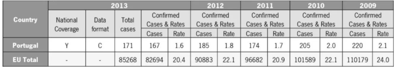
In 2013 it was reported a total number of 85,268 salmonellosis cases in EU. It shows a
decrease of 7.9% when compared with values of 2012. The lowest rates were reported by Portugal
and Greece and the highest notification rates in 2013 were reported by the Czech Republic and
Slovakia, on the other hand the highest proportion of travel-related cases were linked to the Nordic
countries, like Finland, Sweden and Norway (The European Union summary report on trends and
sources of zoonoses, zoonotic agents and food-borne outbreaks in 2013, 2015).
Table 1 | Reported cases and notification rates per 100,000 of human salmonellosis in the EU/EEA, 2009-2013.
Adapted from “The European Union summary report on trends and sources of zoonoses, zoonotic agents and food-borne outbreaks in 2013”.
*Y – yes. C – case-based data
1.2.1 Salmonella spp.
This organism is characterised to be Gram-negative, non-spore forming rod-shaped and
flagellated facultative anaerobic bacterium, that can be classified by its O, H, and Vi antigens. It is a
member of the family
Enterobacteriaceae (Hocking, 2003) and the genus is divided in two species:
S. enterica and S. bongori (Cianflone, 2008). The first is divided into six subspecies: enterica,
salamae, arizonae, diarizonae, houtenae and indica (Pitt & Simpson, 2006).
In terms of growth and survival characteristics, the growth and survival of Salmonella spp. is
affected by some factors like temperature, pH, water activity and preservatives (Podolak et al.,
2010).
Salmonella spp. presents a temperature range for growth of 5.1 – 46.2ºC and an optimal
temperature between 35 – 43ºC. Heat resistance of these organisms in food is dependent on the
composition, pH and water activity of the food. For pH they present a broad range where they can
grow and survive (3.8 – 9.5) nevertheless the optimum pH range for growth is 7 – 7.5. Water
activity (a
w) also influences Salmonella spp. growth. The optimum value for a
wis 0.99 and the lower
limit is 0.93. Presevatives have some impact in the growth of Salmonella spp., for example benzoic
acid, sorbic acid or propionic acid can inhibit its growth (Hocking, 2003; Podolak et al., 2010).
2. B
ACTERIOPHAGES
Bacteriophages (phages) are viruses that specifically infect bacteria and are harmless to
humans, animals and plants (Brown, 2010; Park & Nakai, 2003). They have been used for the
prevention and control of bacterial pathogens. They are among the simplest and most abundant
organisms on earth (it is estimated that there are 10 phages for each bacterial cell) (Park & Nakai,
2003).
Phage structure is very simple having mainly a DNA molecule (sometimes can also be an RNA
molecule) that transport a certain number of genes that are responsible for phage replication and
are protected by a protein capsid.
Figure 3 | Image of a typical bacteriophage. It can be seen a DNA molecule surrounded by a protein coat (Microbial
Food Webs, 2010)
2.1 Bacteriophages Discovery
Bacteriophage discovery is not exactly recent and all information that is currently known is the
result of several worldwide researches.
From Ernest Hanking observations in 1896 who noted the existence of high antibacterial
activity against
Vibrio cholerae and reported the presence of a substance responsible for the
decrease in cholera epidemics (Leverentz et al., 2001) to Frederick W. Twort’s experiments with the
varicella virus which led to a publication of an article in 1915 about the discovery of a virus capable
of lysing bacterial cells (Levine, 1991). The 'real' discovery of phages however commonly is
associated with d'Hérelle’s work during an outbreak of hemorrhagic dysentery in 1915. D'Hérelle
was the first to call this group of viruses bacteriophages (Leverentz et al., 2001). Then continuing his
research he was able to isolate other phages for bacteria which cause diseases such as cholera,
diphtheria, bubonic plague among others, and he was also responsible for developing the major
virus quantitation method that allows the analysis of the phage replication cycle (Levine, 1991).
Associated with this theme, in 1969, Max Delbrück, Alfred Hershey and Salvador Luria won
the Nobel Prize in Physiology and Medicine for their studies about phages, more specifically the
discovery of the replication of viruses and their genetic structure (The Nobel Prize in Physiology or
Medicine 1969, 2014).
2.2 Bacteriophages Classification
There are several types of phages, with some displaying greater specificity and others
displaying a wider host range. They are obligatory parasites and can only replicate in host cells by
controlling their cellular machinery. They are composed mainly of DNA - which contains genes that
encode proteins involved in the replication of the phage - surrounded by a protein capsid (Brown,
2010). Phages can consist of double-stranded or single-stranded DNA or RNA (Ackermann, 2011).
Morphologically, phages can be tailed, polyhedral, filamentous and pleomorphic and there are
17 recognized families. Tailed phages belong to the order of
Caudovirales and can be divided into
three families: Myoviridae, Siphoviridae, and Podoviridae that vary in the characteristics of their tails:
contractile, long and noncontractile, or short tails respectively. Tailed phages represent over 96% of
all isolated phages (Ackermann, 2011).
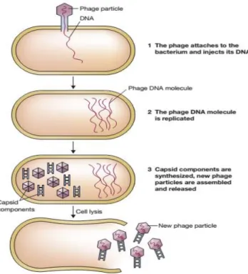
2.3 Bacteriophages Infection Cycles
The overall pattern of infection cycle of a phage is the same for all tailed phage and consists
in three steps: adsorption and penetration, viral eclipse and maturation phase, and extrusion (Figure
4). In the first step, phage particles connect to receptors located in the host cell surface and the
DNA is injected into the cell. Then, in the second phase, replication of phage DNA molecules occurs.
Finally, new phage particles are accumulated and freed from the host through the action of enzymes
holin and lysin (Brown, 2010).
Figure 4 | The general pattern of infection of a bacterial cell by a bacteriophage (Brown, 2010)
There are two types of cycles that phages can adopt: the lytic cycle and the lysogenic cycle.
The first is characterized by the insertion of the virus genetic material into the cell followed by
replication and simultaneous synthesis of the proteins that constitute the capsid. After this, the viral
particles are organized and cause cellular lysis, by which they are released (Brown, 2010). On the
other hand, the lysogenic cycle is based on the incorporation of viral genome into the cell genetic
material without cell death, thus being able of using intrinsic cell mitosis for its replication and
ensuring its propagation to future generations. When the phage is in this state it is called prophage.
By behaving in this manner, when phage proteins finally start to be translated (i.e. when the phage
switches from the lysogenic cycle to the lytic cycle), their effect on bacterial population will be cell
lysis (Brown, 2010).
2.4 Bacteriophages and their applications
The study of phage genomes has brought many advantages in the utilization of these viruses
for a large number of applications. Today, we live in a time of scientific revolution where increasingly
faster and more efficient molecular methods arise. Therefore it is not surprising that phage genome
sequencing has seen an increase in the last years (>1500 phage genomes have been already
sequenced) (Genomes Pages – Phage, 2014). New sequencing methods are being developed in
order to allow the control of pathogens and also study their impact on a global scale (WHO:
Antimicrobial resistance, 2013)
.
The scientific community has been aware of phages’ ability to kill antibiotic resistant bacteria
for a long time and this explains the main importance of studying these entities nowadays (WHO:
Antimicrobial resistance, 2013). However, in spite of these advantages, the use of phages at the
level of food safety is dependent on various regulatory agencies since there are only a few
phage-based products approved due to lack of scientific support through controlled clinical trials supervised
by the competent authorities (The use and mode of action of bacteriophages in food production,
2009). The existence of several genes in the genome of phages with unknown functions worsens the
situation since these genes may be virulence factors or toxin genes (Bru et al., 2004; Sillankorva et
al., 2011; S. Sillankorva et al., 2012). Furthermore, bacteria that survive a phage infection can
create new phenotypes with different receptors that phages do not recognize,
and in this way evade
the phage action and eventually survive (Pires et al., 2011). Despite the large genomic information
available, routine genetic manipulation of phages falls only in the engineering of vectors used for
phage display (Lu & Collins, 2009; Pande et al., 2010; Weber & Fussenegger, 2012).
Food products contamination may happen at any time since the moment they are produced
until they finally are consumed (Lewis, 2008). Treatment of foods with phages has seen an increase,
since they can help to prevent food deterioration as well as the spread of bacterial diseases. It is a
sound strategy in both animal and vegetal food maintenance. Phages are therefore good tools that
can be used for various purposes in the food industry such as treatment of infected animals, as
prophylactic agents to prevent animal illness, food biocontrol agents, and biosanitizers for food
contact surfaces.
2.4.1 Bacteriophage as biocontrol agents in food
It has been experimentally shown that phages are very effective in acting against growing
bacteria but they lose effectiveness in nongrowing bacteria (Snyder & Champness, 2007). In the
latter situation, control could be achieved by applying high concentrations of phages to fight
pathogens by “lysis form without” (it is possible to control infection by administration of a high
phage dose that will overwhelm the pathogen and lyse the bacterial cells) (Berchieri et al., 1991;
Goode et al., 2003).
The utilization of phages as biocontrol agents brings a number of advantages, such as high
specificity to a certain host determined by bacteria cell wall receptors which allows fighting a specific
microorganism while leaving other microbiota unharmed; self-replication and self-limiting, meaning
low dosages are the minimum necessary to affect big bacteria populations and they will continue as
long as there is a host present to allow virus reproduction; phages’ capacity of adapting in response
to defence mechanisms developed by bacteria; since they consist mostly in nucleic acids and
proteins the risk to have contamination is too low (low inherent toxicity); cheap and easy to isolate
and propagate; resistant to food processing and environmental stresses and long shelf life
(Sillankorva et al., 2012). Along the food chain it is possible to use phages to promote food
protection at three different phases that are as therapy, as biosanitizers of food contact surfaces and
as biopreservation agents (Sillankorva et al., 2012).
2.4.2 Bacteriophage as therapy agents
The fact that bacterial pathogens have the capability to evolve resistance to antibiotics has led
to their increasing ineffectiveness. Given this state of events, some small pharmaceutical companies
and several laboratories worldwide are starting to focus on the use of phages as a viable alternative
(Pirnay et al., 2011).
Nowadays, phage therapy is already used to promote food safety, but also used in animals
against the most common foodborne pathogens to prevent and treat experimentally induced
infections (Sillankorva et al., 2012). The capacity of isolating phages and finding phages for
antibiotic resistant bacteria makes them the perfect candidates to be used in therapy (Pirnay et al.,
2015) along with their specificity characteristics and lack of animal and plant toxicity. Furthermore,
their production is simple, fast and inexpensive (Azeredo, 2008).
The use of phages as therapy agents presents some advantages such as: phages can be
bactericidal, during the treatment phage can grow and raise their number, influence only a few
bacteria from the normal flora, effective against antibiotic-sensitive and antibiotic-resistant bacteria,
low inherent toxicities and are easily noticed. As disadvantages it is important to note that not all
phages are good for therapeutics, some phages possess a small host range, and phages with
one of the main weaknesses of phage therapy which receives apprehension of health authorities is
the potential emergence of phage-resistant mutant bacteria and since they are viruses, the public
opinion recognizes them as invaders by the immune system and create a response to be eliminated
from systemic circulation (Teng-hern & Kok-gan, 2014).
It is therefore important to develop tests to guarantee public safety. So, to modern phage
therapy evolve the main goal will be not to forget that before applying a product, validation and
licence of competent authorities is needed to ensure quality and safety in the process (Pirnay et al.,
2015).
2.4.3 Bacteriophage as biosanitizers of food contact surfaces and biopreservation
agents
Phages can be used on food surfaces to reduce bacterial colonization of foods such as meats,
seafood, milk, or processed foods (Atterbury et al., 2003; Hsu et al., 2002; Kennedy et al., 1986;
Suárez et. al, 2002). The importance of phage use in equipment surfaces is further inflated due to
pathogen´s capacity of adhering to inert surfaces and forming biofilms that allow them to persist on
foods and food contact surfaces (Lewis, 2008). Biofilms on equipment are a common problem in
food industry, especially in sites that are not easy to clean or sanitize. Phage use on biosanitation is
promising as they have been shown to significantly reduce viable microbial cells in biofilms under
ideal conditions, although it has to be noted that their specificity may limit their usefulness in this
kind of environments where there is great bacterial diversity (S. M. Sillankorva et al., 2012).
Phages have been reported to lyse hosts at temperatures as low as 1ºC (Greer, 1982, 1988),
which makes them excellent as food biopreservation agents since they can limit bacterial growth on
refrigerated foods (which is especially important when facing psychotropic bacteria) (Bigwood et
al.,2008).
2.5 Bacteriophage infection of
Salmonella
The use of bacteriophages when dealing with foodborne pathogens can be performed through
two main strategies: postharvest and preharvest. Postharvest strategy is defined to be the usage of
phages when animals are already dead in order to prevent the proliferation of pathogens. Preharvest
strategy on the other hand relies on the administration of phages to live animals as a way to both
avoid illness and reduce pathogen presence in the gastrointestinal tract, thus diminishing the
amount of pathogens that can later be present in food. Optimization of both strategies has been the
main focus of investigation regarding the use of phages against foodborne pathogens (Sillankorva et
al., 2012).
The majority of Salmonella outbreaks can be traced back to the Salmonella enterica serovars
Enteritidis and Typhimurium. Eggs and poultry are the main source of the first, while pork and beef
are the main source of the latter (EFSA, 2011). Under artificial laboratory conditions, both poultry
and swine animals experimentally infected with Salmonella phages saw a significant decrease of the
bacterium in major tissues, such as ileum and cecal tonsils. Despite this results, only studies on
poultry farms can truly determine the success of this postharvest strategy in decreasing the risk of
cross-contamination (Sillankorva et al., 2012).
The vast majority of recent in vivo experiments were carried out using cocktails of two to six
phages (Table 2). The gastrointestinal tract of poultry is characterized by different pH values in
distinct segments (2.5 in proventriculus/gizzard up to 8.0 in the colon). Particularly acidic conditions
have a significant negative impact on phage viability (Sillankorva et al., 2012). In order to overcome
this, Ma and colleagues (2008) have encapsulated phage Felix01 in chitosan-coated Ca-alginate
spheres and discovered that in in vitro studies this approach is capable of increasing phage viability
when exposed to lower pH. It should be noted, however, that
in vivo experiments are lacking in
regards to this encapsulated phages’ resilience in such conditions (Ma et al., 2008).
Regarding experiments involving postharvest strategies (Table 3), the use of a single phage as
oppose to a cocktail has been the preferred choice. As of 2012, all
Salmonella phages had been
able to decrease the number of live pathogens present on processed and ready-to-eat foods, raw
meats, and fresh produce (Guenther et al., 2012). Moreover, the combined use of phage and the
Salmonella antagonistic bacteria Enterobacter asburiae is a remarkably auspicious approach as a
way of decreasing the presence of
Salmonella on mung bean sprouts, tomatoes, and alfalfa seeds
without the need of chemicals (Guenther et al., 2012). In some cases, however, phages were found
to become immobilized by the food matrix which rendered them unable to diffuse and consequently
infect cells (Guenther et al., 2012).
Various studies performed shortly after the discovery of phages presented negative results
due to improper use of phages, namely the treatment of viral and unknown agent diseases. With the
increasing use of phages it becomes important not to repeat the same errors (Sillankorva et al.,
2012). It is essential to take note of several parameters (e.g. concentrations and timings of
application) upon administration of phages, since their effectiveness depends on the phage-host
systems. When studying phage effectiveness, it is also important to faithfully simulate the conditions
in which they are going to be used when in the industrial setting (Sillankorva et al., 2012).
The increase of phage-resistant phenotypes should also be always taken into account
although it is possible to overcome this obstacle by using other phages which target these resistant
phenotypes. Nevertheless, the full understanding of the resistance mechanisms obtained by the
hosts to the phages used and the percentage of disposal of the phages by the animal body are still
scarcely studied (Sillankorva et al., 2012).
3. O
BJECTIVES
The main objective of this work was to analyse the genome sequence of phage vB_SenS_Φ38 to
identify all unknown and non-essential genes. In order to do that, common bioinformatics tools
currently used in genome annotation and analysis were used.
This task was divided in four parts: the first one was dedicated to the annotation of the genome
through myRAST (automatic annotation software) and the identification of all genes. The second part
focuses on the use of other bioinformatic tools for annotating transmembrane domains, promoters,
terminators, tRNAs and also to perform comparative analysis of the phage vB_SenS_Φ38 genome
with those of similar phages existing in the genome database. The third part will be focused on the
expression of vB_SenS_Φ38 proteins in its host. Finally, in order to promote a more thorough
evaluation and discussion of the discovered features, all identified hypothetical proteins will be
analysed using HHpred; their effect on Salmonella growth will be determined; and proteins that are
shown to have no effect will be deleted from the genome through an in silico study performed with
Geneious.
C
HAPTER
II
1. B
ACTERIAL STRAINS AND PLASMIDS
In this study
E. coli TOP 10 was used to keep the newly done plasmidic constructions and
Salmonella enterica Serovar Enteritidis 821 was used to perform all the other assays. These two
strains, which belong to the Department of Biological Engineering (DEB) of University of Minho, were
grown on solid LB medium (400 ml of H
20, 10 g of LB and 6 g Agar) supplemented with 100 µg/ml
ampicillin, or in liquid LB medium. They were cryopreserved at -80ºC in glycerol 15% (v/v).
Table 2 | Microorganisms and vectors used in work development
Microorganisms
Vectors
E.coli
TOP 10
pZE11G
Salmonella
enterica
Serovar Enteritidis 821
pZE11G
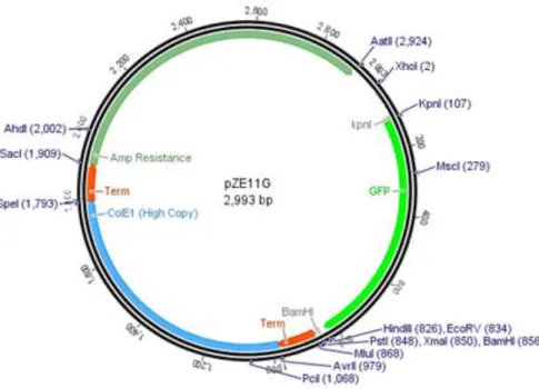
The studies of expression of proteins were carried out with pZE11G, a vector expressing the
green fluorescent protein (GFP) (Figure 1).
Figure 2 | Representation scheme of pZE11G cloning vector used to clone each of unknown gene sequences of
2. DNA
ISOLATION AND
G
ENOME SEQUENCING
To isolate genomic DNA from phage vB_SenS_Φ38, it was added 2.5 volumes of absolute
ethanol and sodium actetate 3M to 500 µl of purified phage, being the DNA precipitated, air-dried
and resuspended in MilliQ water (Sambrook, 2001) .Genome sequencing was performed on a 454
sequencing platform (pyrosequencing) (Plate-forme d'Analyses Genomiques at Laval University,
Quebec, Canada) to 50-fold coverage. Sequence data was assembled using SeqMan NGen4
software (DNASTAR, Madison, WI, USA). Protein pI and molecular mass were predicted using
Compute pI/Mw (Gasteiger et al., 2005).
3. P
RIMERS CONSTRUCTION FOR CLONING
Primers presented in Table 2 were designed for amplification of each gene fragment with
unknown function. Through OligoAnalizer 3.1 (OligoAnalyzer 3.1, 2014) it was possible to predict
T
mºC, GC content and the presence of secondary structures like primer dimers, hairpins, self-dimers,
hetero-dimers. Restriction enzymes are also described in Table 2 for each gene. HindIII/BamHI were
used to most of the fragments, except for vB_SenS_CEB2_0052 and vB_SenS_CEB2_0054
fragments, in which PstI/BamHI were used.
Table 2 | Sequences of forward and reverse primers with restriction enzymes used. Where red nucleotides are HindIII
restriction sequence and purple nucleotides are BamHI restriction sequence
Fragments Restriction Enzymes
Primer Fw Primer Rv
vB_SenS_CEB2 _0007
HindIII/BamHI 5' GATC AAGCTT GTG AAG CGG GCG TCA CAA - 3' (59ºC)
5' - GC GGATCC TTA AAC TCG TAT ATC TGT AAA CCT CAT ACG GCG - 3' (59ºC)
vB_SenS_CEB2 _0009
HindIII/BamHI 5' - GATC AAGCTT ATG GCA TTG CCA AGT AGT GAT G - 3' (56ºC)
5' - GC GGATCC CTA TTC CAG AAT ATT CCT TAT GTA CTC CTG - 3' (55ºC)
vB_SenS_CEB2 _0013
HindIII/BamHI 5' - GATC AAGCTT ATG GCA GAT AAT TAT GTA GTA CGG GA - 3' (55ºC)
5' - GC GGATCC TTA TTC CTC TAC ACT TTT ACG GCG - 3' (55ºC) vB_SenS_CEB2
_0016
HindIII/BamHI 5' - GATC AAGCTT ATG AGC ACC GCT TTT AGT AAA CG - 3' (56ºC)
5' - GC GGATCC TCA TTT TCT CAC CTG AAT AAA GTA TGC C - 3' (55ºC)
vB_SenS_CEB2 _0028
HindIII/BamHI 5' - GATC AAGCTT ATG GTT TCA TCC CCT CTT GTT G - 3' (55ºC)
5' - GC GGATCC TTA CGG AAA TAC CGT AGT AAT ATT GAT GAG - 3' (55ºC)
vB_SenS_CEB2 _0029
HindIII/BamHI 5' - GATC AAGCTT ATG ACT AAC AAA TAC AAT CGC ACA ATG - 3' (55ºC)
5' - GC GGATCC TCA CTC ATC AAT ATT ACT ACG GTA TTT CC - 3' (55ºC) vB_SenS_CEB2
_0030
HindIII/BamHI 5' - GATC AAGCTT ATG GTC ACT CGC AAA ATA ACA GAT - 3' (55ºC)
5' - GC GGATCC TCA TCC CAG CAT CTC CG - 3' (54ºC)
vB_SenS_CEB2 _0031
HindIII/BamHI 5' - GATC AAGCTT ATG AGC GTT TTT ATC GGT ATT ATC GC - 3' (56ºC)
5' - GC GGATCC TTA TTT ACC ATT AAC ATT AGA GTT ACA GC - 3' (55ºC) vB_SenS_CEB2
_0032
HindIII/BamHI 5' - GATC AAGCTT GTG GTT CCC GCC TGG CG - 3' (63ºC)
5' - GC GGATCC CTA CTT TTC CTG CAA TTG ACG CAA CTG TTC GTG - 3' (63ºC)
vB_SenS_CEB2 _0033
HindIII/BamHI 5' - GATC AAGCTT TTG TTT CAG TTT GCC CGT TG - 3' (54ºC)
5' - GC GGATCC TCA GCC ATC GTT TCT ACC C - 3' (54ºC)
vB_SenS_CEB2 _0035
HindIII/BamHI 5' - GATC AAGCTT GTG TGG CGA GTA GAC CTG - 3' (55ºC)
5' - GC GGATCC TCA TTG CAT TGG GTC CCA AAT T - 3' (55ºC)
vB_SenS_CEB2 _0037
HindIII/BamHI 5' - GATC AAGCTT ATG AAT ATT AAT GAC TAC ACC GGT CTG - 3' (55ºC)
5' - GC GGATCC CTA GCG CCA AAA TTC AAT CTC C - 3' (54ºC)
vB_SenS_CEB2 _0040
HindIII/BamHI 5' - GATC AAGCTT ATG AGT AGC ATC GAA AAA GCT ATA GAA G - 3' (55ºC)
5' - GC GGATCC TCA TTT AGC GCC CCT CTC - 3' (54ºC)
vB_SenS_CEB2 _0042
HindIII/BamHI 5' - GATC AAGCTT ATG AAA CAC GAA TAT GAC CGC AAG - 3' (56ºC)
5'- GC GGATCC TCA TTT CGC CAC CAG AAC C - 3' (56ºC)
vB_SenS_CEB2 _0045
HindIII/BamHI 5' - GATC AAGCTT ATG GGA ATC AAA CTT AAT CTT CGT AAA G - 3' (54ºC)
5' - GC GGATCC TTA ACG GTT ACG ACG GCG - 3' (55ºC)
vB_SenS_CEB2 _0046
HindIII/BamHI 5' - GATC AAGCTT ATG ACT TTA AAA GCT AAA GAT AGG AGC G - 3' (55ºC)
5' - GC GGATCC TCA AGC CTC TAA ATC GTC TTC AG - 3' (54ºC) vB_SenS_CEB2
_0047
HindIII/BamHI 5' - GATC AAGCTT ATG ATT GAC CAG GTG GGG GGG - 3' (62ºC)
5' - GC GGATCC TCA GGC GAG CGC CGC TTC - 3' (63ºC)
vB_SenS_CEB2 _0048
HindIII/BamHI 5' - GATC AAGCTT ATG ATG TTA GAA CAA TTT ATT AAA TTA TTT G - 3' (50ºC)
5' - GC GGATCC TTA TTC TTC TTC GAA ATA CTT GTT C - 3' (50ºC)
vB_SenS_CEB2 _0052
PstI/BamHI 5' - GATC CTGCAG ATG ACG CAG AAT GAA GTA GCT - 3' (54ºC)
5' - GC GGATCC TCA CTT CAC ATT CTC CCT AAT CC - 3' (54ºC) vB_SenS_CEB2
_0053
HindIII/BamHI 5' - GATC AAGCTT ATG AAA CCA AAT GAC CTC GTA ACC - 3' (56ºC)
5' - GC GGATCC CTA TTT TAC GAT TAA TTT ATC CTG GCA CAT CA - 3' (56ºC)
vB_SenS_CEB2 _0054
PstI/BamHI 5' - GATC CTGCAG ATG ACT AGC GTA CTA TTC ATC TGG G - 3' (56ºC)
5' - GC GGATCC TCA CTT CTC CTG TTT ACC ACC AA - 3' (56ºC) vB_SenS_CEB2
_0055
HindIII/BamHI 5' - GATC AAGCTT ATG AGT CTC GCA ACC GAT ATA C - 3' (54º)
5' - GC GGATCC TTA CCT GAA CGT ATA AAG TCG AAT AGA TG - 3' (55ºC) vB_SenS_CEB2
_0056
HindIII/BamHI 5' - GATC AAGCTT ATG ACT ACT ATA GCT TTT GAC GGT G - 3' (55ºC)
5' - GC GGATCC TTA TCC TAG TCG ATG TGC TAT CAC T - 3' (55ºC) vB_SenS_CEB2
_0059
HindIII/BamHI 5' - GATC AAGCTT ATG AAC TTA CAA AGC GAT AAA GTT TTT TAC C - 3' (55ºC)
5' - GC GGATCC TCA TTT CTT CTC TCC TGC ATA AGC - 3' (55ºC) vB_SenS_CEB2
_0060
HindIII/BamHI 5' - GATC AAGCTT ATG AAA GTA TAT ATC ATA TCT GGA TGG TAT TAC G - 3' (55ºC)
5' - GC GGATCC TCA TTC CTG CAC CTC CC - 3' (54ºC)
vB_SenS_CEB2 _0061
HindIII/BamHI 5' - GATC AAGCTT ATG AGC GAC AAC GGG CA - 3' (58ºC)
5' - GC GGATCC TCA TTT CTT CTT GCC CCA CAT TCG - 3' (58ºC) vB_SenS_CEB2
_0063
HindIII/BamHI 5' - GATC AAGCTT ATG TCA CTA GCG TAT CGC C - 3' (54ºC)
5' - GC GGATCC TTA GCA ATG CTC GTC TTT ATA AAC C - 3' (54ºC) vB_SenS_CEB2
_0064
HindIII/BamHI 5' - GATC AAGCTT ATG GGC ACT AAA TTT GAA GTA ATA G - 3' (52ºC)
5' - GC GGATCC TTA CAG TTC TAC AGA TGA TAA ATG GT - 3' (52ºC)
4. B
IOINFORMATICS
4.1 Genomic Properties
Phage vB_SenS_Φ38 genome was annotated using MyRAST (Aziz et al., 2008) and Glimmer
(Salzberg, Deicher, Kasif, & White, 1998). MEME (Bailey et al., 2009) helped to identify putative
promoter regions searching 150 bp upstream of each identified ORF followed by manual verification,
and was complemented with PHIRE (Lavigne et al., 2004) and the promoter sequence logos were
built with WebLogo (Crooks et al., 2004). ARAGORN (Laslett & Canback, 2004) and tRNAscan (Lowe
& Eddy, 1997) were used to predict tRNAs. ARNold (Gautheret & Lambert, 2001; Macke et al.,
2001) was used to find rho-independent terminators and the folding structure of putative terminators
and their secondary structures were calculated using Mfold (Zuker, 2003).
4.2 Compartive Genomics
BLASTN and BLASTX (Altschul et al., 1990; Programs available for the BLAST search, n.d.)
were used to compare phage vB_SenS_Φ38 sequence with similar phages sequences deposited in
database. Phage genome sequences resembling vB_SenS_Φ38 were Salmonella phage SETP7
(accession no. KF562865.1),
Salmonella phage vB_SenS-Ent1 (accession no. HE775250.1),
Salmonella phage SETP3 (accession no. EF177456.2) and Salmonella phage vB_SenS-Ent3
(accession no. HG934470.1). Using progressive MAUVE (Darling et al., 2004) and EMBOSS
Stretcher (Rice et al., 2015) it was possible to compare DNA homology between vB_SenS_Φ38
genome and other phage genomes. With CG View (Grant & Stothard, 2008) a graphical map was
built to compare all similar sequences against vB_SenS_Φ38 genome and ClustalW2 (Larkin et al.,
2007) was used to perform progressive alignments based on phylogenetic tree for phage DNA or
proteins.
4.3 Protein Analysis
Transmembrane domains were projected using TMHMM (TMHMM Server v. 2.0 -- Prediction
of transmembrane helices in proteins, n.d.) and Phobius (Käll et al., 2007). vB_SenS_Φ38 protein
sequences were queried against non-redundant protein databases using HHpred (Söding et al.,
2005). The sequences of predicted ORFs and potential alternative start codons were checked by
BLASTP (Altschul et al., 1990; Programs available for the BLAST search, n.d.).
4.4 Deletion of Hypothetical Proteins
Geneious is a bioinformatic tool that allows manipulation of genomic sequences. This tool
reads genomic maps, allows manipulation of repeated regions and is able to perform comparative
analysis of the phage genomes. In this study, it was used to deletion of each hypothetical protein
without antimicrobial properties and to comparison of the initial phage genome with the phage
genome obtained after each deletion (Geneious, 2015).
5. T
RANSFORMATION
5.1 Electrocomptent cells
To produce electrocompetent cells an inoculum of one colony from a fresh plate of the strain
E.coli TOP 10 was done in LB broth supplemented with 100 μg/ml of ampicillin and incubated at
37ºC, 120 rpm agitation (Environmental Shaker incubator ES- 20/60), overnight. Then, a dilution of
1:100 from the overnight culture was made in fresh LB medium and incubated at the same
conditions of temperature and rotation until the culture reached OD
600nm≈0.5. The culture was
transferred to 2 falcons of 50ml and kept on ice for 20 min. Then, a centrifugation step was made
(7000 x g, 10min, 4ºC). This step was repeated three times however the pellet was ressuspend in
different solutions and quantities and the supernatant was always discarded. The first pellet was
ressupended in 50 ml of cold sterile H
2O
d, the second pellet was resuspended in 8 ml of cold sterile
glycerol 10% solution and the last on in 1 ml of sterile glycerol 10% (v/v). Aliquots of 80 μl were
stored at -80ºC. Along this process, ice was always used to prevent thermal shock and to maintain
the efficiency of competent cells.
The procedure used to make S. Enteritidis 821 electrocompetent cells was the same as the
one adopted for E. coli TOP 10.
5.2 Electroporation
To E. coli TOP 10 cells, 2 μl of ligation (section 7.c below) was used
.
This step was performed
using Gene Pulser Xcell (Bio-Rad) with 1 mm cuvettes at 1800 V, 25 μF and 200 Ω. The ligation
was added to 80 μl of chemicompetent cells, mixed, and added to a cuvette. After an electric pulse
the cuvette was removed and 800 μl of LB was added. The final solution was passed to an
eppendorf and incubated at 37ºC with 120 rpm agitation for 1 hour for recovery and cellular growth.
Finally, the cells were plated in LB plates supplemented with ampicillin.
To Salmonella cells 1 μl of plasmid construction previously done was used. The rest of the
protocol is similar to E. coli TOP 10 cells.
6. DNA
PHAGE EXTRACTION
Before cloning, it was necessary to digest the vector and also the amplified fragments that
code for proteins. DNA phage Extraction Protocol was used.
Phage DNA extraction was carried out
with 500 µl of phage (1.2x10
12pfu/ml) according to Sambrook (Sambrook, 2013).
To analyse the
integrity of DNA, an agarose gel electrophoresis was performed (1% (w/v)) and concentration
measured with NanoDrop 1000™ (Thermo Scientific). Phage DNA stock was stored at -18 ˚C.
7. C
LONING
Phage DNA was used to amplify each gene fragment. Specific primers were used for each
fragment. All reagents used in the PCR are described in Table 3 and the conditions presented in
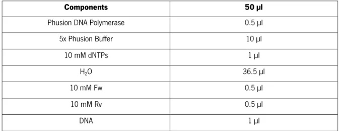
Table 4.
Table 3 | PCR components for amplification of each fragment. The volumes were calculated for 50 µl of final reaction
volume
Components
50 µl
Phusion DNA Polymerase
0.5 µl
5x Phusion Buffer
10 µl
10 mM dNTPs
1 µl
H
2O
36.5 µl
10 mM Fw
0.5 µl
10 mM Rv
0.5 µl
DNA
1 µl
Table 4 | PCR Program conditions for amplification of each fragment using Phusion DNA Polymerase
Step
Temperature
Time
Initial denaturation
98˚C
30 sec
Denaturation, primer annealing,
extension (34 cycles)
98˚C
10 sec
*T
mºC + 3ºC
15 sec
72˚C
30 sec
Final Extension
72˚C
5 min
4˚C
∞
*Annealing temperature was 3ºC higher than the melting temperature of the primers, since this is the recommended by the manufacturer (New England Biolabs).
An agarose gel was run, the DNA fragment was excised and each fragment was cleaned
through Zymoclean Gel DNA Recovery Kit (ZymoPURE™).
The vector pZE11G and the gene fragments were double digested with either
HindIII/BamHI
or
PstI/BamHI. 0.5 µl of each enzyme and 2 µl of Cut Smart buffer (10x) were used in a final
volume of 20 µl.
T4 DNA Ligase (New England Biolabs) was used to catalyse the ligation between the digested
pZE11G and each fragment that code for proteins of unknowns function described in Table 3.
Colony PCR was used to confirm correct insertion of the fragments in the vector. From a
transformation plate, colonies were selected, picked into 50 µl of sterile H
20. From this, 1 µl was
pipeted into a PCR tube and then PCR master mix components described in Table 5 were added.
Amplification was performed as described in Table 6.
Table 5 | Colony PCR Components used
Components
25 µl
Kapa Taq DNA Polymerase
0.1 µl
Buffer 10x
2.5 µl
10 mM dNTPs
0.5 µl
H
2O
18.9 µl
10 mM Fw
1 µl
10 mM Rv
1 µl
DNA
1 µl
Table 6 | PCR Program Steps
Step
Temperature
Time
Denaturation
95˚C
3 min
Denaturation, primer annealing,
extension (34 cycles)
95˚C
30 sec
*T
mºC – 5ºC
30 sec
72˚C
30 sec
Final Extension
72˚C
5 min
4˚C
∞
* Annealing temperature was 5°C lower than than the melting temperature of the primers, since this is the
The correct sizes of the amplified products was checked by electrophoresis in 1% or 2%
(w/v)
agarose gels depending on the size of the PCR product. SYBR Safe (Invitrogen) was used as DNA
stain for visualization of DNA and the agarose gel visualized in ChemicDoc XRS (BioRad).
After Colony PCR, each positive clone was grown overnight in 20 ml of LB supplemented with
ampicillin (100 μg/ml) at 37ºC, 120 rpm. Plasmid DNA was isolated with the Plasmid Miniprep Kit
(ZymoPURE™). In the end, to ensure integrity of DNA an electrophoresis was performed and the
concentration of plasmid measured in NanoDrop 1000™ (Thermo Scientific). 700 µl of the
overnight culture was added to 300 µl of glycerol, to a final concentration of 15% (v/v) of glycerol
and the sample stored at -80ºC.
S. Enteritidis 821 transformed cells were prepared according to section 5.2 and after
overnight growth on 20 ml LB supplemented with ampicillin (100 μg/ml), cultures were
cryopreserved at -80ºC (700 µl of culture and 300 µl of glycerol, to a final concentration of 15%
(v/v) of glycerol).
8. E
XPRESSION OF
P
HAGE PROTEINS
To evaluate the effect of each hypothetical protein on the growth of
S. Enteritidis 821, two
methodologies were used (solid and liquid media) and two controls were used: a negative control (S.
Enteritidis 821 cells with pZE11G) and a positive control (S. Enteritidis 821 cells with a pZE11G
expressing an endolysin).
The induction of protein expression was accomplished by adding Isopropyl
β-D-1-thiogalactopyranoside (IPTG). Expression of phage proteins was performed both in solid and liquid
media.
8.1 Solid Medium
Overnight cultures of the transformed
S. Enteritidis 821 cells carrying each hypothetical
protein were spotted (5 µl) on petri dishes with: LB, LB + Amp (100 μg/ml) and LB + Amp (100
μg/ml) + IPTG (2mM). In petri dishes with IPTG were used different dilutions of the strain (overnight
culture, dilution 1:100 and dilution 1:10000 (Figure 2)). After the spots were completely dried, they
were incubated at 37ºC overnight.
Figure 2 | Petri dish scheme used to analyse expression of phage proteins (Simões, 2015)
8.2 Liquid Medium
A dilution of an overnight culture of the transformed
S. Enteritidis 821 cells carrying each
hypothetical protein were introduced in three different medium: LB, LB + Amp (100 μg/ml) and LB
+ Amp (100 μg/ml) + IPTG (2mM). From each overnight inoculum 100 µl were pipetted and then
added to the different media already done (5 ml). The cultures were incubated at 37ºC (120 rpm)
and to determine if there was a decrease in cell mass, a sample (100 µl) was hourly taken for eight
hours and the optical density measured (OD
620nm).
C
HAPTER
III
1. G
ENOMIC PROPERTIES
vB_SenS_Φ38 has a linear dsDNA genome of 42 439 bp, with a GC content of 49.9%, which is
lower than 52% that was described for serovars of S.enterica (McClelland et al., 2001; Thomson et
al., 2008). The phage vB_SenS_Φ38 genome was run in myRAST and Glimmer was used to
annotate automatically the genes. Glimmer was used as an additional tool to search for potential
genes which myRAST could have missed. Crossing the results of both programs (Attachment I and
Attachment II), 60 ORFs were found. The putative proteins were blasted against InterPro
database
and 32 were found to correspond to proteins with known function and other 28 to hypothetical
proteins. Using Compute pI/Mw, the theoretical isoelectric point (pI) and molecular weight (Mw) of
the phage (Gasteiger et al., 2005) were determined and resulted in a pI of 4.22 and a Mw of
3522895.31.
Promoter presence was searched with three different programs: PHIRE, MEME and WebLogo.
The first one was used to find their possible localization.
In order to confirm PHIRE results, MEME
was used to complete this information, which was further manually verified. Candidate promoter
sequences were identified by searches of 150 bp sequences upstream of ORFs and 5 possible
promoters were identified. Then WebLogo was used to build the promoter consensus sequence
(Figure 1). Each logo consists in a stack of symbols, one letter for each position in the sequence and
the higher the height of the letter, higher will be its relative frequency (Crooks et al., 2004). The
promoter consensus sequence obtained was AxAxATAATxxxxxxCxxTTx.
Figure 3 | Weblogo multiple sequence alignment of consensus sequence
The search for tRNAs with ARAGORN and tRNA-scan showed that no tRNAs were found in the
genome sequence of phage vB_SenS_Φ38.
With the help of TMHMM and Phobius the presence of transmembrane domains was verified and
resulted in no transmembrane domain found for phage vB_SenS_Φ38.
At last, ARNold was used to find rho-independent terminators in nucleic acid sequences and
Mfold to complete the analysis of terminators presence. ARNold predicted that there were 28
transcription terminators and after this all the sequences with a score of free energy value less than
-9 were excluded. The remaining sequences were analysed using Mfold to assure that only
terminators with a loop were chosen. A cross-check of terminators with the genome sequence was
carried out to understand the terminators position in the sequence and only the better positioned
terminators were chosen. At the end of this analysis, only 6 terminators were found in the genome.
2. C
OMPARATIVE GENOMICS
The genome alignment was based on the BLASTN search results and the remaining
conclusions were confirmed by a progressive Mauve alignment. With BLASTN it was possible to find
the most similar phage genome sequences to the phage in study. Four genome sequences with
homologies ≥ 90% are apresented in Table 1.
Table 2 | NCBI sequences of phages with more homology with Salmonella phage vB_SenS_Φ38